Reading...
![]()
Play button
![]()
Play button
![]()
Use LEFT and RIGHT arrow keys to navigate between flashcards;
Use UP and DOWN arrow keys to flip the card;
H to show hint;
A reads text to speech;
26 Cards in this Set
- Front
- Back
|
How many mammary glands and ducts per gland do cows, ewes, does, and mares have?
|
Cow
Four mammary glands or quarters One duct system per quarter Ewe, doe Two glands One duct system per gland Mare Two complexes 2 (to 3) duct systems per complex |
|
|
What is a quarter when referring to a cow's reproductive system?
|
Refers to the four mammary glands
|
|
|
How much blood courses through the udder to produce 1L of milk?
How much does one udder weight? |
350-500L
60kg |
|
|
What are the parts of the suspensory apparatus of the udder? What are they composed from and where do they course?
|
Lateral laminae
Dense collagenous tissue. From the symphyseal tendon caudal to the udder to the surface of the udder. Medial laminae Separate right and left halves of the udder. Composed mostly of elastic tissue from tunica flava abdominis. Right and left medial laminae are separated by a small amount of loose connective tissue. |
|
|
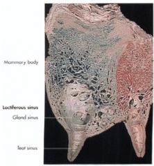
What does each bovine mammary gland consist of? How long are the teats?
|
Each gland or quarter consists of a body and a teat
Each teat is 7-8 cm in length |
|
|
Can you amputate just one quarter of the bovine udder?
|
It would be difficult. You can amputate either the cranial or caudal half as the blood supply is separate (see infused picture).
|
|

Where is milk produced in the teat? Where does the milk course through to reach the outside?
|
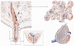
Milk is produced by the alveoli (12) in the gland
Milk courses through lactiferous ducts (13 & 14) to the lactiferous sinus composed of the gland sinus (9) and teat sinus (9’’) Milk leaves the teat through the teat canal (papillary duct, streak canal) (5’) and teat orifice (5’’) |
|
|
What is a milk knot?
|
Palpable lactiferous ducts filled with milk
|
|
|
What do you call the mucosal ridges at the proximal end of the teat canal between the teat sinus and teat canal?
What is the clinical siginifigance? |
Fürstenberg’s rosette
This area can become proliferative and need to be cut back or dilated. |
|
|
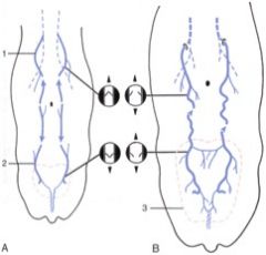
What is the primary blood supply to the bovine udder?
|
The external pudendal a.
Gives off the cranial and caudal mammary aa. After the cranial mammary dives down to the udder, it becomes the caudal superficial epigastric a. |
|
|
What are the three venous drainages of the bovine udder?
|
External pudendal v.
Cranial mammary v. to subcutaneous abdominal “milk” v. Caudal mammary v. to dorsal labial v. to internal pudendal v. |
|
|
What happens to venous return in a heifer's first pregnancy?
|
The blood draining the udders to the caudal superficial epigastric v. to cranial superficial epigastric to the internal thoracic v. overwhelms the venous valves and basically just flows freely to the internal thoracic v. This creates what is known as a milk vein.
|
|
|
When a cow lays down, she occludes some of the venous returns from the udder. How can she do this without causing blood to back up into the udder?
|
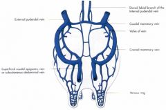
The other drainage sites: external pudendal v, cranial mammary v., caudal mammary v.
|
|
|
How any areas of sensation to bovine udders have?
What are they? |

3
Ventral brs. of L1 and L2 spinal nn. (a. & b.) Cranial parts of the forequarters Genitofemoral n. (c.) Middle section of the udder Pudendal n. (mammary brs.) (f.) Caudal aspect of the hind quarters |
|
|
What is the nerve supplying sensory information from the teat wall and gland of the bovine udder?
|
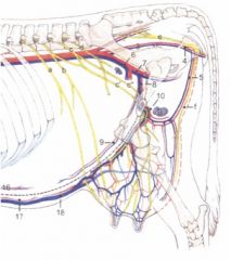
Genitofemoral n. (c.)
Afferent fibers – sensory Sympathetic fibers to: Smooth muscle of the teats and blood vessels Myoepithelial cells of the gland tissue |
|
|
Typically, who has a larger udder - goats or sheep?
|
Goats
|
|
|
What happens when a cow or small ruminant have extra teats?
|
They get removed because of the milking machine not being set up for that.
|
|
|
How many duct systems do equine udders have? Are the glandular systems from the same system?
|
Each complex has 2 (or 3) glands and completely separate duct systems
External groove separates right and left halves |
|
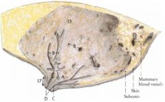
Name A-D on this equine udder
|

Lactiferous ducts (B)
Lactiferous sinus (A) -Gland sinus -Teat sinus Papillary duct (teat canal, streak canal) (C) Teat orifice (D) |
|
|
What are the cutaneous innervations of the equine udder?
|
Ventral brs. of L2-L4 spinal nn.
Pudendal n. (S2-S4) |
|
|
What supplies the innervation of the mammary gland of the equine udder?
|
Genitofemoral n. (L3-L4)
|
|
|
Where does lymph drain from the mare udders? Where does it drain to?
|
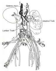
Lymph from the mammary glands drains to the superficial inguinal (mammary) lnn.
Efferent vessels from the superficial inguinal lnn. course through the inguinal canal to the deep inguinal lnn. (6) Lymph flows to the medial iliac lnn. (2) to the lumbar trunk to the cisterna chyli |
|
|
How many pairs of mammary glands do sows have? How many teat orifices per gland?
|
7 pairs of mammary glands
2 teat orifices and duct systems per teat |
|
|
What are the blood supplies of the mammary glands of the sow? What are her lymph drainages?
|
Blood supply – external thoracic and cranial and caudal superficial epigastric arteries and satellite veins
Lymph drainage – superficial cervical and sternal lymph nodes for first two pairs of glands and via the superficial inguinal lymph nodes for the five caudal pairs |
|
|
How many pairs of mammary glands do camelids have? How many teat orifices per gland?
|
Four teats – each teat composed of double non-communicating glands
|
|
|
Where are the mammary lymph nodes? Where do the efferents course to?
|
Superficial inguinal (mammary) lnn.
Usually 2 on each side - 1 palpable and 1 non-palpable Efferent vessels course through the inguinal canal to the deep inguinal ln. Lymph flows to the medial iliac lnn. to lumbar trunk to cisterna chyli |