Reading...
![]()
Play button
![]()
Play button
![]()
Use LEFT and RIGHT arrow keys to navigate between flashcards;
Use UP and DOWN arrow keys to flip the card;
H to show hint;
A reads text to speech;
22 Cards in this Set
- Front
- Back
|
What values can you get a direct measurement of on a Peripheral Blood (PB) smear? Indirect measurements?
|
Direct= RBC, MCV, and Hb (can directly obtain measurement from smear).
Indirect= Hematocrit, MCHb, and MCHConc., measured by plugging into formula (less accurate). |
|
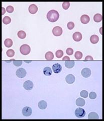
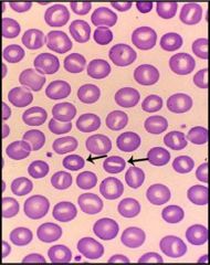
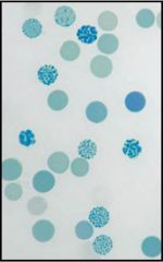
What is the central dot in the cells shown on the bottom (stained with methylene blue)?
What are these cells an indicator of? |
Reticulocyte RNA (that continues to synthesize Hb). No nucleus.
They estimate RBC production of Bone marrow. If Retic count is low following blood loss for instance --> concern about BM problem. |
|
|
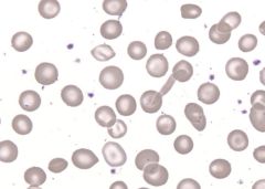
What is anisocytosis? What clinical measurement indicates this?
|
Variation in the size of RBCs
indicated by ↑ RDW (Red cell distribution width) |
|
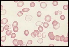
This condition is seen in Iron deficiency anemia when a person is being treated with Fe supplementation. It can also be seen in post transfusion syndrome.
|
Dimorphism = 2 distinct RBC populations are present (one is normal the other is hypochromic)
*note this is anisochromasia (variable degree of staining of RBCs) |
|
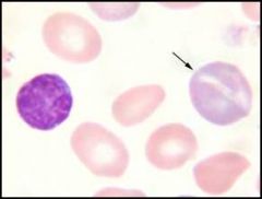
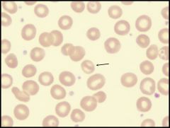
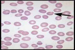
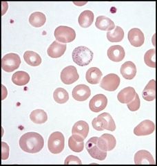
What type of cell is the arrow pointing to? What makes the cell this color?
Exposure to what can cause increased number of these cells? If you don't see the production of these cells in a patient with Sickle cell, what do you suspect? |
Reticulocyte, it is polychrompatic because Hb is eosinophilic (red) and RNA is basophilic (blue).
Hypoxia exposure --> ↑ retics. ↓ production of retics suggests Parvovirus B19 invasion of RBCs --> aplastic crisis |
|
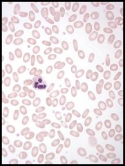
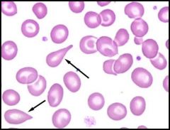
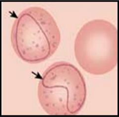
What is poikilocyte?
What type of cell is seen above? |
Poikilocyte = RBC of abnormal shape
Above= elliptocyte (also gets confused with Ovalocyte) |
|
What cell is seen above and what does it suggest?
|
Tear drop cell (Dacrocyte), suggests some event in bone marrow or spleen that encourages squeezing of RBC (infection- TB, hypersplenia, fibrosis, etc.)
|
|
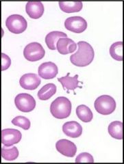
What type of cell is seen here? What does it NOT have the propensity to do?
|
Acanthocyte- spiculated cell, has throny projections
Cannot form rouleaux |
|
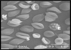
What type of cells are seen here?
|
Echinocyte (sea urchin cell, tiny evenly spaced spicules)
Burr cell "crenated cell" (uneven distribution of spicules) |
|
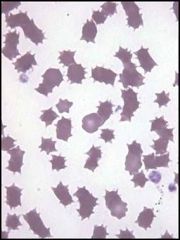
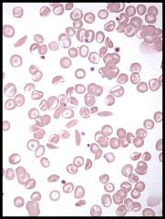
What types of cells are seen here? What major conditions are associated with such cells?
What are the precursors to these cells? |
Keratocytes (horn cells- from mechanical damage to RBCs)
Schistocytes (fragmented RBCs, from damage) - DIC -Microangiopathic hemolytic anemia - TTP Blister cell = Pre-Keratocyte (membrane can still be seen). |
|
What condition are the following cells seen in? Why do the cells get this shape?
|
Bite cells (Degmacytes)- because Heinz bodies form (abnormal precipitated Hb that protrides from cell. Macrophages bite it causing defective membrane).
|
|
What type of cells are shown here?
|
Stomatocytes
Uniconcave RBCs with slit-like central pallor. |
|
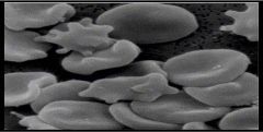
What cells are shown here? What is the viscosity of this individual's blood?
|
Sickle cell (Drepanocytes). Curved, irregular cells get stuck. Due to abnormal polymerization of Hb within cell.
High viscosity of blood. |
|
What type of cell is this and what broad category of anemia is this seen in?
|
Pinch Bottle Cells (Knizocytes)- seen in Hemolytic Anemia, including hereditary spherocytosis.
|
|
What is the shape of this cell? What condition is it associated with?
|
Mushroom shape (called a Pincer Cell)
Due to Oxidant induced hemolysis |
|
What is seen in this smear? What are two causes of this condition?
These two causes also form a loop like inclusion in RBCs called _____. |
Basophilic Stippling (either fine punctate granules or coarse granules, RNA ribosomes)
Indicates impaired erythropoiesis. Usually seen in Lead poisoning and Thalassemias. Cabot Ring (also seen in these conditions) |
|
What is this inclusion body called and what does it consist of? Where do you see it?
|
Pappenheimer Bodies = secondary lysosomes with ferritin (larger granules that sit in one area of RBC)
Sideroblastic anemias, or post-spelenctomy state |
|
What type of Hemoglobinopathy is shown here?
|
Hemoglobin C crystal (crystal precipitates of Hb C can be seen)
|
|
What type of Hemoglobinopathy is shown here (stained with supravital stain)?
|
Hemoglobin H (small aggregates of Beta Hb B4 tetramers seen).
|
|
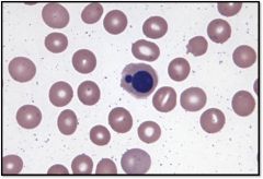
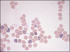
Dyserythropoiesis as shown here might suggest what condition in which abberant RBCs are produced by the bone marrow?
|
Myelodysplastic syndrome (sick bone marrow)
*this was a picture of abnormal nuclei budding. Normally nucleated blood cells might be seen after acute bleeding in which cell is trying to compensate. |
|
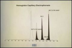
2 month old boy with some target cells, odd elongated cells, and cells with dense areas of Hb on PB smear had a gel electrophoresis. What condiiton does he have?
|
Hemoglobin SC disease
|
|
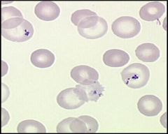
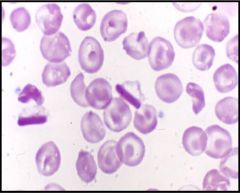
8 year old boy from Rhode Island presents with fever and thrombocytopenia. What organism might you see?
What if it was a 40 year old man from India with chronic cyclic fevers? |
Babesiosis
Malaria parasite (both affect the RBCs, the context is key). |