![]()
![]()
![]()
Use LEFT and RIGHT arrow keys to navigate between flashcards;
Use UP and DOWN arrow keys to flip the card;
H to show hint;
A reads text to speech;
63 Cards in this Set
- Front
- Back
- 3rd side (hint)
|
Periodontium tissues? |
Gingiva, cementum, PDL, alveolabone |
|
|
|
Provide a tissue seal around the cervical portion(neck) of tooth, cover alveolar process of jaw, hold tissue against tooth during mastication |
Gingiva |
|
|
|
Suspend & maintain tooth in socket |
PDL |
|
|
|
-Anchors end of PDL fibers to tooth so tooth stay in socket -protect dentin of root |
Cementum |
|
|
|
Surround & support tooth root |
Alveolar bone |
|
|
|
Protect underlying supporting structures of periodontium from oral environment |
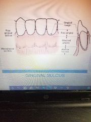
Gingiva
4sections - free gingiva -attached gingiva - gingival sulcus - interdental papilla |
|
|
|
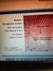
Free gingiva/Marginal gingiva |
Located coronal to CEJ Encircle tooth Attach to too by JE Can be separated from tooth w/periodontal instrument Forms soft tissue lateral wall of gingival sulcus |
|
|
|
Attached gingiva |
-Continuous with free gingiva -Tightly bound to underlying cementum on cervical 3rd of root & periosteum of alveolar bone -Extend apically from free gingival groove to mucogingival jux (MJG) |
|
|
|
Range of attach gingiva |
Max Incisior = 3.5 - 4.5mm Mand incisor = 3.3 - 3.9mm Max PM = 1.9mm Mand PM = 1.8mm |
|
|
|
Gingival sulcus |
Bordered by tooth surface & epithelial lining of free gingiva in health. - if bleeding, it's 'pocket' not sulcus! Health: 1-3mm Measure free gin margin to epithelial attachment |
|
|
|
Free + Attached = Keratinised tissue (KT) |
|
|
|
|
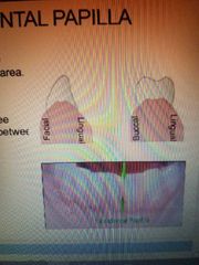
Interdental papilla |
-Interproximal space frm alveolar crest - contact area -Stippled extension of free gingiva that fills space btw 2 teeth
2 papillae 1 facial + 1 lingual |
|
|
|
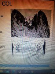
Col |
-saddle like depression btw facial&lingual papillae -locared directly apical to contact area -non-keratinized tissue ( very susceptible to disease*) |
|
|
|
Histological Gingiva components |
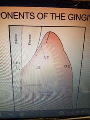
Oral Epithelium Sulcular Epithelium Junctional Epithelium |
|
|
|
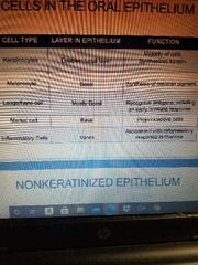
Cells in oral epithelium |
1.Keratinocyte- deepest basal layer(majority cell, synthesize keratin) 2.Melanocyte- basal(멜라닌색소합성) 3.Langerhans- mostly basal(recognize antigens, initiating early immune response) 4.Merkel- basal(proprioceptive cell) 5.Inflammatory- varies (associated w/inflammatory response in mucosa) |
|
|
|
Non-keratinized epithelium |
Associated w/lining of oral cavity Stratum corneum&Stratum granulosum layers are absent Surface cells have nuclei |
|
|
|
Oral epithelium |
-Covers outer surface of free gingiva/attached gingiva -Extend from crest of GM to MGJ -Keratinized / parakeratinized 10-12 day cell turnover time Barrier btw oral environment &deeper tissue |
|
|
|
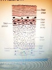
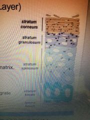
Cellular structure of oral epithelium |
Stratum Corneum Stratum Granulosum Stratum Spinosum Stratum Basale |
|
|
|
Stratum corenum( keratinized layer) |
outermost, flat devoid of nuclei keratin filament surround by matrix continuously being sloughed & replaced by epithelial cell that migrate frm underlying layer |
|
|
|
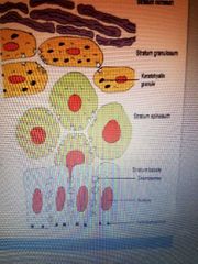
Stratum granulosum |
Cells are flat Found in layers about 3-5 cells thick Prominent in keratinized epithelium Absent in non-keratinized epithelium |
|
|
|
Stratum Spinosum (prickle cell layer) |
Cells are polyhedral & large Cells of basal & prickle cell layers attach to each other w/desmosomes Stratum basale&1st layer of s.spinosum are referred to as stratum germinativum |
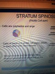
Facet= 다이몬드컷처럼 flat side |
|
|
Stratum basale |
Single later of cuboidal cells Round / ovoid nucleus Located @jux of epithelium & lamina proproa Made up of 2 type cells 1) serrated & specialized for attachment to basement membrane (hemidesmosomes) 2)non-serrated & specialized for dividing / multiplication. |
|
|
|
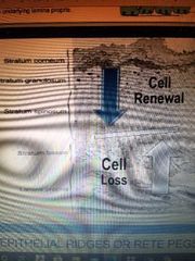
Process of keratinzation |
4. Stratum Corenum : most superficial layer where nuclei degenerate/ at this stage, cells are lost into mouth 3. Stratum Granulosum : where cells flatten 2. Stratum Spinosum : cells appear as "prickle" under microscope 1. Stratum Basale : new cells are formed to replace ones that have been shed / next to underlying lamina propria |
|
|
|
Cells loss increase = cell renewal decrease |
|
|
|
|
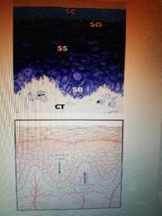
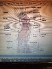
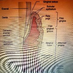
Epithelial ridges / rete pegs |
In health, oral epithelium joins with connective tissue in a WAVY INTERFACE w/epithelial ridges - deep extensions of epithelium that reach down into connective tissue - this epithelial layer show projection into underlying connective tissue know as rete pegs |
|
|
SE GS. OE. JE. ERidge enamel space |
구조 |
|
|
|
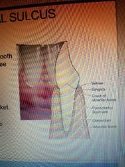
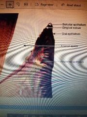
Sulcular epithelium |
-elithelial lining of gingival sulcus -extend frm crest of ging.margin to coronal edge of JE -non-keratinized stratified squamous epithelium -in health, sulcular epithelium joins CT @ smooth interface w/no epithelial ridges |
|
|
|
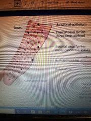
Junctional Epithelium |
-band of epithelial cell(이빨주위) -forms base of sulcus -attach gingiva to tooth -nonkeratinized stratified squamous epithelium -has no rete pegs in health but MAY have rete pegs in disease |

|
|
|
|
|
|
|
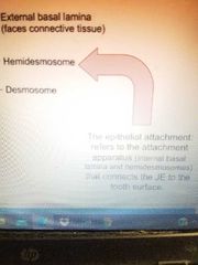
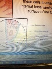
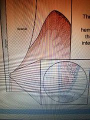
Attachment apparatus(internal basal lamina & hemidesmosomes) connect JE to tooth surface |
Hemidesmosome |
|
|
|
JE cell next tooth surface frm hemidesmosome that enable these cells to attach to internal basal lamina & tooth surface |
|
|
|
|
|
|
|
|
GCF |
Sulcular / gingival fluid Seep into sulcus frm CT thru sulcular wall LIittle / no fluid is found in healthy gingival sulcus Fluid flow increase in plaque biofilm & resulting in gingival inflammation |
특징 Cleansing material for sulcus Contain plasma protein to help JE adhere to tooth Antimicrobial properties Help activate antibody defense |
|
|
gingiva CT a.k.a Lamina Propria |
Hold marginal ging firmly against tooth Provide ridgidness needed to withstand forces of mastication Connect free gingiva to root cementum & adjacent attached gingiva |
|
|
|
Dentogingival unit |
JE & ging.Fiber Provide structural support to gin.tissue Combined length of dentogingival unit is supracrestal tissue attachment (biologic width) |
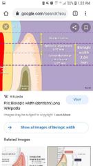
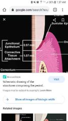
Biologic width = CT+ JE 2-3mm |
|
|
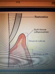
Supracrestal tissue attachment |
JE & gin.fiber attachment to collectively known as supracrestal tissue attachment (biological width) 2-3mm |
|
|
|
크라운같이 restoration 잇어 소프트티슈가 부엇으면 부은곳빼고 재야 biological width |
|
|
|
Large tissue cell release inflammatory substances when damaged. Also contain histamine (promote inflm reaction) |
Mast cell |
|
|
|
Derived from WBC, usually in lymph node(important in developing immunity) ? |
Plasma cell |
|
|
|
Found in CT, synthesize collagen? |
Fibroblast |
|
|
|
Large tissue cell, remove damaged tissue, cell, bacteria thru phagocytosis? |
Macrophage |
|
|
|
Identified by their location & capacity to differentiate into other cell types like smooth muscle cell in formation of new arteries, phagocyte in inflammatory process and bone cell in formation of new bone? |
Undifferentiated Masenchymal cell |
|
|
|
3 arteries supply blood to CT |
-supraperiosteal arteries: Facial&Lingual/ Interdental / PDL |
|
|
|
콜라겐 fiber 덩어리 connect tooth(cementum) to bone? 이빨뿌리주위 티슈? Primary attachment of tooth to alveolar bone? NO relationship w/Gingiva? |
PDL |
|
|
|
Resist force of luxation & tipping From crest of interraducular septum extending to cementum in furcation? |
Interraducular fiber |
|
|
|
Resist lateral force & tipping teeth? Originate from apex cementum spread apically & laterally to bone? |
Apical fiber |
|
|
|
Oblique fiber |
Absorb occlusal forces, Vertical pressure Located in middle 3rd root apical to horizontal fiber. Originate from cementum runs coronally & diagonally to bone |
|
|
|
Horizontal fiber |
Resist horizontal pressure against crown Originate from cementum & runs@ right angle, insert to bone |
|
|
|
Alveolar crest fiber |
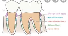
Resist horizontal force & keep tooth in socket Originate from cementum runs apically to alveolar crest |
|
|
|
Sharpey's fibers |
Terminal end of principle fiber Embedded in cementum on one end & into bone on the other end |
|
|
|
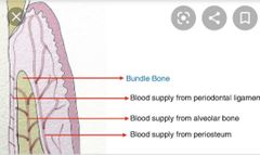
Blood supply to PDL |
come from apical vessel & vessel in interproximal bone Nerve supply is from Trigeminal pathways. |
|
|
|
PDL 기능 |
Supportive (suspend & maintain tooth in socket) Formative (build & maintain cementum & alveolar bone) Nutritive (nourish cementum&bone) Remodeling (remodel alveolar bone in response to pressure) Sensory (proprioception)- transmit tactile pressure & pain perception |
|
|
|
Calcified tissue that cover root // Provide a mean of attachment for PDL fibers |
Cementum |
|
|
|
Cementum |
Primary = Acellular Locate on coronal 2/3 of root about 1mm thick Responsible for attaching tooth to alveolar bone Secondary = Cellular Cover apical 1/3 root about 5mm thick Can be laid down on top of acellular cementum |
|
|
|
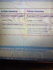
Acellular cementum vs cellular cementum |
Acellular = form before teeth are in occlusion, devoid cells, cover cervical 2/3 root, tooth support Cellular = form after teeth have reached occlusion, contain cementocyte, cover apical 1/3 root, compensate for active eruption & normal tooth wear by continuous deposition |
|
|
|
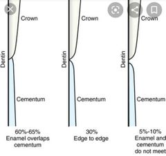
Cementum / Enamel relationship |
Overlap (60-65%), Butt (30%), Exposed(5-10%) |
|
|
|
Bone where sharpey's fibers terminate? |
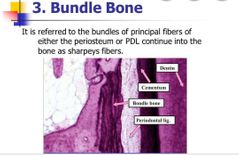
Bundle bone (Found # inner socket surface) |
|
|
|
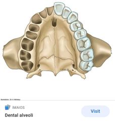
Lining of tooth socket ( space where tooth sit)? |
Alveoli |
|
|
|
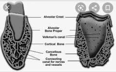
Cribriform plate Alveolar bone Proper Lamina Dura |
Perforated by numerous opening ~carry blood /nerve from bone to PDL~ |
|
|
|
Cancellous/ Trabeculae bone |
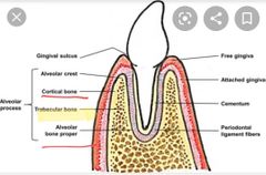
Spongy bone btw cortical plate & alveolar bone proper |
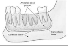
바깥 cortical plate 중간 cancellous(trabeculae) 안쪽 alv.proper |
|
|
Compact bone on facial lingual alveolar process ? |
Cortical plate |
|
|
|
Fenestration |
Marginal bone is still intact 잇몸창문..잇몸에구멍 Still cover w/periosteum & overlying gingiva Where roots are prominent & overlying bone is thin |
|
|
|
Dehiscence |
Marginal bone btw fenestration & alveolar crest may disappear altogether & produce defect 봉합터짐 |
|