Reading...
![]()
Play button
![]()
Play button
![]()
Use LEFT and RIGHT arrow keys to navigate between flashcards;
Use UP and DOWN arrow keys to flip the card;
H to show hint;
A reads text to speech;
142 Cards in this Set
- Front
- Back
|
Sensory Input
|
Senses going from the outside to the central nervous system
|
|
|
Integration
|
interpretation of sensory input
|
|
|
Motor Output
|
response to stimuli by activating effector organs
|
|
|
Peripheral Nervous System (PNS)
|
Sensory (afferent) division
Motor (efferent) division |
|
|
Sensory Divison
|
somatic sensory fibers
visceral fibers |
|
|
Motor Division
|
somatic nervous system
autonomic nervous system |
|
|
Somatic Sensory Fibers
|
carry impulses from skin, skeletal muscles, and joints to the brain
|
|
|
Visceral Fibers
|
transmit impulses from visceral organs tot he brain
|
|
|
Somatic Nervous System
|
conscious control of the skeletal muscles
|
|
|
Autonomic Nervous System
|
regulates smooth muscle, cardiac muscle, and glands
|
|
|
Neurons (Nerve Cells)
|
Structural units of the nervous system
Composed of a body, axon, and dendrites |
|
|
Neuron Cell Body
|
contains the nucleus
|
|
|
Axon
|
generates and transmits action potentials and secretes neurotransmitters from its terminals; its fibers from the nerve
|
|
|
Dendrites
|
the receptive or input regions of the neuron
|
|
|
Myelin Sheath
|
Whitish segmented sheath around most long axons
It functions in: protection of the axon increasing the speed of nerve impulse transmission |
|
|
Depolarization
|
the inside of the membrane becomes less negative
|
|
|
Repolarization
|
the membrane returns to its resting membrane potential
|
|
|
Hyperpolarization
|
the inside of the membrane becomes more negative than the resting potential
|
|
|
Synapses
|
A Junction that mediates information transfer from one neuron to another neuron, and effector cell
|
|
|
Presynaptic Neuron
|
Conducts impulses toward the synapse
|
|
|
Postsynaptic Neuron
|
transmits impulses away from the synapse
|
|
|
Neurotransmitters
|
acetylcholine, biogenic amines(epinephrine), amino acids, peptides(endorphins)
classifications: excitatory inhibitory some have both effects |
|
|
Excitatory
|
causes depolarization
|
|
|
Inhibitory
|
causes hyperpolarization
|
|
|
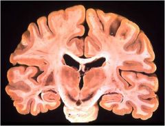
Central Nervous System
|
brain and spinal cord
they contain white matter and gray matter |
|
|
White Matter
|
dense collections of axon fibers
|
|
|
Gray Matter
|
Mostly soma and unmyelinated fibers
|
|
|
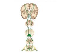
The Brain
|
cerebral hemispheres
cerebellum brain stem |
|
|
Ventricles of the Brain
|
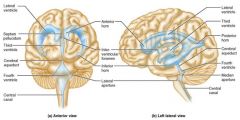
paired C-shaped lateral ventricles
third ventricle fourth ventricle |
|
|
Cerebral Hemispheres
|
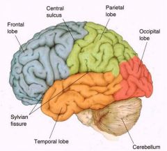
make up most of its mass
contain deep grooves which divide the hemispheres into five lobes frontal, parietal, temporal, occipital, insula |
|
|
Cerebral Cortex
|
superficial gray matter
each hemisphere acts contralaterally (controls opposite side of the body) hemispheres are not equal in function |
|
|
Primary Motor Cortex
|
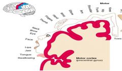
located in the precentral gyrus
allows conscious control of precise, skilled, voluntary movements |
|
|
Primary Somatosensory Cortex
|
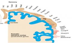
located in the postcentral gyrus
receives information from the skin and skeletal muscles |
|
|
Primary Visual Cortex
|

located on the extreme posterior tip of the occipital lobe
receives visual information from the retinas |
|
|
Primary Auditory Cortex
|
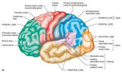
located at the superior margin of the temporal lobe
receives information related to pitch, rhythm, and loudness |
|
|
Prefrontal Cortex
|

located in the frontal lobe
involved with intellect, cognition, recall, and personality necessary fro judgment, reasoning, persistence, and conscience |
|
|
Cerebral White Matter
|
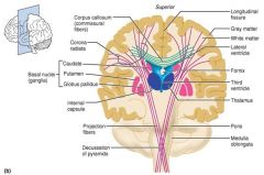
responsible for communication between the cerebral cortex and lower CNS center
corpus callosum - connects hemispheres |
|
|
Basal Nuclei
|
masses of gray matter found deep within the white matter
|
|
|
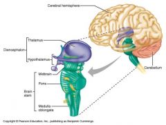
Diencephalon
|

central core of the forebrain consisting of three structures - epithalamus, thalamus, hypothalamus
|
|
|
Epithalamus
|
pineal gland - makes melatonin
choroid plexus - cerebrospinal fluid |
|
|
Thalamus
|
where all sensory information is received and sorted out to the proper areas of the cerebral cortex
|
|
|
Hypothalamus
|
brain of the brain
controls pituitary gland controls maintenance of body temperature regulates sleep cycle |
|
|
Brain Stem
|
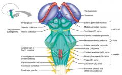
consists of three regions - midbrain, pons, medulla oblongata.
signal to heart and diaphram begin here cranial nerves begin here as well. |
|
|
Cerebellum
|
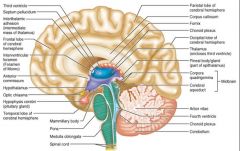
Provides precise timing and appropriate patters of skeletal muscle contraction(coordination
|
|
|
Protection of the Brain
|
bone (cranium)
meninges (three connective tissue membrane layers) cerebrospinal fluid (CSF) |
|
|
Meninges
|

dura mater
arachnoid mater pia mater |
|
|
dura mater
|
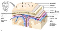
composed of two tough connective tissue layers
|
|
|
Arachnoid Mater
|

the middle mater
separated from dura mater by the subdural space beneath the arachnoid is the subarachnoid space filled with CSF and blood vessels |
|
|
Pia Mater
|

composed of this tissue that clings tightly to the brain
|
|
|
Cerebrospinal Fluid (CSF)
|
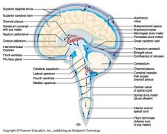
Forms a liquid cushion that gives buoyancy to the CNS organs
Protects CNS from blows and other trauma Nourishes the brain and carries chemical signals throughout it and its ventricles |
|
|
Cerebrovascular Accidents (Strokes)
|
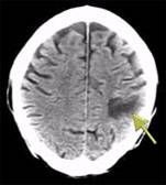
caused by insufficient blood flow to the brain leading to brain tissue death
transient ischemic attacks (TIA's) - temporary episodes of reversible brain tissue injury |
|
|
Spinal Cord
|
CNS tissue enclosed within the vertebral column from skull to L1
Provides two-way communication to and from the brain Protected by bone, meninges, and CSF |
|
|
General organization of gray matter
|
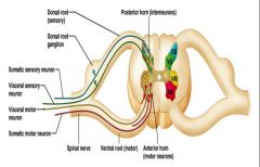
somatic sensory (ss)
visceral sensory (vs) visceral motor (vm) somatic motor (sm) |
|
|
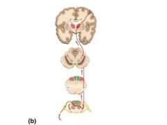
Ascending Pathways
|
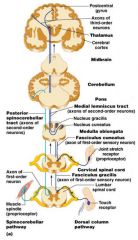
1st order - detects and sends sensory signals to CNS
2nd order project to thalamus 3rd order - projects to higher centers (cerebral cortex) |
|
|
Spinothalamic Tract
|
|
|
|
Major Descending Tracts
|
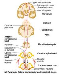
2 neron pathway - upper motor neuron, lower motor neuron
corticospinal tract - coordinated voluntary limb movements, fibers decussate in lower medulla |
|
|
Corticospinal
|
|
|
|
Spinal Cord Injury
|
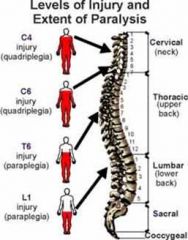
results in motor and sensory loss in regions inferiorly
paraplegia - injury between T1 and L1 quadriplegia - injury in cervical area |
|
|
Spinal Cord Injury
|

results in motor and sensory loss in regions inferiorly
paraplegia - injury between T1 and L1 quadriplegia - injury in cervical area |
|
|
Amyotrophic Lateral Sclerosis
|
a.k.a. lou gehrig disease
degeneration of motor neurons lead to muscle atrophy muscle weakness leading to progressive paralysis intelligence and sensory functions are intact |
|
|
Peripheral Nervous System
|
all neural structures outside the brain and spinal cord
includes: sensory receptors, peripheral nerves, associated ganglia and motor endings |
|
|
Sensory Receptors
|
realization of these stimuli, sensation and perception, occur in the brain
different types: mechanoreceptors, thermoreceptors, photoreceptors, chemoreceptors, nociceptors, proprioceptors |
|
|
Mechanoreceptors
|
respond to touch pressure, vibration, stretch, and itch
|
|
|
Thermoreceptors
|
sensitive to changes in temperature
|
|
|
Photoreceptors
|
respond to light energy
|
|
|
Chemoreceptors
|
respond to chemicals (smell, taste, changes in blood chemistry)
|
|
|
Nociceptors
|
sensitive to pain-causing stimuli
|
|
|
proprioceptors
|
respond to "stretch" giving info on one's position and movements
|
|
|
Nerves
|
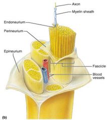
types: sensory, motor, or mixed, the most common type
damages to the nerve tissue are permanent |
|
|
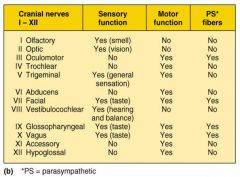
Cranial Nerves
|
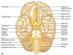
twelve pairs of cranial nerves arise from the brain/brainstem
|
|
|
Cranial Nerve List
|
|
|
|
Cranial Nerve I: Olfactory
|
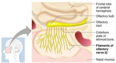
functions solely by carrying afferent impulses for the sense of smell
|
|
|
Cranial Nerve II:Optic
|
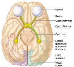
sensory fibers only
functions solely by carrying afferent impulses for vision |
|
|
Cranial Nerve III: Oculomotor
|
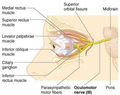
innervates eye muscles
motor nerve only carries parasympathetic nervous system fibers functions in raising the eyelid, directing the eyeball, (ps): constricting the iris and controlling lens shape |
|
|
Cranial Nerve IV: Trochlear
|
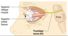
innervates eye muscle
motor nerve that directs the eyeball |
|
|
Cranial Nerve V: Trigeminal
|
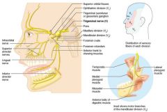
Conveys sensory impulses from various areas of the face, and supplies motor fibers for chewing
|
|
|
Cranial Nerve VI: Abducens
|
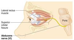
motor nerve innervating muscle that directs the eye
|
|
|
Cranial Nerve VII: Facial
|
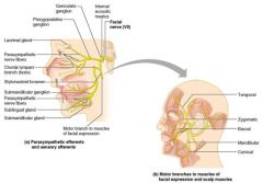
motor functions include facial expressions
sensory function is in taste carries PS fibers |
|
|
Cranial Nerve VIII: Vestibulocochlear
|
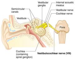
two divisions - cochlear(hearing) and vestibular(balance)
functions are solely sensory for the sense of balance and hearing |
|
|
Cranial Nerve IX: Glossopharyngeal
|

carries PS fibers
motor- innervates tongue and muscle around the neck in the back of throat sensory - information of taste going to the brain |
|
|
Cranial Nerve IX: Glossopharyngeal
|
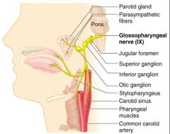
carries PS fibers
motor- innervates tongue and muscle around the neck in the back of throat sensory - information of taste going to the brain |
|
|
Cranial Nerve X: Vagus
|
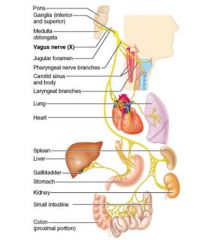
only cranial nerve that extends beyond head and neck
most motor fibers are PS fibers to the heart, lungs, and visceral organs (involuntary) sensory function is in taste |
|
|
Cranial Nerve XI: Accessory
|
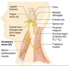
motor nerve supplying muscles in neck and back of throat
|
|
|
Cranial Nerve XII: Hypoglossal
|
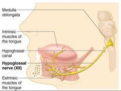
innervates muscles of the tongue
motor only |
|
|
Spinal Nerves
|
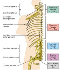
They are named according to their point of origin
8 cervical(C1-C8) 12 thoracic(T1-T12) 5 lumbar (L1-L5) 5 sacral (S1-S5) 1 coccygeal |
|
|
Cervical Spinal Nerves
|
carries motor and sensory information through the upper extremity
|
|
|
Thoracic Spinal Nerves
|
supply the chest, some muscles of the back and parts of the abdomen(trunk)
|
|
|
Lumbar Spinal Nerves
|
supply the lower parts of the abdomen and the back the buttocks, some parts of the external genital organs, and upper leg area
|
|
|
Sacral Spinal Nerves
|
supply motor and sensory information from and to the lower leg
|
|
|
Spinal Nerves: Roots
|
each spinal nerve runs through the intervertebral foramen and connects to the spinal cord via two roots: ventral root and dorsal root
|
|
|
Ventral Root
|
arises from the anterior horn and contains motor fibers
|
|
|
Dorsal Root
|
arises from sensory neurons in the dorsal root ganglion and contain sensory fibers
|
|
|
Dorsal Ramus
|
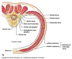
innervates dorsal muscles and joints in region
|
|
|
Ventral Ramus
|

innervates ventral and lateral skin and muscle
|
|
|
Nerve Plexuses
|
interlacing nerve networks called plexuses are found along the spinal column except at T2-T12
found in: cervical, brachial, lumbar, sacral |
|
|
Cervical Plexus
|
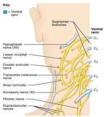
formed by C1-C4
most branches innervate the neck, ear, back of head, and shoulders phrenic nerve - motor and sensory nerve of diaphragm |
|
|
Brachial Plexus
|
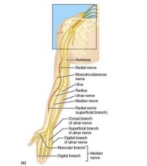
formed by C5-C8
gives rise to the nerves that innervate the upper limb radial, musculocutaneous, ulnar, median nerves |
|
|
Lumbar Plexus
|

arises from L1-L4
major nerves are the femoral and the obturator |
|
|
Sacral Plexus
|
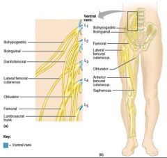
arises from L4-S4
major nerve is the sciatic, longest and thickest nerve of the body |
|
|
Somatic Reflexes
|
4 properties: require stimulation
quick: minimal delay involuntary stereotypic: predictable response |
|
|
Two Divisions of the Autonomic Nervous System
|
sympathetic and parasympathetic
|
|
|
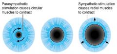
Sympathetic
|
mobilizes the body during extreme situations
|
|
|
Parasympathetic
|
performs maintenance activities and conserves body energy
|
|
|
Role of the Parasympathetic Division
|
concerned with keeping body energy use low
activity is illustrated in a person who relaxes after a meal blood pressure, heart rate, and respiratory rates are low gastrointestinal tract activity is high skin is warm and the pupils are constricted originates from the brain stem and sacral areas |
|
|
Role of the Sympathetic Division
|
"fight-or-flight" system
activates when someone is scared, excited, and nervous located in the thoracic spinal cord |
|
|
Adrenal Medulla
|
an extension of the ANS
produces and releases norepinephrine and epinephrine stimulated by the sympathetic system |
|
|
Photoreceptors
|
sense and encode light patterns
|
|
|
Conjunctiva
|
transparent membrane that lines the eyelids
lubricates and protects the eye |
|
|
Lacrimal Apparatus
|
consists of the lacrimal gland and associated ducts
lacrimal glands secrete tears which enters the eye and drains from it through ducts |
|
|
Lens of the Eye
|
separates the internal cavity into anterior and posterior segments
|
|
|
Sclera (posterior)
|

protects the eye and anchors extrinsic muscles
|
|
|
Cornea (anterior)
|
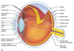
lets light enter the eye
|
|
|
Vascular Tunic
|
has three regions
choroid ciliary body iris |
|
|
Choroid
|

supplies blood to eye(layers)
|
|
|
Ciliary Body
|

ring of smooth muscle surrounding lens
anchors ligaments that hold the lens in place |
|
|
Iris
|
colored part of the eye, and pupil
regulates the amount of light entering the eye |
|
|
Sensory Tunic: Retina
|

contains
photoreceptors ganglion and other cells macula lutea contains the fovea - greatest cone concentration |
|
|
The Retna: Photoreceptors
|
where rods and cones are located
|
|
|
Rods
|
respond to dim light
are used for peripheral vision |
|
|
Cones
|
respond to bright light
have high-acuity color vision |
|
|
Ganglion Cell Axons
|
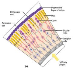
leave the eye as the optic nerve
|
|
|
Optic Disc
|
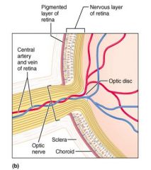
is the site where the optic nerve leaves the eye
|
|
|
Blood Supply to the Retina
|

retina receives its blood supply from two sources
choroid central artery |
|
|
Posterior Segment
|
filled with clear gel called vitreous humor that
transmits light supports the retina and posterior lens |
|
|
Anterior Segment
|
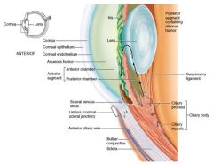
filled with aqueous humor
a plasmalike fluid that drains via the canal of schlemm supports, nourishes and removes wastes |
|
|
The Lens
|

a biconvex, transparent, flexible structure
allows precise focusing of light onto the retina with age, the lens becomes more dense and loses its elasticity |
|
|
pathway of light entering the eye
|
cornea, aqueous humor, lens, vitreous humor, retinal photoreceptors
|
|
|
Emmetropic Eye
|
normal eye with light focused properly
|
|
|
Myopic Eye (nearsighted)
|
the focal point is in front of the retina
|
|
|
Hyperopic Eye (farsighted)
|
the focal point is behind the retina
|
|
|
Photoreception
|
process by which the eye detects light energy
|
|
|
Photopigments
|
rods and cones contain visual pigments that absorb light
|
|
|
Outer Ear
|
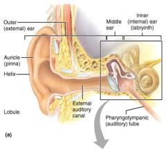
the auricle
helix(rim) lobule(earlobe) external auditory canal tympanic membrane (eardrum) |
|
|
Middle Ear (Tympanic Cavity)
|
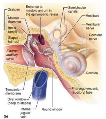
small air filled cavity
pharyngotympanic tube ear ossicles |
|
|
Pharyngotympanic Tube
|
connects middle ear to nasopharynx
equalizes pressure in the middle ear cavity with external air pressure |
|
|
Ear Ossicles
|
three small bones: malleus, incus, and stapes that transmit vibratory motion of the eardrum to the inner ear
|
|
|
Inner Ear
|
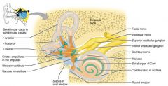
bony labyrinth
channels within the temporal bone contains: the vestibule, the cochlea, and the semicircular canals |
|
|
The Vestibule
|

central egg-shaped cavity of the bony labyrinth
|
|
|
Semicircular Canals
|

lie in three planes of space
|
|
|
Cochlea
|
a spiral, conical, bony chamer that
contains the organ of corti(hearing receptor) |
|
|
Conduction Deafness
|
something hampers sound conduction to the inner ear (e.g., impacted earwax, perforated eardrum, osteosclerosis of the ossicles
|
|
|
Sensorineural Deafness
|
results from damage to neural structures at any point from the cochlear hair cells to the auditory cortical cells
|