![]()
![]()
![]()
Use LEFT and RIGHT arrow keys to navigate between flashcards;
Use UP and DOWN arrow keys to flip the card;
H to show hint;
A reads text to speech;
233 Cards in this Set
- Front
- Back
|
ocular anatomy |
the study of the structures that constitute the eye and, for completeness, usually includes the study of related structures of the brain that make up the visual pathway |
|
|
physiology |
the study of the function of the structures |
|
|
What are the 6 anatomical directions? |
superior |
|
|
The orbit is, and contains: |
-the bony socket, a round opening in the skull |
|
|
What are the 7 orbital bones? |
frontal |
|
|
sutures |
lines where the orbital bones are fused together |
|
|
foramens
|
eight different openings allowing arteries, veins, and nerves that serve the orbital contents and parts of the face to enter and leave the orbit
|
|
|
Specifically, each orbit contains...
|
the eyeball, orbital fat, fascia, levator muscle & Mueller's muscle of the upper lids, lacrimal gland, optic nerve, EOMs, nerves, and circulatory supply for the orbital contents
|
|
|
fascia
|
connective tissue sheaths
|
|
|
Which 3 orbital bones form the orbital rim at the front?
|
maxilla
frontal zygomatic |
|
|
Which orbital bone forms the apex at the back?
|
sphenoid
|
|
|
Which orbital bone is the thinnest?
|
ethmoid - located on the medial side of the orbit and may be penetrated by infections
|
|
|
sinusitis
|
produces eye pain, especially when the eye moves
|
|
|
Where are the thickest bones located?
|
at the lateral wall of the orbit
|
|
|
diplopia
|
double vision - can be caused by injury or disease to the orbital bones resulting in the eye becoming displaced relative to the other eye
|
|
|
sinuses
|
air spaces within the bones
*named specifically for the cones that contain them: frontal, maxilla, ethmoid, & sphenoid sinuses* |
|
|
The superior wall/roof of the orbit consists of:
|
the sphenoid (2%)
the frontal bone (98%) |
|
|
The medial wall of the the orbit consists of:
|
(from front to back)
frontal process of the maxillary bone lacrimal bone ethmoid bone a small part of the sphenoid bone |
|
|
ethmoiditis
|
infection of the ethmoid sinuses - major cause of infections of the orbit and orbital contents
|
|
|
lacrimal fossa
|
2 depressions in the orbit, one in the medial wall contains the lacrimal sac allowing drainage of the tears from eye to nose
|
|
|
The floor of the orbit consists of:
|
(front to back)
maxilla zygomatic a small portion of the palatine *the floor is .5-1 mm thick* |
|
|
"blowout fracture"
|
blunt trauma resulting in a fracture of the orbital floor
|
|
|
Laterally, the orbit is made of:
|
-the zygomatic bone and the greater wing of the sphenoid bone
-is the thickest wall |
|
|
Which is the only wall not associated with paranasal sinuses?
|
the lateral wall
|
|
|
palpebrae
|
eyelids - folds of skin that protect and reinforce the eyes and orbits
|
|
|
The eyelids serve what 3 basic functions?
|
-protects from small foreign bodies and light
-replenish/spread tear film across front surface -pumps tears through lacrimal sac (regulating amount of tear fluid |
|
|
What two muscle groups control blinking?
|
orbicularis
levator |
|
|
reflex blinking
|
rapid forceful closure of the eyes
|
|
|
blepharospasm
|
condition in which the eyes are so tightly closed they cannot be opened
|
|
|
palpebral aperture/fissure
|
the space between the eye lids
measures ~10mm at the widest point |
|
|
ptosis
|
when the palpebral aperture is too small (i.e. drooping lids) blocking the pupil
|
|
|
exophthalmos
|
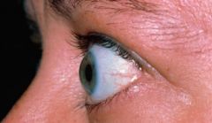
when the palpebral aperture is too large from retracted eyelids
~> often found in patients with Grave's disease |
|
|
canthi
|
the point at which the upper and lower lids meet
|
|
|
lateral canthus
|
the canthus closer to the ear
|
|
|
medial canthus
|
the canthus closer to the nose
|
|
|
What are the two fleshy mounds located in the medial canthus area?
|
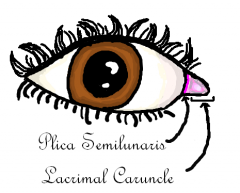
the plica semiluminaris (the deeper of the two) and the caruncle (contains sweat and oil glands and sometimes hair)
|
|
|
entropion
|
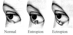
eyelashes directed toward the eye by the lids turning inward (resulting from traums, scarring 2ndary to inflammation, or a loss of elasticity of the lid tissues
|
|
|
ectropion
|

when eyelid tissues lose their elacticity they may droop away from the eye; tears can't drain effectively; patient may report "crying" as a symptom
|
|
|
gray line
|
the junction of the skin and the conjunctiva
|
|
|
How many layers are the eyelids constructed of?
|
7
skin subcutaneous areolar layer orbicularis oculi submuscular areolar layer levator palpebrae superioris tarsal plate conjunctiva |
|
|
subcutaneous areolar layer
|
-normally contains no fat
loosely connected over lid -2nd layer of the eye lid |
|
|
orbicularis oculi
|
-occupies entire length of the eyelid
-responsible for eyelid closure -controlled by the facial nerve (CN VII) |
|
|
submuscular areolar layer
|
-a layer of connective tissue
-similar to subcutaneous layer -contains most of the major nervous and circulatory supplies for the lids -fourth layer of the eyelid |
|
|
levator palpebrae superioris
|
-another muscle under the areolar tissue
-extends from its origin @ the back of the orbit on the sphenoid to its insertions in the lid -is the major muscle responsible for lid retraction -fifth layer of the eyelid |
|
|
tarsal plate
|
-is smaller in the lower lid than the upper lid (about half the size)
-comprised of dense fibrous and elastic connective tissue -responsible for shape/rigidity of the eyelids -sixth layer of the eyelid |
|
|
meibomia/tarsal glands
|
large, parallel sebaceous glands, in the tarsal plates, running down the length of the tarsal plates
produce the oil that floats on the watery layer of the tear film to impede evaporation of the tears & prevent tear overflow |
|
|
chalazion
|
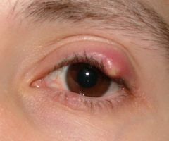
-lipogranuloma of the gland
-result of a blocked meibomian gland -internal hordeolum (an infection/inflammation of the gland) |
|
|
Mueller's muscle
|
-a muscle lying below the orbital septum
-origin from levator in the upper lid & an extension of the inferior rectus muscle in the lower lid -accounts for only 2 mm of lid elevation |
|
|
conjunctiva
|
-the deepest layer of the palpebrae
-the mucous membrane that covers the inside of the lids and the outside of the globe |
|
|
marginal conjunctiva
|
where the conjunctiva begins at the gray line
|
|
|
tarsal conjunctiva
|
the part of the conjunctiva lining the inside of the lid and tarsal plates
|
|
|
orbital conjunctiva
|
the part of the conjunctiva above (or below in the lower lid) the tarsal plates
|
|
|
palpebral conjunctiva
|
the marginal , tarsal and orbital portions of the conjunctiva covering the inside of the lids
|
|
|
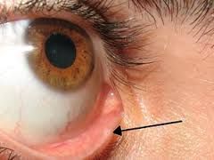
bulbar conjunctiva
|
the conjunctiva covering the eyeball
|
|
|
chemosis
|

the edema of the conjunctiva
|
|
|
conjunctivitis
|
inflammation of the conjunctiva because of blood vessels becoming engorged because of irritation or infections
|
|
|
goblet cells
|
produce mucous that covers the entire surface of the conjunctiva and cornea (located in the epithelium of the conjunctiva)
|
|
|
glands of Krause and Wolfring
|
accessory glands that produce the watery layer of the tear film in conjunction with the lacrimal gland
|
|
|
glands of Zeis
|
are attached to the follicles of the eyelashes producing oil that protects the hair from drying out
|
|
|
glands of Moll
|
sweat glands located at the lid margin
|
|
|
blepharitis
|
hair shafts become infected causing lid margin to become inflamed
|
|
|
blepharoconjunctivitis
|
in the conjunctiva and lid margin both become infected/inflamed
|
|
|
lacrimal system
|
responsible for production, maintenance, and removal of the tear film
|
|
|
The tear film is comprised of how many layers?
|
-3
--the superficial oily layer --the middle tear fluid/watery layer --the mucin/mucous layer |
|
|
What does the tear layer do?
|
it brings oxygen, nutrition, and natural anti-infective chemicals to the anterior surfaces of the eyeball
|
|
|
epiphora
|
tears spilling onto the cheek; can result from the overproduction of tears with the drainage system being normal, or normal tear production with problems draining
|
|
|
the globe
|
-is the eyeball itself and is essentially 3 concentric spheres: fibrous tunic, vascular tunic, and nervous tunic
-divided into anterior/posterior by the iris |
|
|
fibrous tunic
|
-comprised of the cornea and the sclera
--cornea = anterior 1/6 --sclera = posterior 5/6 |
|
|
vascular tunic
|
-a.k.a. the uvea
-is the middle layer of the globe -consists of the iris, ciliary body, and the choroid |
|
|
nervous tunic
|
-innermost layer of the globe
-consists of the retina and retinal pigment epithelium (RPE) |
|
|
cornea
|
-the major refracting surface of the eye
-unique because it's transparent -thinner in the center vs. the edges -has 5 layers and normally has no blood vessels --epithelium, Bowman's, stroma, Descemet's, and endothelium |
|
|
corneal neovascularization
|
when a condition exists that doesn't allow adequate circulatory support for the cornea (i.e. edema, inflammation, or inadequate tear film oxygen content), new vessels will grow into the cornea which disturs its transparency
|
|
|
What covers the front surface of the cornea?
|
epithelial cells
|
|
|
epithelium
|
-first layer of the cornea
-cells that prevent water from entering the front of the cornea and provide several layers of protection from abrasion from foreign matters |
|
|
Bowman's membrane
|
-second layer of the cornea
-once this layer is damaged, the risk for scar tissue development increases drastically |
|
|
stroma
|
-third layer of the cornea
-the largest layer of the cornea -comprised of collagen |
|
|
collagen
|
a specific type of connective tissue that is very tough and doesn't stretch
|
|
|
Descemet's membrane
|
-fourth layer of the cornea
-very thin -doesn't have the capacity to regenerate if damaged -if damaged may decrease transparency -involved in maintaining the integrity of the corneal endothelium |
|
|
endothelium
|
-fifth layer of the cornea
-a single-thickness layer of cells consisting of about 500,000 cells @ birth that don't regenerate -transports nutrients from the aqueous humor to the cornea |
|
|
keratoconus
|
the stroma of the cornea is thinned and the IOP causes the cornea to bulge forward
|
|
|
sclera
|
-makes up the posterior 5/6 of the fibrous tunic along with the episcleral connective tissue
-made of collagen fibers |
|
|
episclera
|
is a layer of connective tissue lying between the sclera and the bulbar conunctiva
|
|
|
choroid
|
-the layer just interior to the sclera and it provides some of the vascular support
-represents the majority of the vascular tunic -a network of blood vessels that provides vascular support |
|
|
What are the 3 parts of the vascular tunic?
|
the choroid, the ciliary body, and the iris
|
|
|
trabecular meshwork
|
-located at the junction of the uvea
-acts as a drain for the intraocular fluid that is continually produced in the eye |
|
|
How does the iris get its color?
|
from the amount of melanin/pigment it contains
|
|
|
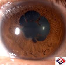
iris
|
-divides the eye's internal space into the anterior chamber (infront of the iris) and the posterior chamber (behind the iris)
-the "colored part" of the eye |
|
|
What are the 3 layers of the iris?
|
1) the stroma - made of loosely structured collagen fibers
2) the non-pigmented epithelium 3) the pigmented epithelium |
|
|
The stroma layer of the iris contains:
|
the melanocytes/pigment cells, the sphincter/constrictor muscle, and the blood vessels
|
|
|
sphincter muscle
|
-a.k.a. the constrictor muscle
-makes the pupil get smaller in bright light |
|
|
non-pigmented epithelium
|
is basically the dilator muscle, which is responsible for opening the pupil in the dark
|
|
|
How are the 2 muscles in the eye controlled?
|
-non-voluntary
-part of the autonomic (non-voluntary) nervous system |
|
|
mydriatic drugs
|
make the pupil larger by stimulating the dilator muscle
*acts as the sympathetic nerves to* |
|
|
cycloplegic drugs
|
makes the pupil larger by paralyzing the sphincter muscle
*"knocks out" the parasympathetic system* |
|
|
posterior synechia
|
when the iris sticks to the lens as a result of inflammation to the iris
|
|
|
pigmented epithelium
|
-the deepest layer of the iris
-very darkly pigmented, extending around the edge of the pupil (pupillary frill) |
|
|
Where is the ciliary body located?
|
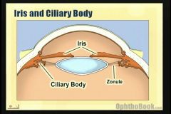
immediately behind the iris and just inside the sclera
|
|
|
What are the layers of the ciliary body?
|
the ciliary muscle (makes up the bulk of the ciliary body) and the stroma (contains the blood vessels and ciliary processes)
|
|
|
ciliary processes
|
responsible for the production of aqueous humor
|
|
|
How does the aqueous humor flow in the eye?
|
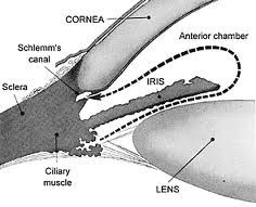
enters posterior chamber in front of the lens, flows through the pupil into the anterior chamber where it comes into contact with and filters through the trabecular meshwork to the canal of Schlemm, then empties into the aqueous veins of Ascher in the sclera and mixed with blood, then removed from the eye
|
|
|
aqueous humor
|
-fills front part of the posterior chamber between the lens and the iris, and the entire anterior chamber
-provides nutrients for the lens and posterior cornea and carries away waste products, and maintains IOP since it's the only fluid continually produced in the eye |
|
|
ocular hyptertension
|
if the rate of IOP production is too high or the rate of drainage is too low, pressure increases
|
|
|
ocular hypotension
|
usually due to penetrating injury, but can result as a decrease in aqueous production because of inflammation in the ciliary body
|
|
|
What are the functions of the ciliary body?
|
-produces aqueous humor
-accommodation -secretion of one component of vitreous humor -prevents passage of material into the aqueous humor -helps control the flow of the aqueous humor into the trabecular meshwork |
|
|
presbyopia
|
lack of focusing ability usually caused as the eye loses its elasticity with age
|
|
|
ophthalmic artery
|
-all of the blood supply to the eye comes through this artery
-bloody supply fills the choroid through 2 sets of smaller vessels |
|
|
central retinal artery
|
is the ophthalmic artery (just renamed) after the long and short ciliary arteries have branched off into the eye
|
|
|
Haller's layer
|
outermost layer of the choroid, made up of larger vessels
|
|
|
Sattler's layer
|
2nd layer of the choroid, vessels are considered to be medium sized
|
|
|
How are the vessels of Haller's and Sattler's layers classified?
|
arterioles and venules, because of the construction of their walls
|
|
|
choriocapillaris
|
3rd layer of the choroid, vessels are primarily capillaries
|
|
|
Bruch's membrane
|
thin layer of connective tissue separating the choriocapillaris and the retina - most internal layer of the choroid
|
|
|
What is the retina's general function?
|
to convert the light energy falling onto it into electrical impulses that can be analyzed by the brain
|
|
|
What are the 10 layers of the retina?
|
"In New Generation It Is Only Ophthalmologists Examine Patient's Retina" (inner to outer)
10)inner limiting membrane 9)nerve fiber layer 8)ganglion cell 7)inner plexiform 6)inner nucleus 5)outer plexiform 4)outer nuclear 3)external limiting membrane 2)photoreceptors 1)RPE |
|
|
retinal pigment epithelium (RPE)
|
most external retinal layer
provides vitamin A to the photoreceptors transports nutrients into the retina phagocytize ("eat") the used-up portions of the photoreceptor cells prevents the intraocular reflection of stray light |
|
|
photopigments
|
the chemicals that are used to collect the light energy in the photoreceptors
|
|
|
retinitis pigmentosa
|
a genetic defect that results in the RPEs not "eating" up the used portions of the photoreceptor cells; starts as night blindness; can progress to total blindness
|
|
|
axon
|
one end of a nerve cell
~> specialized for transmitting neural impulses |
|
|
dendrite
|
one end of a nerve cell
~> specialized to receive neural impulses |
|
|
synapse
|
a small gap located between the axon of one cell and the dendrite of the next
|
|
|
neurotransmitter
|
the chemical released from the axon that floats across the synapse and stimulates the dendrite of the next nerve cell in line
|
|
|
photoreceptor layer
|
2nd layer of the retina
2 types of photoreceptors: rods and cones |
|
|
How do rods and cones differ?
|
anatomically, in their physiologic response characteristics, and in their distribution throughout the retina
|
|
|
cones
|
-have pointed (cone shaped) outer segments called receptors
-responsible for color discrimination -require much higher light levels to be stimulated than rods |
|
|
rods
|
-have flat outer segments called receptors
-response of the rods is based on the presence/absence of light energy |
|
|
Conditions of color deficiency are predominantly:
|
errors of the cones
|
|
|
Problems seeing in dim illumination are usually because of:
|
rod dysfunctions
|
|
|
macula lutea/macula
|
-central 1 cm of the retina
-called the macula because it has a pigment that gives it a yellow color -photorecedptors (mainly cones) are the most dense in this area |
|
|
fovea
|
a depressed area in the center 1.5 mm of the macula
|
|
|
foveola
|
-the area in the fovea that contains ONLY cones
-responsible for fine discriminations and high VA |
|
|
external limiting membrane
|
-3rd layer of the retina
-not really a membrane; it's the point at which the photoreceptors are joined together by a specific type of cellular junction called zonula adherens -marks the midpoint of the length of rods and cones |
|
|
outer nuclear layer
|
-4th layer of the retina
-where the cell bodies of the photoreceptors are located -these cell bodies contain the nuclei |
|
|
outer plexiform layer
|
-5th layer in the retina
-contains the first synapses in the visual pathway in this layer -the last layer to be supported by the choroidal circulation |
|
|
plexiform
|
the term given to layers without cell bodies that contain synaptic sites
|
|
|
bipolar cells
|
-provide for the transmission of the visual signal, up the visual pathway, toward the brain
-acts as a 'relay' or 'wire' -this is called a "vertical" transmission |
|
|
horizontal cells
|
-connect receptors to other receptors and horizontal cells
-these connections are involved in integrating the input from groups of cells into receptive fields -represent horizontal processing of visual information |
|
|
Which cell bodies are found in the 6th layer of the retina?
|
-the cell bodies of bipolar and horizontal cells, Mueller's cells, and amacrine cells
-6th retinal layer=inner nuclear layer |
|
|
Muller cells
|
are nutritional support cells that are scattered throughout all layers internal to the outer nuclear layer, specifically layers 5-9
|
|
|
inner nuclear layer
|
-6th layer of the retina
-is the first (most external) layer that is completely supported by the circulation of the retina |
|
|
amacrine cells
|
have no apparent axons or dendrites, they have processes that allow bi-directional transmission of neural signals ("horizontal" processing)
|
|
|
inner plexiform layer
|
-7th layer of the retina
-where the amacrine cells' processes and the axons of the bipolar cells connect to the ganglion cells |
|
|
ganglion cells
|
-another step in the transmission of visual impulses to the brain
-visual information is carried via the axons of the ganglion cells out of the eye ("vertical" processing) |
|
|
ganglion cell layer
|
-8th layer of the retina
-consists of the cell bodies of the ganglion cells |
|
|
optic nerve (CN II)
|
the axons of ganglion cells collect as a bundle to create the optic nerve (CN II)
|
|
|
nerve fiber layer
|
-9th layer of the retina
-on their way to form the optic nerve (CN II), the axons of the ganglion cells run across the inside of the retina which constitutes this 9th layer |
|
|
internal limiting membrane
|
-10th layer of the retina
-separates the ganglion cell layer from the face of the vitreous -comprised of collagen fibers that connect the Mueller cells |
|
|
cortex
|
the elongated cells/fibers in the lens are collectively called the cortex
|
|
|
Because of what process allows the lens to grow throughout life?
|
-a single layer of epithelium
-some cells of which elongate to form new cortical fibers, surrounds the cortex, thus allowing continuous growth |
|
|
capsule
|
is a tough elastic membrane that surrounds the lens
|
|
|
zonular fibers
|
-a.k.a. zonules
-is what the lens is suspended by from its edge to the ciliary muscle |
|
|
lens
|
-is a structure comprised mostly of elongated epithelial cells that are arranged in concentric layers (like an onion)
-is the 2nd most powerful refracting/light focusing component of the eye |
|
|
What is the simplified explanation of the process by which people focus their eyes?
|
the ciliary muscle tightens, allowing the zonules to loosen, and the elasticity of the capsule makes the lens bulge in the center so that it becomes thicker and more powerful
|
|
|
vacuole
|
-a space between the lens fibers
-can be caused by mechanical swelling of the lens, damage from radiation, or a metabolic disturbance |
|
|
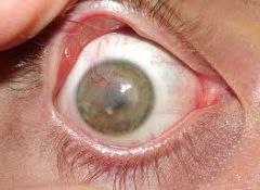
Vacuoles usually lead to what?
|
a clouding of the lens (cataracts)
|
|
|
How are cataracts named?
|
location, cause, size, density, or time of occurence
|
|
|
The first part of the visual pathway involves:
|
the structures from the rods and cones to the optic nerve
|
|
|
The second part of the visual pathway involves:
|
from the eye, specifically, the optic nerve, to the visual cortex
|
|
|
The visual pathway runs:
|
a horizontal path from the front of the head to the back of the head
|
|
|
visual cortex
|
is the part of the brain responsible for analyzing the neural signals representing sight
|
|
|
visual field
|
the expanse of vision that a person sees
|
|
|
perimetry
|
-visual field testing
-measures the visual field in each eye -can help determine where in the visual pathway a disease exists |
|
|
How are the "sectors" of the retina created?
|
-quadrants
-by dividing the retina into halves with imaginary lines vertically and horizontally through the fovea |
|
|
optic chiasm
|
is formed by the fusion of the optic nerves inside the skull near the pituitary gland
|
|
|
left optic tract
|
-consists of a bundle of fibers including TEMPORAL retinal fibers from the LEFT eye and NASAL retinal fibers from the RIGHT eye
-they continue from the left side of the chiasm -represents the RIGHT side of the visual field |
|
|
right optic tract
|
-consists of a bundle of fibers including TEMPORAL retinal fibers from the RIGHT eye and NASAL retinal fibers from the LEFT eye
-they continue from the right side of the chiasm -represents the LEFT side of the visual field |
|
|
scotomas
|
loss of sensitivity/visual sensation in part of the field of both eyes
|
|
|
quadrantanopsia/quadrantanopia
|
a field defect that affects about 1/4 of the VF
|
|
|
hemianopsia/hemianopia
|
a field defect that affects about 1/2 of the VF
|
|
|
homonymous
|
when a field defect exists in both eyes and affects the same field in both eyes
|
|
|
prechiasmal lesions
|
lesions involving the retina or the optic nerve, which results in a unilateral (one eye) field defect
|
|
|
chiasmal/postchiasmal lesions
|
lesions involving the retina or the optic nerve resulting in a bilateral (often homonymous) field defects
|
|
|
binasal hemianopsia
|
-field defect resulting from damage to the temporal fibers
-NOT homonymous |
|
|
bitemporal hemianopsia
|
-field defect resulting from damage to the nasal fibers
-NOT homonymous |
|
|
lateral geniculate nucleus (LGN)
|
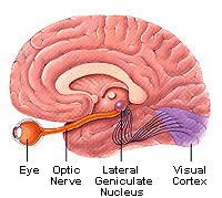
-where the optic tract ends
-a pyramid shaped mass of cells where a synapse occurs between the terminal ends of the ganglion cell axons and the dendrites of the nerve fibers, which carry the visual information to the visual cortex |
|
|
congruous
|
identical
|
|
|
optic raditations
|
the axons of the cells of the LGN continue to the visual cortex
|
|
|
Brodmann's area 17
|

-located at the very back tip of the brain
-the part of the occipital cortex that receives the fibers of the optic radiation from the LGN and is the primary receptive area for vision |
|
|
macular sparing
|
-an unusual feature of he congruous homonymous hemianopsia
-the term given to a VF when a complete hemianopsia exists except for the central 2-5 degrees where the field is intact |
|
|
association fibers
|
distribute the visual information to the cortex for analysis and for association with other sensory data and motor coordination
|
|
|
Brodmann's area 8
|
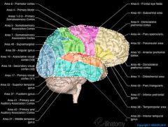
-responsible for planning eye movements
-located in the frontal lobe -one of the major pathways of the association is from the visual cortex to this area |
|
|
How are the extraocular muscles unique from others in the body?
|
the number of muscle fibers controlled by each nerve fiber is very small, allowing very accurate control of eye movements
|
|
|
striated muscles
|
voluntarily controlled
|
|
|
common tendinous ring/annulus of Zinn
|
-the common tendon from which arise the four recti muscles of the eye
-it surrounds the optic foramen and a part of the medial end of the superior orbital fissure |
|
|
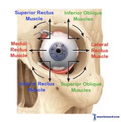
medial rectus
|
-most powerful of the EOMs
-runs from its origin at the optic foramen, along the medial wall, to its insertion onto the sclera -has only one action adduction -controlled by the inferior branch of the oculomotor (CN III) |
|
|
adduction
|
turns the eye inward toward the nose
|
|
|
inferior rectus
|
-runs along the floor from the orbital apex to its insertion
-makes a 23 degree angle with the medial wall -primary action is depression; also adducts and extorts slightly -controlled by the inferior division of the oculomotor nerve (CN III) |
|
|
depression
|
turns the eye downward
|
|
|
extorsion
|
turns top of eye out toward the temple and the bottom of eye in toward the nose
|
|
|
lateral rectus
|
-runs along the lateral wall
-has only one action abduction -controlled by the abducens nerve (CN VI) |
|
|
abduction
|
moves eye away from the midline, toward the temple
|
|
|
superior rectus
|
-makes a 23 degree angle with the medial wall
-primary action is elevation; also abducts and slightly helps with intorsion -controlled by a branch of the oculomotor nerve (CN III) |
|
|
elevation
|
turns eye upward
|
|
|
intorsion
|
turns bottom of eye toward nose and bottom toward the temple
|
|
|
superior oblique
|
-originates on or very near the annulus of Zinn on the sphenoid bone and runs along the superior wall to a small bony loop at the front of the orbit called the trochlea
-makes a 51-53 degree angle with the medial wall -primary action is depression, abduction, and intorsion |
|
|
inferior oblique
|
-the only EOM that originates at the front of the orbit, beginning at a fossa in the maxillary bone and runs between the eye and the inferior rectus
-primary action is elevation and abduction, and extorsion -controlled by a division of the oculomotor nerve (CN III) |
|
|
antagonist
|
something, such as a muscle, disease, or physiological process, that neutralizes or impedes the action or effect of another
|
|
|
agonist
|
a contracting muscle that is resisted or counteracted by an antagonist muscle
|
|
|
strabismus
|
-"squint"
-the condition present when both eyes cannot be directed to an object of regard at the same time -"lazy eye" |
|
|
heterophoria
|
the condition in which there is a tendency for the eyes to misalign, but the person can overcome that tendency so that both eyes do point at objects of regard
|
|
|
ductions
|
eye movements tested monocularly
|
|
|
versions
|
binocular eye movements in the same direction
|
|
|
vergence testing
|
testing with the eyes moving in opposite directions
|
|
|
What are the 3 types of vergences?
|
convergence
divergence vertical divergence |
|
|
convergence
|
both eyes turn toward the nose
|
|
|
divergence
|
both eyes turn away from the nose
|
|
|
vertical divergence
|
one up turns up the other turns down
|
|
|
antagonist muscle pairs in one eye
|
muscle antagonist
medial rectus lateral rectus lateral rectus medial rectus superior rectus inferior rectus inferior rectus superior rectus superior oblique inferior oblique inferior oblique superior oblique |
|
|
agonist muscle pairs in two eyes
|
right eye left eye
medial rectus lateral rectus lateral rectus medial rectus superior rectus inferior oblique inferior rectus superior oblique superior oblique inferior rectus inferior oblique superior rectus |
|
|
The thinnest bone of the orbit is:
|
ethmoid
|
|
|
The floor of the orbit is composed of which three bones?
|
maxilla
zygomatic palatine |
|
|
The condition in which the eyes are so tightly closed they can't be opened is called:
|
blepharospasm
|
|
|
When the eyelid turns in toward the globe, the condition is called:
|
entropion
|
|
|
Which muscle is responsible for eyelid closure and is controlled by the facial nerve (CN VI)?
|
obicularis oculi
|
|
|
When a meibomian gland becomes blocked and a red, painful bump appears on the lid, it is called:
|
chalazion
|
|
|
Any interruption in the innervation to Mueller's muscle results in:
|
ptosis
|
|
|
The bulbar conjunctiva covers what?
|
the eyeball
|
|
|
The __________ system is responsible for the production, maintenance, and elimination of the tear film.
|
lacrimal
|
|
|
Tears spilling onto the cheek is called:
|
epiphoria
|
|
|
The anterior chamber and posterior chamber in front of the lens are filled with :
|
aqueous
|
|
|
The vascular tunic/uvea consists of (from front to back):
|
iris
ciliary body choroid |
|
|
The innervation of the cornea is mainly sensory branches of what nerve?
|
cranial nerve V (trigeminal)
|
|
|
What condition is the most common cause of red eyes?
|
conjunctivitis
|
|
|
What is one of the major functions of the ciliary body?
|
accommodation
|
|
|
The bloody supply to the eye comes through what?
|
ophthalmic artery
|
|
|
The ______________ is the area of the retina that is responsible for fine discriminations and high VA.
|
fovea
|
|
|
Which structure is the second most powerful refracting component of the eye?
|
the lens
|
|
|
In the visual pathway, if a lesion occurs at the chiasm, the resultant field defects are usually:
|
bilateral
|
|
|
Which of the rectus muscles in innervated by cranial nerve VI
|
lateral
|
|
|
Which rectus muscle's primary action is depression?
|
inferior
|
|
|
Which rectus muscle is the strongest and is responsible for adduction of the eye?
|
medial
|
|
|
The tertiary action of the superior oblique is:
|
intorsion
|
|
|
Extorsion is described as rotating the top of the eyeball ______ and the bottom ______.
|
out, in
|
|
|
Which EOMs are involved when the patient's gaze is far left and up?
|
left superior rectus and right inferior oblique
|