![]()
![]()
![]()
Use LEFT and RIGHT arrow keys to navigate between flashcards;
Use UP and DOWN arrow keys to flip the card;
H to show hint;
A reads text to speech;
196 Cards in this Set
- Front
- Back
|
What are the 4 types of basic tissue? What are some of the structures they form?
|
1. Epithelial
- Epidermis - Hollow viscera internal surface lining 2. Connective - Dermis + subcutaneous tissue - Muscle sheaths & tendons 3. Muscular - Smooth, skeletal, cardiac 4. Nervous - Nerve cells |
|
|
What are the 3 general body systems?
|
1. Somatic
- body walls and limbs 2. Visceral - organs and associated neurovascular supply 3. Supply - nervous/arterial/venous/lymphatic systems |
|
|
What does the coronal plane divide the body in to?
|

Ventral & Dorsal
|
|
|
What does the saggital plane divide the body in to?
|

Left & Right Halves
|
|
|
What does the transverse plane divide the body in to?
|
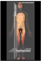
Superior & Inferior
|
|
|
What do medial and lateral refer to?
|
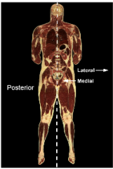
Medial = closer to midline
Lateral = further from midline |
|
|
What do superior and inferior refer to?
|
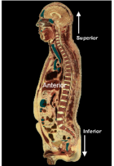
Superior = above
Inferior = below |
|
|
What do anterior and posterior refer to?
|
Anterior = front
Posterior = behind |
|
|
What do flexion and extension refer to?
|
Bends in joint angle
- Flexion = decrease - Extension = increase |
|
|
What do abduction and adduction refer to?
|
Change in angle wrt. midline
- Abduction = increase - Adduction = decrease |
|
|
What do internal and external rotation refer to?
|
Internal = rotation towards axis of body
External = rotation away from centre of body |
|
|
What do proximal and distal refer to?
|
Terms of comparison in distance
- Proximal = closer - Distal = further away |
|
|
What do superficial and deep refer to?
|
Terms of comparison for depth below skin
- Superficial = closer to skin surface - Deep = further beneath skin |
|
|
What do ventral and dorsal refer to?
|
Terms of comparison in the coronal plane
- Ventral = in front - Dorsal = at back |
|
|
What is the taxonomic hierarchy?
|
K - Kingdom
S - Superphylum P - Phylum S - Subphylum C - Class O - Order F - Family G - Group S - Species |
|
|
What taxonomic classification is 'coelomate'? What are the 4 distinguishing properties of coelomates?
|
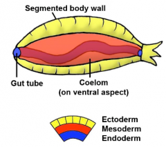
Coelomate is a superphylum type
1. Segmented body wall => Independent movement of segments 2. Hollow fluid filled cavity (coelom) 3. Gut tube 4. 3 Germ layers |
|
|
What taxonomic classification is 'chordate'?
What are the 3 distinguishing properties of chordates? |
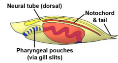
Chordate is a phylum type
1. Hollow nerve cord / neural tube => Ectoderm derived 2. Notochord => Mesoderm derived 3. Pharyngeal pouches => Endoderm covered by Ectoderm |
|
|
What taxonomic classification is 'verterbrate'?
What are some distinguishing vertebral features? |
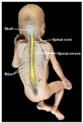
Vertebrate is a subphylum type
1. Spine, skull and skeleton 2. Spinal cord with associated nerves 3. 4 limbs with 5 digits (homologous structures between basic tetrapods) |
|
|
What taxonomic classification is 'Mammalia'?
What are the 4 distinguishing properties that mammals posses? |
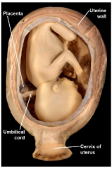
Mammalia is a class type
1. Skin and appendages i) Hair ii) Sweat + sebaceous glands iii) Mammary glands 2. Placental (except. monotrema and marsupials) 3. Warm blooded => 4 Chambered heart 4. Expanded forebrain |
|
|
What taxonomic classification is 'Primate'?
What are the 4 distinguishing properties that primates posses? |
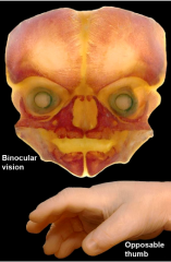
Primates are an order type
1. Binocular vision (forward facing eye sockets) 2. Opposable thumb 3. Freedom of arm rotation 4. Nails not claws |
|
|
What taxonmic classification is 'Hominidae'?
What are the 3 distinguishing properties that hominidae posses? |
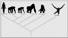
Hominidae are a family type
1. No tail 2. Large body size 3. More upright posture |
|
|
What taxonomic classification is 'Homo sapiens'?
What are the 4 distinguishing properties that Homo sapiens posses? |
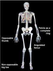
Homo sapiens is a species type
1. Completely upright posture 2. Non-opposable big toe 3. Line of gravity running along spine 4. Speech |
|
|
What are the 3 germ layers?
|
1. Ectoderm
2. Mesoderm 3. Endoderm |
|
|
What are the 4 ectorderm derivatives?
|
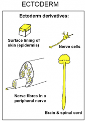
1. Brain + Spinal cord
2. Epidermis 3. Nerve Cells 4. Nerve Fibers |
|
|
What are the 6 mesoderm derivatives?
|
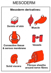
1. Dermis
2. Muscle 3. Connective Tissue 4. Vessels 5. Solid Viscera 6. Fibrous Sheaths of Nerves |
|
|
What does the endoderm form?
|
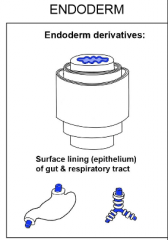
Epithelium of respiratory & gut tract
|
|
|
What does polarity refer to?
|
The positioning of segments from cranial to caudal
|
|
|
What function do the Gluteus maximus and minimus possess in bipedal locomotion?
|
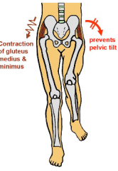
Contraction holds the unsupported leg up during 'stance phase' and it also prevents pelvic tilt
|
|
|
What evolutionary feature enables humans to speak?
|
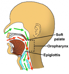
Epiglottis sits back and down
=> Air can enter/exit pharynx through mouth => Allows complex manipulation of air and sound |
|
|
What taxonomic classification is 'Animal'?
What is the main distinguishing feature of animals? |
Animals are a Kingdom type
- Get energy from an external source (also have cell membrane and no rigid wall structure) |
|
|
What do rostral and caudal refer to?
|
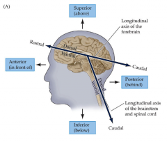
The neuraxis
- Rostral = further up - Caudal = further down |
|
|
What composes the CNS, and what composes the PNS?
|
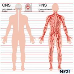
CNS = Brain + Spinal Cord
PNS = Nerves connecting body to CNS (everything else) |
|
|
What are meninges and their function?
What are the 3 layers of meninges? |
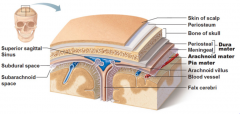
Meninges are membranes that envelop the brain + spinal cord, protecting the CNS
1. Dura Mater 2. Arachnoid Mater 3. Pia Mater |
|
|
What is the subdural space?
|
A potential space between the dura and arachnoid mater that can open due to injury
|
|
|
Which is the thickest of the 3 meninges layers?
|
The Dura Mater
|
|
|
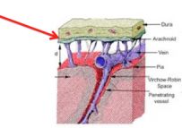
What is the main feature of the arachnoid mater?
|
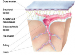
It has web-like filament projections which extend across the subarachnoid space, attaching the arachnoid mater to the pia mater that is deep
|
|
|
What function does the pia mater serve?
|

Acts as a fluid impermeable barrier between CSF and brain, whilst allowing bloody vessels to pass through
|
|
|
Where is the superior saggital sinus located and what is its function?
|

It sits on top of the 'Falx Cerebri'
- Surrounded by dura mater - Allows blood from blood to drain back in to circulation - Has pia mater invaginations => CSF drained through arachnoid granulations and returned to venous circulation |
|
|
What is the 'Falx Cerebri'?
|
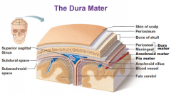
A dura mater invagination that descends vertically in the longitudinal fissure between brain hemispheres
|
|
|
What are the 3 functions of CSF?
|
1. Provides cushioning to CNS
2. Excretes harmful metabolites produced in to the blood stream for detoxification 3. Can be an endocrine medium (transports hormones to other areas of brain) |
|
|
What is the pathway of CSF circulation, starting with its production and ending in its exit to the blood stream?
|
1. Choroid Plexus
- production/filtration of CSF 2. Lateral Ventricles (2) 3. Third Ventricle 4. Fourth Ventrile 5. Subarachnoid Space (circulated around CNS) 6. Arachnoid Villi 7. Venous Sinuses 8. Blood Stream |
|
|
What happens when there is a blockage in CSF drainage? How does this affect children in comparison to adults?
|
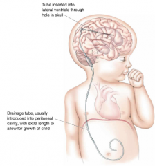
- If drainage path is blocked, an accumulation of CSF builds (hydrocephalus)
- As the meninges is rigid in adults, no volume change causes an increase in pressure => pressure pushes against brain causing neurological damage => pushing down on brain stem can cause loss of respiratory ability - Children have non-rigid bone structure allowing increase of volume and large CSF filled heads (catheter typically used to drain) |
|
|
What are the 2 types of dural haematomas and what happens with each?
|
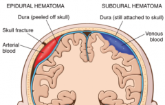
1. Epidural Haematoma
- shearing of arteries causes bleeding in between skull and dura (dura peels off skull) - build of blood puts pressure on the intracranial space =>brain shift 2. Subdural Haematoma - tearing of bridging veins in the subdural space - dura still attached to skull |
|
|
How is white and grey matter arranged in the brain? In the spinal cord?
Why are they arranged this way? |
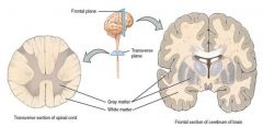
Brain
- Grey matter = exterior - White matter = interior Spine - Grey matter = interior - White matter = exterior Grey matter is exterior in the brain as it only travels inwards and through the brain stem, whilst information in the spinal cord integrates in the centre and projects outward |
|
|
What are white and grey matter? What are their functions?
|
Grey = cell bodies = neural processes
White = glial cells & myelinated axon fibres = signal projection |
|
|
What is a myelin sheath? What's its purpose?
|
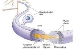
- Fatty phospholipid layer surrounding neuron axon
- Allows insulation & saltatory conduction - It is a membraneous projection of glial cells - Two types of glial cells produce: 1. Oligodendrocytes (CNS) 2. Schwaan Cells (PNS) |
|
|
How are nerve fibres arranged within a nerve?
|
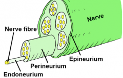
1. Individual axons surrounded by endoneurium
2. Several axons arranged in a fascicle surrounded by perineurium 3. Several fascicles surrounded by epineurium to form single nerve |
|
|
How many vertebrae are there in each spinal section? In total?
|
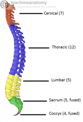
Cervical = 7
Thoracic = 12 Lumbar = 5 Sacral = 5 (fused) Coccyx = 4 (fused and variable between 3-5) Total = 33 (+- 1) |
|
|
How many spinal nerves are there in each spinal region? In total?
|
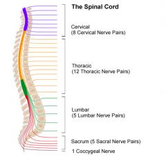
Cervical = 8
Thoracic = 12 Lumbar = 5 Sacral = 5 Coccygeal = 1 Total = 31 |
|
|
What is the 'cauda equina' and where does it start?
|
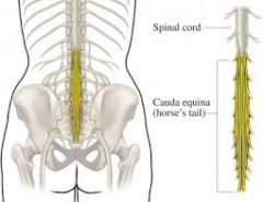
As the spinal cord stops growing at the T12 level, the rest of the spinal nerves branch out in a horse tail shape to reach the rest of the spinal segments. Starts at L1 and goes downwards.
|
|
|
Which spinal nerve roots have sensory functions and which have motor functions?
|
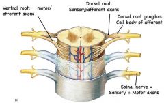
Dorsal (posterior) =
sensory Ventral (anterior) = motor |
|
|
How many cranial nerves are there?
What are their general functions? |
12 Pairs of Cranial Nerves
(innervate head and neck) - Special sensory function - General sensory function - Motor function - Autonomic function |
|
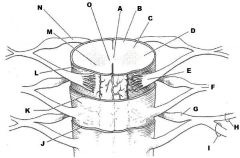
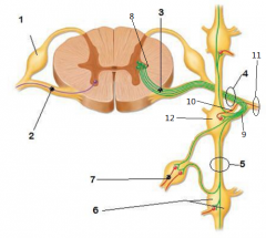
What structure does each label refer to?
(ignore O) |
A. Central Canal
B. White Matter C. Dorsal Horn D. Lateral Horn E. Anterior (ventral) Root F. Mixed Spinal Nerve Plexus G. Dorsal Root Ganglion H. Posterior (dorsal) Rammus I. Anterior (ventral) Rammus J. Dura Mater K. Arachnoid Mater L. Pia Mater M. Ventral Horn N. Posterior (dorsal) Root |
|
|
With respect to the nervous system, what is a nucleus?
|
A cluster of neurons in the CNS
|
|
|
What are ganglia?
|
Collections of cell bodies in the periphery
|
|
|
What is an interneuron?
|
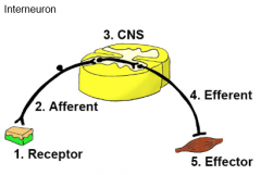
A neuron that connects a transmission pathway without processing the information itself
- Can have thousands of receptors and effectors |
|
|
What is a projection neuron?
|
An interneuron that carries info to the brain for processing
|
|
|
What is a nerve plexus?
|
Bundles of nerves with a mix of dorsal and ventral origins
|
|
|
What is the autonomic nervous system and it's 2 divisions?
|
ANS is the effector system of the CNS/PNS
=> mediates homostatic and unconscious organ/body control 1. Sympathetic Nervous System (SNS) 2. Parasympathetic Nervous System (PNS) |
|
|
What are the respective functions of the SNS & PNS?
|
SNS = fight or flight
PNS = rest and digest or breed and feed |
|
|
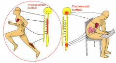
What spinal regions do sympathetic nerves come from?
|
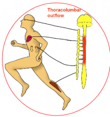
Thoracic & Lumbar (thoracolumbar outflow)
|
|
|
What spinal regions do parasympathetic nerves come from?
|
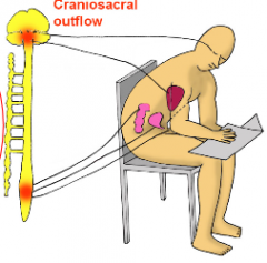
Cranial & Sacral (craniosacral outflow)
|
|
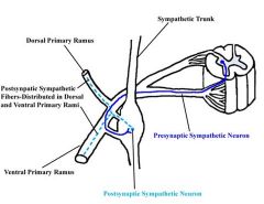
Label each of the
following structures |
1. Dorsal root ganglion
2. Anterior root of spinal nerve 3. Pre-ganglionic sympathetic fibre 4. Nerve Plexus 5. Sympathetic trunk 6. Sympathetic chain ganglion 7. Post-ganglionic sympathetic fibre 8. Lateral horn 9. White communicating ramus 10. Grey communicating ramus 11. Mixed spinal nerve (later branches in to dorsal/ventral rami) 12. Paravertebral sympathetic ganglion |
|
|
Where do nerves from the anterior horn of the spinal nerve lead to?
|
Anterior horn goes to both dorsal and ventral rami to carry out its efferent function
|
|
|
What regions do the anterior ramus of the spinal nerve lead to?
|
Antero-lateral aspects of the trunk + limbs
|
|
|
What regions do the posterior ramus of the spinal nerve lead to?
|
Skin + muscles of back
|
|
|
What is the function of the white ramus?
|

White rammus carries myelinated pre-ganglionic sympathetic axons to the paravertebral sympathetic chain ganglion where the signal can travel to 1 of 3 destinations
|
|
|
What is the function of the grey ramus?
|
Contains postganglionic sympathetic fibres which have synapsed in the paravertebral sympathetic ganglion
Postganglionic sympathetic fibres can go to either dorsal or ventral ramus |
|
|
What is a paravertebral sympathetic ganglion?
|
A sympathetic chain ganglion located just lateral to its corresponding vertebra
|
|
|
What are the 3 pathways a preganglionic sympathetic fibre can take after passing the white ramus?
|
1. Synapses in paravertebral ganglion
=> travels through gray ramus of adjacent level => either goes through ventral or dorsal ramus to effector organ 2. Bypasses paravertebral ganglion of that level => travels along sympathetic trunk until it reaches a paravertebral ganglion of a different level => exits through gray ramus of that level => goes to the ventral or dorsal ramus closer to its effector organ 3. Bypasses paravertebral ganglion => travels long way through sympathetic nerve until it reaches a prevertebral ganglion => synapses there, allowing postganglionic fibre to innervate viscera nearby |
|
|
What is a prevertebral sympathetic ganglion?
|
A sympathetic ganglion located between the paravertebral ganglion and the target organ
|
|
|
How is the parasympathetic nerve pathway arranged?
|
1. Exits through lateral horn
2. Preganglionic parasympathetic nerves travel through anterior root 3. Travel through either ventral or dorsal rami 4. Go a long way till they hit parasympathetic ganglion (adjacent to viscera to be innervated) 5. Synapse in ganglion then postganglion fibres innervate viscera |
|
|
What is the enteric nervous system?
What is it influenced by? |
Nervous system with neurons in the brain and the GI tract
- influenced by SNS/PNS - allows for peristalsis and secretions of GI tract |
|
|
What is the mesoderm?
|
The middle germ layer of the embryo
|
|
|
What are the 3 primary divisions of mesoderm in the embryo?
|
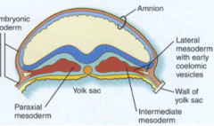
1. Paraxial mesoderm
2. Intermediate mesoderm 3. Lateral mesoderm |
|
|
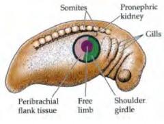
What is the name for the general structures that the paraxial mesoderm forms?
How does it form these structures? |
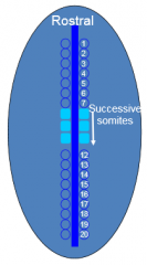
Paraxial Mesoderm forms the 'somites'
1. Swellings appear at stages along paraxial mesoderm => somitomeres 2. Starting with the 8th somitomere, somitomeres are replaced with somites caudally |
|
|
What happens to somitomeres that don't form somites?
|
The rostral somitomeres (1-7) + the neural crest cells form the head
|
|
|
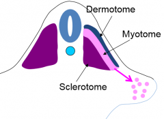
What are the 3 general divisions of the somites and the structures they form in the body?
|
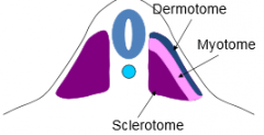
1. Sclerotome
- Axial skeleton except skull 2. Dermatome - Dermis of skin 3. Myotome - Torso muscles |
|
|
What fate do the medial myotome somitic cells have?
What about the lateral myotome? |

Medial
1. intrinsic back muscle Lateral 1. limb muscle 2. ventrolateral body wall muscles |
|
|
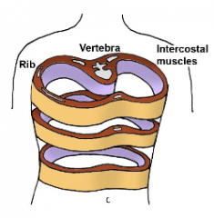
What fate do the medial sclerotome somitic cells have?
What about the lateral sclerotome? |
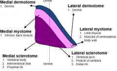
Medial
1. Vertebral body 2. Intervertebral disc 3. Proximal rib Lateral 1. Vertebral arch 2. Pedicle of vertebra 3. Distal rib |
|
|
What is the difference in fates of the medial and lateral dermotome?
|
None!
They both form the dermis of skin. I tricked you. Don't let me trick you. |
|
|
What is the general fate of the intermediate mesoderm?
|
Forms the urogenital system (exc. bladder & urethra)
- Urinary tract forms before the genitalia do |
|
|
What are the 3 stages of kidney tissues?
From this, describe the kidney or reproductive structures formed by each. (where necessary, describe the differences between males and females) |
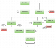
http://imgur.com/tqe2UjD
|
|
|
What are the 2 divisions of lateral plate mesoderm?
What do they form? |
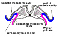
1. Splanchic Mesoderm (interior)
+ endoderm = Viscera 2. Somatic Mesoderm (exterior) + ectoderm = Body Wall |
|
|
How is the coelom of the embryo formed?
|
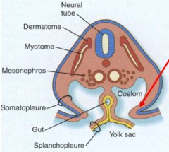
Coelom space is enclosed by the splanchnic & somatic mesoderm
*Somatopleure = somatic mesoderm + ectoderm *Splanchopleure = splanchnic mesoderm + endoderm |
|
|
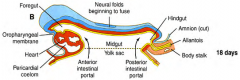
How is the yolk sac separated from the embryo?
|
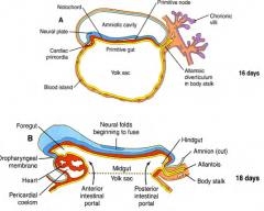
Via a 'purse string suture'
|
|
|
What is formed from the pinched off lumen space, following a purse string suture of the embryo?
|
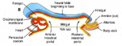
The Gut:
1. Foregut (anterior) 2. Midgut (centre) 3. Hindgut (posterior) |
|
|
How is the oral cavity formed?
|
- Prior to 3 weeks, oropharyngeal membrane (oral plate) separates stomadeum & foregut
- Breaks down allowing ectoderm and endoderm to meet |
|
|
How is the anal cavity formed?
|
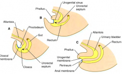
1. Anus sealed by cloacal membrane (endo + ecto)
2. Allantois is currently a branch of hind gut 3. Urorectal septum extends to divide allantois and hind gut 4. Hindgut now becomes rectum and allantois forms the bladder and urogenital tract 5. Urogenital membrane covers allantois, perineal membrane covers rectum, both break later |
|
|
What is the allantois?
|
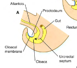
The allantois is initially a branch of the hindgut
=> stores nitrogenous waste in some species (vestigial in humans) => later gives rise to bladder + urogenital tract |
|
|
What arteries supply the:
i) Foregut ii) Midgut iii) Hindgut |
i) Coeliac Artery
ii) Superior Mesenteric Artery iii) Inferior Mesenteric Artery |
|
|
What is GIT herniation?
What are 2 ways in which this can fail? |
- Gut grows too quickly for the body cavity (at 6/7 weeks)
- Gut herniates in to the umbilical cord to allow for more room - Gut retracts back in to body at week 9 when the body cavity is larger 1. Omphalocele = failure to retract 2. Umbilical hernia = retraction but failure to seal |
|
|
At the what point of development is the GIT still connected to the yolk sac?
|
At around 1 month
|
|
|
How does growth of the GIT occur?
|
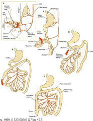
1. Midgut buckles around the yolk stalk attachment region
2. Large intestine folds across the small intestines around the axis of yolk stalk 3. Small and large intestines then further elongate to give characteristic folding |
|
|
Where do the viscera develop from?
|
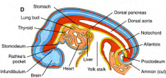
Endodermal outpockets which interact with the mesoderm
|
|
|
How does the liver develop?
|
1. Starts as growth from the ventral wall of GIT
2. Grows within the 'septum transversum' (diaphragm precursor) 3. Anchored to ventral aspect of body wall by 'falciform ligament' 4. Rotates right as the gut folds 5. Connective tissue and hepatic capsule formed by mesoderm 6. Liver then starts production of fetal blood cells |
|
|
How does the lung develop?
|
1. Endoderm forms outpockets between liver and pharyngeal arches
2. Formation of trachea 3. Splitting in to primary bronchi 4. Spilitting in to secondary bronchi 5. Total of 23 successive splits |
|
|
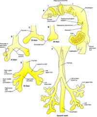
How does the pancreas develop?
|
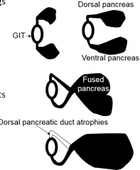
1. 2 Outpockets of endoderm form dorsal and ventral pancreas
2. The two fuse with each other 3. Dorsal pancreatic duct degrades |
|
|
What are the pharyngeal arches?
|
- Gill remnants
- Arise from neural crest cells - Gives rise to various facial structures |
|
|
What are the pharyngeal pouches?
|
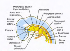
Pouches which form on the endodermal side of the pharyngeal arches
- 4 pouches with each forming specific structures |
|
|
What are the structures that each pharyngeal pouch forms (4 total) ?
|
Pouch 1.
i) Eustachian (auditory) tube Pouch 2. i) Palatine tonsil Pouch 3. i) Parathyroid (inferior) ii) Thymus (epithelial layer) Pouch 4. i) Parathyroid (superior) ii) Thymus iii) Postbranchial bodies |
|
|
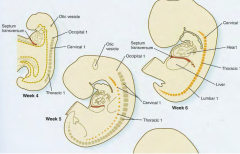
In what period of time do the limbs form?
|
4-8 Weeks
|
|
|
What sections of the germ layers form the limb bud?
|
Lateral myotome (paraxial mesoderm) = Muscle
Lateral plate mesoderm =Bone + connective tissue |
|
|
What is the limb field?
|
A patch of mesoderm (per limb) which has the capacity to form limb
- composed of 'free limb' mesoderm patch and surrounding mesodermal tissue - removal of free limb patch usually does not affect limb development as surrounding tissue can compensate |
|
|
How does a limb develop from a limb bud?
How is this regulated? |
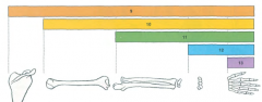
1. Ectoderm overlaying limb bud forms 'apical ectodermal ridge'
2. Ridge allows control of limb growth 3. Hox genes regulate segmentation and sequencing of limb 4. As Hox is expressed successively in the growing limb bud, loss of one hox gene typically results in loss of single corresponding limb segment |
|
|
What abnormality results from the incorrect splitting of the ectodermal ridge in a developing limb?
|
Diplopodia
|
|
|
What does thalidomide do?
|
Originally used to treat morning sickness
- indentified as a teratogen (inteferes with development) - disrupts limb development within the first trimester |
|
|
How do the digits develop?
|
1. Hands and feet start off as symmetrical discs
2. Sculpted by apoptosis to form digits 3. Other mechanisms determine digit number (1-5) |
|
|
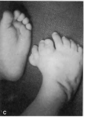
What can happen with incorrect sculpting of the digits?
|
1. Webbing of the fingers
2. Syndactly (fused fingers) |
|
|
Name 3 general functions of muscles
(6 in total) |
1. Produce movement
2. Provide stability 3. Contorl body openings 4. Produce heat 5. Assist circulation 6. Aids digestion |
|
|
What are the 3 types of muscle?
Are they: i ) voluntary/involuntary ii) striated/non-striated iii) somatic/visceral |
1. Skeletal
i) Voluntary ii) Striated iii) Somatic 2. Smooth i) Involuntary ii) Non-striated iii) Visceral 3. Cardiac i) Involuntary ii) Striated iii) Visceral |
|
|
What are the 4 layers of connective tissue that surround the layers of the muscle?
|
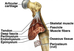
1. Single muscle fibre surrounded by
endomysium 2. Several muscle fibres surrounded by perimysium to form fascicle 3. Several fascicles surrounded by epimysium to form the entire muscle 4. The epimysium is then continuous with the deep fascia which is similar but thicker, allowing attachment to the tendon (and subsequently the bone) |
|
|
What are deep fascia?
|
-Dense layers of fibrous connective tissue
-Can form intermuscular septa separating different muscles in to functional fascial compartments |
|
|
What is a retinaculum?
What does it prevent? |
Deep fascia that forms a tight band, holding tendons in place
=> prevents bow stringing of tendons |
|
|
What is a tendon?
|
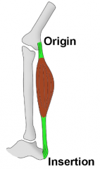
Tough bands of connective tissue which allow muscles to attach to bones
|
|
|
What is an aponeurosis?
|
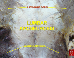
A large connective tissue sheet similar in function to the tendon
|
|
|
What is a raphe?
|
Fibrous connective tissue where two muscles meet at the midline
|
|
|
What is a fascial sheath?
|
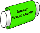
Connective tissue which envelops a neuromuscular bundle loosely
|
|
|
What is a synovial tendon sheath?
How would an infection in the synovial sheath spread? |
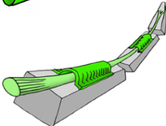
Connective tissue that surrounds tendon
- forms double layered tube - prevents tendons from getting sheared by bone Infection would be confined to the sheath unless it is continuous with a bursa in which case it would spread there |
|
|
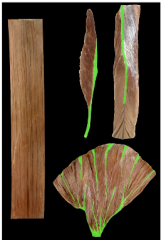
What are the respective features of:
1) Fusiform muscles 2) Parallel muscles 3) Convergent muscles |
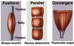
1. Fusiform (subdivision of parallel)
i) Wide belly ii) Narrow tendons iii) Quick action 2. Parallel i) Long parallel fibres to force generating axis ii) Powerful and fast contractions 3. Convergent i) Many origins converge to tendonous insertion |
|
|
What are the defining features of:
1) Unipennate muscles 2) Bipennate muscles 3) Multipennate muscles 4) Circular muscles |
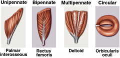
1. Unipennate
- muscles that run along 1 side of a tendon 2. Bipennate - muscles that run along both sides of a tendon 3. Multipennate - there are multiple bipennate muscles, which run at different angles 4. Circular - circular in shape - typically sphincter or orifice muscles |
|
|
What are the 3 types of muscle levers? How do they act?
|
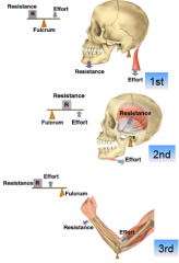
1. First Order
2. Second Order 3. Third Order |
|
|
What is the difference between parallel and oblique muscles?
|
Parallel
- range of motion proportional to length Oblique - power proportional to mass |
|
|
What are some ways muscles are named by?
|
1. Size (e.g. pec major)
2. Shape (e.g. deltoids) 3. Location (e.g. rectus abdominis) 4. Number of heads (e.g. biceps brachii) 5. Action (e.g. flexor carpi ulnaris) 6. Quirky (e.g. soleus) |
|
|
What is a reflex contraction?
|
A very fast involuntary contraction that is integrated in the spinal cord
- e.g. diapgragm movement or knee-jerk |
|
|
What is the difference between tonic contraction and phasic contraction?
|
Tonic contraction is the normal sustained contraction (typically unconscious)
Phasic contraction is the voluntary tension induced to perform an action |
|
|
What are the 2 forms of phasic contraction?
|
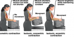
1. Isometric contraction
- Increased muscle tension - No length change or movement - i.e. Muscles act against immovable force 2. Isotonic contraction i) Concentric contraction - High muscle tension - Muscle shortens and movement ii) Eccentric contraction - Lower muscle tension - Muscle lengthens and movement |
|
|
What are the 4 actions that muscles can have?
|
1. Prime Mover
- Main action 2. Synergist - Assists prime mover 3. Antagonist - Acts against the prime mover 4. Fixator - Stabilises the prime mover |
|
|
What are the 3 possible sites of muscle injury?
|
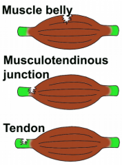
1. Muscle Belly
2. Musculotendinous Junction 3. Tendon |
|
|
Where does a muscle's neurovascluar supply enter from?
|
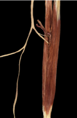
The Neurovascular Hilum
|
|
|
Is the arrangement of nerves to the muscle caudal-cranial or cranial-caudal?
|
Cranial-caudal
|
|
|
From what germ layer is the circulatory system derived from?
What implications does this have? |
Circulatory system is mesoderm derived
=> the circulatory system only goes to similarly mesoderm derived structures * Exception is articular cartilage which is from mesoderm but is avascular |
|
|
What is the general structure of the vessels (3 layers) ?
|
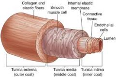
1. Tunica intima
- Endothelium 2. Tunica media - Smooth muscle 3. Tunica adventitia - Fibrous tissue |
|
|
What are the structural differences between the arteries and veins? How do these relate to their function?
|
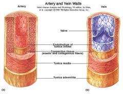
1. Arteries have thicker smooth muscle walls as they have to pump blood for further distances and withstand higher pressure
2. Veins have larger lumens as they don't have to withstand high pressures, and a larger diameter allows greater blood flow 3. Arteries have high elastic fibre content allowing them to stretch and withstand higher pressures 4. Veins have valves to prevent backflow |
|
|
How does the structure of the lymphatics compare to the arteries and veins?
|
In general, they are a lot smaller and have very thin walls. They also contain valves.
|
|
|
What are the 2 types of arteries and their features?
|
1. Elastic arteries
- Closest to the heart - Act as 'conducting vessels' - Large elastic tissue composition - Can stretch in response to each pulse - Allows maintenance of relatively constant BP 2. Muscular arteries - Large amount of smooth muscle in media - Act as 'distribution vessels' - Grow progressively smaller - Branch to somatic structures - Possible redistribution/rechanelling |
|
|
What are arterioles?
|
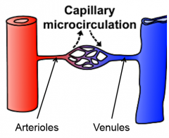
Small branches of the arteries
- controlled by 'vasomotor nerve fibres' - mostly SNS innervation - tonus determines BP |
|
|
What are capillaries?
|

Exchange vessels between the arterioles and venules
- Thinnest walled (1 cell width) - Arranged in capillary beds - Exchange mediated by hydrostatic pressure difference |
|
|
How can collateral circulation form?
|
Anastamoses between arteries or blood vessels
|
|
|
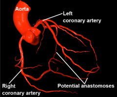
What are the 3 types of anastamoses?
|
1. True anastamoses
= artery + artery 2. Potential anastamoses = arteriole + arteriole 3. AV anastamoses (heat reduction) = artery + vein |
|
|
What are end arteries?
What are the 2 types of end arteries? |
Arteries that don't link up with other arteries
1. Anatomical - Single artery which does not anastamose - Occlusion = necrosis 2. Functional - Artery with potential small-caliber arteriole anastomosis - Occlusion usually leads to dilation of collaterals |
|
|
What is a thrombus?
|
A blood clot that has formed and is localised
|
|
|
What is an embolus?
|
A blood clot that has formed and is delocalised
- travels through body and obstructs a different blood vessel |
|
|
In what direction does the porforating vein valves direct blood?
|
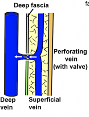
Superficial to deep
|
|
|
What veins don't have valves?
Why is this? |
Deep veins of the trunk
- Thoracic pump sufficient to return blood to heart |
|
|
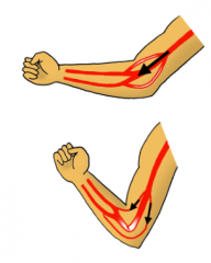
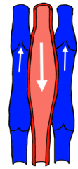
What are the 3 mechanisms (excluding valves) through which veins can return blood to the heart?
|
1. Venae comitantes
2. Thoracic pump 3. Musculovenous pump |
|
|
What is a venae comitans?
Where is typically found? |
A pair of veins wraped wrapped around an artery
=> Found mostly in limbs => Countercurrent heat exchange => Artery pulsations squeeze veins aiding in venous return |
|
|
How does the thoracic pump function?
|
1. During inspiration, the diaphragm is pulled down increasing thoracic volume and decreasing thoracic pressure
=> allows venous blood to flow down concentration gradient in to heart via IVC 2. During expiration, the diaphragm is pulled up decreasing thoracic volume and increasing thoracic pressure => forces venous blood in to heart via SVC |
|
|
What is the musculovenous pump?
|
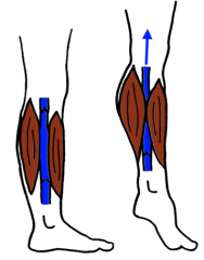
Veins can be wedged between pairs of muscles
=> contraction of muscles forces blood along veins |
|
|
How do varicose veins form?
|
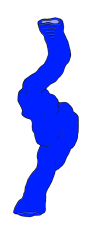
Loss of elasticity in the perforating veins
|
|
|
What is lymph?
|
Fluid similar in composition to blood plasma that circulates through the lymphatic system
|
|
|
Where do lymph vessels carry lymph to?
What happens there? |
Carries lymph to lymph nodes
=> lymph is exposed to defence cells and filtered |
|
|
How is lymph returned to the body?
What are the 2 main structures which drain this lymph and what areas of the body do they drain from? |
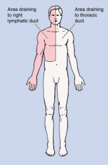
Lymph is returned to the body via venous circulation
1. Right lymphatic duct => drains upper right quadrant 2. Thoracic duct => drain everything else |
|
|
What are the 3 mechanisms that lymph uses to flow across the body?
|
1. Milking effect
=> close contact with veins 2. Squeezing effect => squeezed by surrounding muscles 3. Sucking effect => interthoracic pressure due to respiration |
|
|
What are lymph nodes?
|
Structures composed of lymphoid tissue
- Filter lymph and expose antigens to immune system - All lymph drains through at least 1 lymph node before returning to venous circulation - Can have many pathways in but only 1 out |
|
|
What are the major sites lymph nodes are found?
|
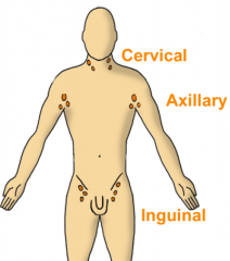
1. Cervical region (i.e. neck)
2. Axillary region (i.e. armpit) 3. Inguinal region (i.e. groin) |
|
|
What sort of autonomic nerve supply do blood vessels receive?
|
- Mostly SNS (except erectile)
=> allows maintenance of partial tone |
|
|
What sort of bloody supply do vessels recive?
|
- Media and adventitia receive their own blood supply via the vasa vasorum
- Endothelium and intima receive nutrition through diffusion |
|
|
What are the 4 roles of the skin?
|
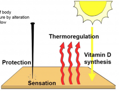
1. Protection
2. Thermoregulation 3. Sensation 4. Vitamin D synthesis |
|
|
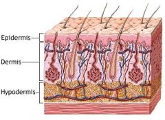
What are the 3 layers of the skin from superficial to deep?
|
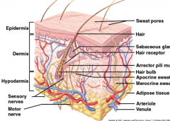
1. Epidermis
2. Dermis 3. Hypodermis |
|
|
What are the 3 cell types of the epidermis?
|
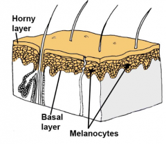
1. Keratinocytes
- Tough protection 2. Melanocytes - Pigmentation 3. Basal Cells |
|
|
What is the stratum corneum?
What is the stratum basalis? |

Corneum = the topmost layer of the epidermis
(keratinocytes + melanocytes) Basalis = the bottom layer of the epidermis (basal cells) |
|
|
What are some structures found in the dermis of the skin?
(7 total) |
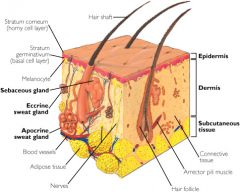
1. Blood Vessels
2. Arrector Pili Muscles 3. Sweat glands 4. Sebaceous glands 5. Hair follicle 6. Nail roots (dead keratenous cells) 7. Sensory nerve endings |
|
|
What are the 2 types of sweat glands (based on location) and where are they found?
|
1. Merocrine
- All around body 2. Apocrine - Armpits and genital regions |
|
|
What are the 2 zones of the dermis? What are their properties?
|
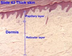
1. Papillary Layer
- Superficial to reticular layer - Collagenous connective tissue - Interface of epidermis & dermis 2. Reticular Layer - Elastic tissue - Provides strength & flexibility |
|
|
What are the properties of the hypodermis? Of what clinical significance is this?
|
Hypodermis is largely composed of subcutaneous tissue which is highly areolar
=> point of insertion for hypodermic needles as fluid can travel and diffuse through holes |
|
|
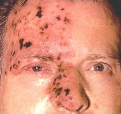
What is the rule of 9s?
|
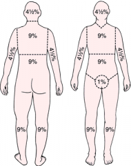
A rule that segments the body in to sections of skin surface area (9% each) that aids in determining the loss of fluid following a burn injury
|
|
|
What is a pilosebaceous unit?
|
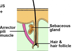
Hair follicle + Sebaceous gland + Arrector pili
|
|
|
Name some types of sweat glands, and the product they secrete
(4 total) |
1. Mammary
=> milk 2. Odoriferous => odour 3. Sebaceous => oil 4. Ceruminous => ear wax |
|
|
What parts of the skin receive a neurovascular supply?
|
Dermis receives rich neural and vascular supply. The rest don't. Vessels travel through the hypodermis but it receives little of the supply itself.
|
|
|
What are the 2 types of sensory nerve fibres in the skin and what do they detect?
|
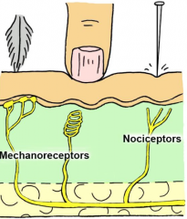
1. Mechanoreceptors
- detect change in pressure 2. Nociceptors - detect potential harm (heat, chemical, etc.) |
|
|
What are the 3 types of skin motor nerve fibres?
|
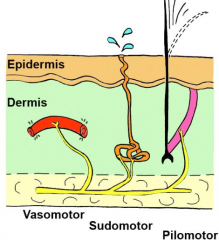
1. Vasomotor
- costriction/dilation of blood vessels 2. Sudomotor - stimulation of sweat glands 3. Pilomotor - control of arrector pili |
|
|
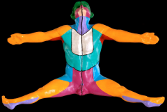
Why do skin infections not cross the midline?
|
Because the dermatome that is infected is linked back to a spinal nerve. These spinal nerves branch out from the midline but never cross. Nerve infections travel along the dermatome.
|
|
|
Is there more overlap for adjacent touch dermatomes or pain & temperature dermatomes?
|
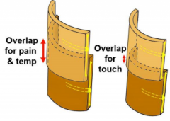
Pain & temperature
=> allows large changes in temperature to be registered as painful |
|
|
What is meant by 'referred pain'?
How is it caused? |
Pain experienced at a site different from the source
=> pain from 1 site has sensory fibres which return to its vertebra => can be mistaken for pain coming from dermatome linked to same vertebra |
|
|
What is a lymphotome?
|
The area of skin drained by a particular lymph node
|
|
|
What is the clinical significance of lymph nodes?
|
Metastatic tumors (cancer) can spread along the lymphatic system to lymph nodes where they aggregate (i.e. they're a site for checking cancer)
|
|
|
What are the 3 steps involved in the formation of the heart tube?
|
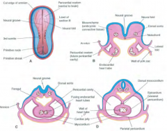
1. Trilaminar embryo forms mesoderm in horseshoe shape
2. Splanchnic mesoderm (lateral plate) forms bilateral endocardial heart tubes 3. Bilateral heart tubes fuse to form heart |
|
|
What 3 mesodermal tissues form the following structures of the heart:
a) Muscle b) Sac c) Epithelial lining |
a) Myocardium
b) Pericardium c) Endocardium |
|
|
How does the heart fold in development?
|
1. Starting from a tubular heart, this fold and elongates in to a U shape
2. Reorganisation of this structure allows a 4 chambered heart to form |
|
|
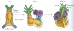
Describe the structure of a tubular embryonic heart
|
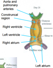
1. A pair of vitelline veins converge to form the left atrium
2. The left atrium then leads on to the right atrium 3. The right atrium is separated from the left ventricle via the sinus venosus 4. The left ventricle is continuous with the right ventricle 5. the right ventricle is separated from the aorta and pulmonary arteries by the truncus arteriosus |
|
|
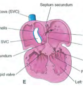
How are the atria divided in embryonic development?
(4 steps) |
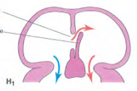
1. Septum primum starts to divide atria (superior to inferior)
2. Septum primum fuses with endocardial cushion, shutting the Foramen primum 3. The Foramen secundum forms from perforations in the septum primum 4. The Septum secundum develops (superior to inferior) to meet septum primum forming the foramen ovale |
|
|
How are the ventricles divided in embryonic development?
|
Ventricular septum grows from the apex of the heart superiorly
=> forms a thick but incomplete septum => final part of septum closed by thin membrane |
|
|
What is the difference between vasculogenesis and angiogenesis?
|
Vasculogenesis = formation of blood vessels from scratch
Angiogenesis = formation of blood vessels from pre-existing ones |
|
|
When does the embryo stop circulating blood through diffusion, and start circulating blood through vessels?
|
Start of 3rd week of development
|
|
|
How does the aortic arch form its asymmetrical structure?
|
Starts off symmetrical and segmented
=> some blood vessels are lost and others are degraded to become asymmetrical |
|
|
What are bursae and what functions do they hold?
|
Double layered pockets of synovial membrane, filled with synovial fluid
- Reduce friction - Provide padding - Can allow communication (and also spread of infection) to joint cavity |
|
|
What are fat pads?
|
Self-explanatory
- Act as space fillers - Intracapsular but extrasynovial (i.e. between the tendon and the articular capsule) - Help spread synovial fluid - Has neurovascular supply |
|
|
What are the 2 types of joint injury
|
1. Dislocation
- Full separation of bones in a joint 2. Subluxation - Partial displacement of bones in a joint |
|
|
What areas of the joint possess a neurovascular supply?
|
- Capsules & ligaments have a rich pain/proprioception fibres
- Articular cartilage is avascular and aneural |
|
|
What is Hilton's Law?
|
States that nerves supplying the muscle surrounding a joint also supply the joint
|
|
|
Where does the blood supply of joints come from?
|
Adjacent muscle
|
|
|
What are the 2 types of arthritis? How are they respectively caused?
|
1. Rheumatoid arthritis
=> autoimmune attack of synovial membrane 2. Osteoarthritis => mechanical degradation of joint over time |
|
|
What is arthroplasty?
|
Replacement of a diseased joint with an artificial device (prosthesis)
|
|
|
What is the thymus? What is special about the thymus? |
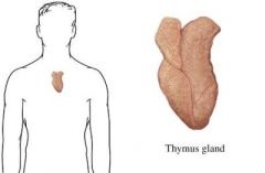
A specialised organ of the immune system, composed of lymph tissue. It reaches its greatest size during puberty and then involutes with age. |
|
|
What are the peyer's patches and where are they located? |
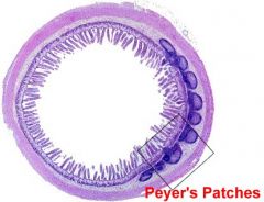
Collections of lymphoid tissues located in the muscosa of the GIT |