![]()
![]()
![]()
Use LEFT and RIGHT arrow keys to navigate between flashcards;
Use UP and DOWN arrow keys to flip the card;
H to show hint;
A reads text to speech;
44 Cards in this Set
- Front
- Back
|
classify bones according to shape: long, short, flat, irregular |
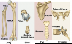
long: humerus, ulna, femur short: carpals, tarsals flat: skull, sternum, scapula, ribs irregular: vertebrae, mandible, pelvis |
|
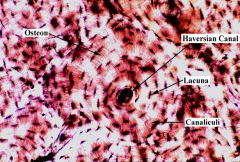
identify structures of compact bone: osteon, central haversian canal, lacunae, canaliculi, osteocytes, lamellae
|
osteon: the basic structural unit of compact bone; consists of central canal, lamellae (rings), lacunae, osteocytes, and canaliculi central haversian canal: in the middle of compact bone; contains blood vessels, connective tissue, nerve fibers, and lymphatic vessels lacunae: small spaces between lamellae; contain osteocytes canaliculi: tiny canals radiating in all directions from lacunae; allow for communication between osteocytes and the passing of materials that make up the matrix osteocytes: bone cells lamellae: concentric rings of bone matrix that radiate out from central canal |
|
|
differentiate types of vertebrae |
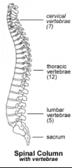
cervical: 7 thoracic: 12 lumbar: 5 sacrum |
|
|
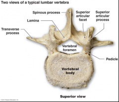
differentiate markings on vertebrae: body, foramina, spinous process, lamina, transverse processes |
|
|
|
identify and differentiate types of bone cells: osteoblasts, osteoclasts, osteocytes |
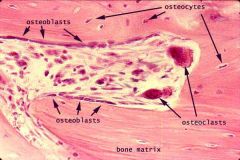
osteoblasts: make bone osteoclasts: destroy bone osteocytes: bone cells |
|
|
know mechanism of movement for C1 and C2 |
C1: atlas; supports the skull where the head attaches to the neck C2: axis; allows the head to rotate from its support atop the C1 vertebra where the skull attaches to the neck |
|
|
know function of cartilage in between each vertebrae |
cushion and soften the forces created by walking and jumping, which might otherwise fracture the vertebrae or jar the brain |
|
|
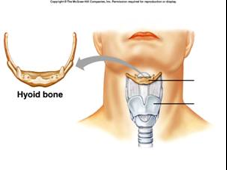
locate the hyoid bone |
|
|
|
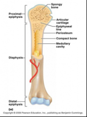
identify regions of a long bone: epiphysis, diaphysis, compact and cancellous bone tissue, epiphyseal plate |
|
|
|
differentiate true, false, and floating ribs |
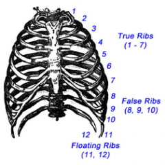
true: 1-7 false: 8-10 floating: 11,12 |
|
|
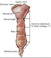
identify parts of the sternum: suprasternal or jugular notch, clavicular notch, sternal angle, manubrium, body, and xyphoid process |
|
|
|
identify the clinical significance of the sternal angle |
marks the location of the second rib. a doctor can count ribs and know where to listen for specific heart sounds and more. |
|
|
identify the clinical significance of the xyphoid process |
used as a landmark during CPR to know where to do chest compressions |
|
|
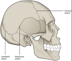
identify skull sutures: coronal, sagittal, lambdoid, squamous |
|
|
|
identify bones of a disarticulated skeleton: carpals, tarsals, metacarpals, metatarsals, calcaneus, talus... |
|
|
|
identify major bones on an articulated skeleton |
*refer to handout |
|
|
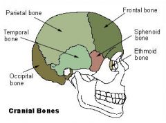
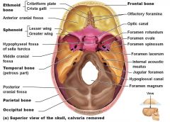
identify major bones of the cranium: frontal, parietal, occipital, temporal, zygomatic, maxilla, mandible |
|
|
|
identify major foramen and features of the skull and match to function: foramen ovale, mental foramen, external auditory meatus, occipital condyles, foramen magnum, styloid process, mastoid process |
|
|
|
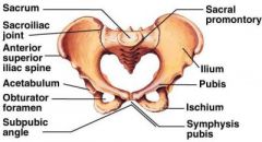
identify pelvic structures: illium, ischium, pubis, symphysis pubis, pelvic/subpubic angle, illiac crest, acetabulum, obturator foramen, ischial tuberosity |
|
|
|
differentiate male and female pelvis |
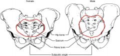
-Because the female pelvis is adapted for childbirth, it is wider than the male pelvis -The female sacrum is wider, shorter, and less curved |
|
|
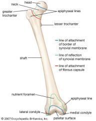
identify features of the femur: head, neck, greater trochanter, lateral/medial epicondyle, lesser trochanter |
|
|
|
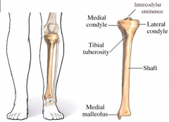
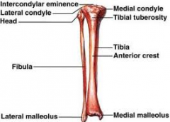
identify features of the tibia: medial/lateral condyle, medial malleolus |
|
|
|
identify features of the fibula: head, lateral malleolus |
|
|
|
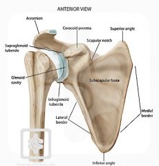
identify scapula structures: acromion process, coracoid process, superior angle, inferior angle |
|
|
|
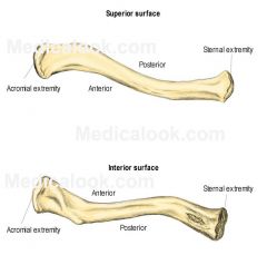
identify clavicular structures: acromial end, conoid tubercle, sternal end |
|
|
|
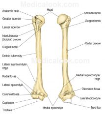
identify features of the humerus: head, greater/lesser tubercle, medial/lateral epicondyle |
|
|
|
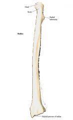
identify features of the radius: head, neck, radial tuberosity, styloid process |
|
|
|
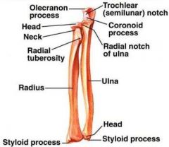
identify features of the ulna: head, olecranon process, styloid process, ulnar notch |
|
|
|
classify joints according to type: fibrous, cartilaginous, synovial |
|
|
|
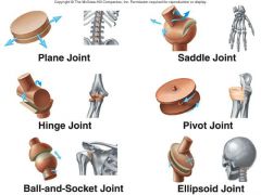
classify joints according to movement: plane, hinge, pivot, ellipsoid, saddle, ball & socket |
|
|
|
define various actions occurring at joints |
monoaxial: occurring around 1 axis biaxial: occurring around 2 axes at right angles to each other multiaxial: occurring around several axes |
|
|
identify structures of a synovial joint |
contains synovial fluid |
|
|
treppe |
the gradual increase in muscular contraction following rapidly repeated stimulation. |
|
|
temporal (wave) summation |
sensory summation that involves the addition of single stimuli over a short period of time |
|
|
incomplete tetany |
summations occur rapidly, but muscle still relaxes slightly between contractions. Muscle is stimulated again and again before it fully relaxes. |
|
|
complete tetany |
Stimuli arrive so rapidly there is NO relaxation that occurs. |
|
|
muscle fatigue |
Decreased ability to do work. POSSIBLE CAUSES: psychologic fatigue: CNS dysfunction. most common. muscular fatigue: depletion of ATP in muscles. second most common. synaptic fatigue: depletion of acetylcholine in the neuromuscular synapse. least common. |
|
|
length-tension relationship |
Tension is the force applied to an object to be lifted when a muscle contracts. The initial length of a muscle has a strong influence on the amount of active tension it produces. As the length of the muscle increases, its active tension also increases, to a point. |
|
|
explain how frequency of stimulation, voltage, and muscle stretch affect muscle tension |
all or none response: a muscle fiber or motor unit contracts with a consistent force in response to each action potential recruitment/multiple motor unit summation: for a whole muscle, a stimulus of increasing magnitude results in graded contractions of increased force as more motor units are recruited |
|
|
explain the myofilament theory of muscle contraction |
actin and myosin myofilaments do not change in length during contraction. actin and myosin myofilaments slide past one another in a way that causes sarcomeres to shorten (cross-bridging). the I band and H zones become narrower during contraction, and the A band remains constant in length. |
|
|
explain the role of ATP in contraction of muscles |
the binding of ATP to the cross bridge results in the cross bridge disconnecting from actin. |
|
|
list, in sequence, the steps of impulse conduction across a neuromuscular synapse |
1. influx of calcium 2. binding of myosin to actin 3. the power stroke of the cross bridge that causes the sliding of the thin filaments 4. the binding of ATP to the cross bridge results in the cross bridge disconnecting from actin 5. the hydrolysis of ATP into ADP and Pi leads to the re-energizing and repositioning of the cross bridge 6. the transport of calcium ions back into the sarcoplasmic reticulum |
|
|
muscle of respiration |
diaphragm |
|
|
insertion and origin of biceps brachii |
origin: scapula insertion: radius |