![]()
![]()
![]()
Use LEFT and RIGHT arrow keys to navigate between flashcards;
Use UP and DOWN arrow keys to flip the card;
H to show hint;
A reads text to speech;
42 Cards in this Set
- Front
- Back
|
crest |
A Crest is a ridge or raised surface on the skeleton. |
|
|
epicondyle |
Epicondyle - A prominence that sits atop of a condyle. The epicondyle attaches muscle and connective tissue to bone, providing support to this musculoskeletal system. |
|
|
foramen |
A foramen (plural: foramina ) is an opening inside the body that allows key structures to connect one part of the body to another. |
|
|
fossa |
A fossa (from the Latin "fossa", ditch or trench) is a depression or hollow, usually in a bone, such as the hypophyseal fossa, the depression in the sphenoid bone. |
|
|
suture |
Skull sutures are immobile joints where cranial bones are connected with dense fibrous tissue. |
|
|
trochanter |
The greater trochanter of the femur is a large, irregular, quadrilateral eminence and a part of the skeletal system. |
|
|
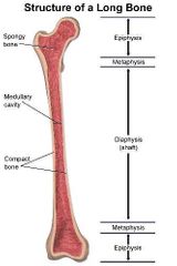
femur |
the proximal bone of the hind or lower limb that extends from the hip to the knee. — called also thighbone. |
|
|
Hematopoiesis |
Hematopoiesis is the process by which immature precursor cells develop into mature blood cells. The currently accepted theory on how this process works is called the monophyletic theory which simply means that a single type of stem cell gives rise to all the mature blood cells in the body. |
|
|
parts of the femur |
|
|
|
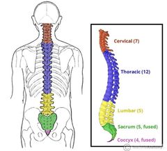
axial skeleton. |
is the part of the skeleton that consists of the bones of the head and trunk of a vertebrate. In the human skeleton, it consists of 80 bones and is composed of six parts; the skull (22 bones), the ossicles of the middle ear, the hyoid bone, the rib cage, sternum and the vertebral column. |
|
|
appendicular skeleton |
The appendicular skeleton is the portion of the skeleton of vertebrates consisting of the bones that support the appendages. The appendicular skeleton includes the skeletal elements within the limbs, as well as supporting shoulder girdle pectoral and pelvic girdle. |
|
|
male verus female pelvis |
The female pelvis is larger and broader than the male pelvis, which is taller (owing to a higher iliac crest), narrower, and more compact. The distance between the ischium bones is small in males. ... This results in the female inlet being large and oval in shape, while the male inlet is more heart shaped. |
|
|
spinal column |
|
|
|
first cervical vertebrae |
In anatomy, the atlas (C1) is the most superior (first) cervical vertebra of the spine. It is named for the Atlas of Greek mythology, because it supports the globe of the head which is the skull. The atlas is the topmost vertebra and with the axis forms the joint connecting the skull and spine. |
|
|
axis vertbra |
In anatomy, the second cervical vertebra (C2) of the spine is named the axis (from Latin axis, "axle") or epistropheus. By the atlanto-axial joint, it forms the pivot upon which the first cervical vertebra (the atlas), which carries the head, rotates. |
|
|
bone cells/ osteoblast |
a cell that secretes the matrix for bone formation. |
|
|
bone cells/ oseoclasts |
Osteoclasts are the cells that degrade bone to initiate normal bone remodeling and mediate bone loss in pathologic conditions by increasing their resorptive activity. |
|
|
bone cells/ osteocytes |
Osteocytes are mature osteoblasts that have become trapped within the very bone matrix they produced. Osteocytes continue to form bone to some degree, which is important for maintaining the strength and health of the bone matrix. |
|
|
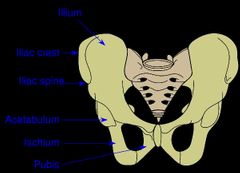
ilium |
the large broad bone forming the upper part of each half of the pelvis. |
|
|
iliac crest |
The iliac crest is the thick curved upper border of the ilium, the most prominent bone on the pelvis. The iliac crest is concave in front, rounding inward, and convex in back, rounding outward. |
|
|
ischium |
the curved bone forming the base of each half of the pelvis. |
|
|
pelvic girdle |
|
|
|
pubis |
A Large Bone Supporting the Hip and Lower ExtremitiesThe pubis , also known as the pubic bone, is located in front of the pelvic girdle. |
|
|
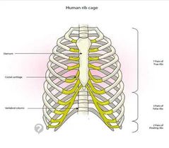
the ribcage |
|
|
|
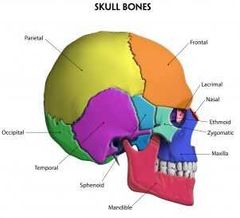
the skull bones |
|
|
|
fontanelles |
There are two fontanelles (the space between the bones of an infant's skull where the sutures intersect) that are covered by tough membranes. The fontanelles include: ... The anterior fontanelle remains soft until about 2 years of age. posterior fontanelle - the junction of the two parietal bones and the occipital bone. |
|
|
synovial |
Our bodies contain six types of synovial joints. Synovial joints are the most movable type of joint found in the human body. Joints are formed where bones come together. The six types of synovial joints are the pivot, hinge, saddle, plane, condyloid, and ball-and-socket joints. |
|
|
x ray |
the use of high energy electromagnetic waves passing through the body |
|
|
catscan |
a painless , non-invasive diagnostic procedure using ionizing radation that provides a crossectional image of the body. |
|
|
MRI |
a noninvasive scanning procedure that provides visualization of fluid, soft tissue, and bony structures |
|
|
arthroscope |
visualization of the interior of a joint using an endoscope |
|
|
bone-scan |
used to detect the spread of cancer in the bone or and pathological fractures |
|
|
osteoarthritis |
degenative disease- joint stiffness and pain |
|
|
rheumatiod arthritis |
2nd most common type |
|
|
bursitis |
inflamation of the bursa (fluid filled sac that cushions tendons) |
|
|
ewing sarcoma family of tumors |
cancer of the bones |
|
|
goat |
body cannot break down uric acid |
|
|
**kyphosis |
exaggerated thoracic curvture also known as hunchback |
|
|
lordosis |
exaggerated curvature of the of the spine known as swayback |
|
|
scoliosis |
lateral curvture of the spine. |
|
|
rickets |
bowing of the legs |
|
|
osteomyelitis |
infection of the bone |