Reading...
![]()
Play button
![]()
Play button
![]()
Use LEFT and RIGHT arrow keys to navigate between flashcards;
Use UP and DOWN arrow keys to flip the card;
H to show hint;
A reads text to speech;
18 Cards in this Set
- Front
- Back
|
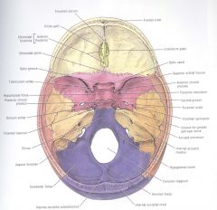
3 fossas of endocranium
|
Anterior
Middle Posterior |
|
|
Boney landmarks of skull
|
|
|
|
Clinoid
|
means bed
- anterior clinoid above the optic canal |
|
|
Between the anterior and middle fossa
|
- lesser wing of sphenoid
- anterior clinoid |
|
|
Between the posterior and middle fossa
|
laterally; superior ridge of petrous portion of temporal bones
anteriorly: dorsum sellae sella: Latin = saddle; adjective - sellar, sella turcica = Turkish saddle. |
|
|
Contents of anterior cranial fossa
|
• Cristae galli: an expansion of the ethmoid bone/ it attaches the anterior aspect of the falx (dura)
• Cribriform plate: penetration of the olfactory nerve • Ethmoidal foraminae: anterior and posterior ethmoidal nerves • Orbital plate of the FRONTAL BONE |
|
|
Cristae galli
|
crista: Latin = crest, crista galli = the (median) crest of a cock.
|
|
|
Contents of The Posterior Cranial Fossa
|
• Foramen magnum: most inferior portion of the p.c.f
• Hypoglossal canal: exit of the hypoglossal nerve (nXII) • Jugular foramen: exit of vagus (nX),glossopharyngeal (nIX), and accessory (nXI) nerves. • Internal auditory meatus: exit of facial (nVII) and vestibulocochlear (nVIII) nerves. • Clivus: basilar portion of the occipital bone and the body of the sphenoid. • Internal occipital crest: attaches the posterior portion of the falx. |
|
|
Clivus
|
clivus: Latin = slope (cf. declivity)
|
|
|
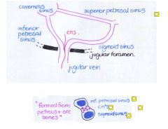
Jubular vein formed from
|
|
|
|
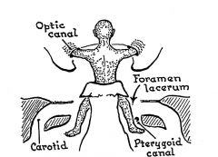
Contents of the middle cranial fossa
|
• 4 clinoid processes: a “bed”
• Optic canal and associated groove: • The Sella Turcica: the “turkish saddle”. • Foramen lacerum: encompasses the apertures of the carotid canal and the pterygoid canal > passing into the foramen is the greater petrosal nerve (derived from the facial nerve (nVII) which then enters the pterygopalatine fossa. • Superior orbital fissure: exit of the ophthalmic division ofthe trigeminal nerve (nVi), oculomotor (nIII), trochlear (nIV) and abducens (nVI) nerves. It is formed from the greater and lesser wings of the sphenoid bone, it enters the orbital cavity. • Foramen rotudum: exit of the maxilliary division of thetrigeminal nerve (nVii) which enters the pterygopalatine fossa • Foramen ovale: exit of the mandibular division of the trigeminal nerve (nViii) and also the lesser petrosal nerve (parasympathetic fibres to otic ganglion). It enters the infratemporal fossa. • Foramen spinosa: exit of the middle meningeal artery, alsoexits in to the infra |
|
|
The anterior surface of the petrous portion of the
temporal bone has |
(i) a depression for the
trigeminal ganglion, has (ii) an arcuate eminence (elevation over the anterior portion of the semicircular canals), (iii) foramina and grooves for the greater and lesser petrosal nerves. Yet another aside……. the squamous portion of the temporal bone has a groove for the middle meningeal artery |
|
|
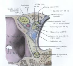
Cavernous Sinus
- contents |
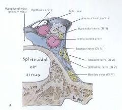
The cavernous sinus lies between 2 layers of
dura (ie the dura is split) Lying BETWEEN these layers (i) Trigeminal nerve (Vi) (possibly a bit of Vii) (ii)Oculomotor n., Trochlear n., Abducens n. (iii) Internal carotid (iv) Sympathetic plexii Lying OUTSIDE BOTH these layers (i) Middle meningeal artery (ii)Petrosal nerves (parasympathetics) Lying INSIDE BOTH these layers (i) Optic chiasm, Optic n., (ii) Pituitary gland (iii) Temporal lobe of cortex |
|
|
Cavernous Sinus
- drains |
- opthalmic vein
- temporal and pituitary |
|
|
BS to to trigeminal ganglia
|
Twigs of the internal carotid “feed” the
trigeminal ganglion………….what happens if blood supply interrupted ??? can completely switch off the trigemenial ganglion - can get positive sensory signs then -ve signs (Ipsilateral) |
|
|
cavernous Sinus near exit of the S>O>F
|
|
|
|
Greater Petrosal nerves
|
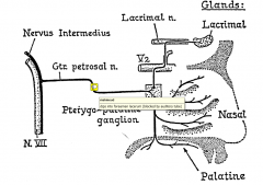
• A branch of the FACIAL nerve, travels below the trigeminal ganglion
• Exits via the foramen lacerum • Travels through the pterygoid canal into the pterygopalatine fossa • Synapses on pterygopalatine ganglion, supplies lacrimal, nasal and palatine glands (tears, mucus, saliva). from the posterior cranial fossa - IAM small branch comes off and gets through petrous temporal bone to get to anterior surface and breaks through to get to middle cranial fossa, travels medial to fopreamen lacrum and then to pterygoid canal, trying to find its ganglion pterygoplaalatine fossa - ganglion to supply lacrimal and nasal and palatine glands... |
|
|
Lesser Petrosal nerve:
|
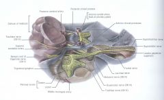
• A branch of the GLOSSOPHARYNGEAL nerve, travels lateral to the greater petrosal nerve.
• Exits via the foramen ovale • Synapses on otic ganglion, supplies parotid gland, via auriculotemporal nerve (saliva). |