![]()
![]()
![]()
Use LEFT and RIGHT arrow keys to navigate between flashcards;
Use UP and DOWN arrow keys to flip the card;
H to show hint;
A reads text to speech;
80 Cards in this Set
- Front
- Back
|
Define anatomy |
The study of structure of body parts and their relationships to one another. |
|
|
The two major types of anatomic study |
Microscopic- Examines the body structures that can't be observed by the naked eye. Gross anatomy (Macroscopic)- Examines the structure and relationships of large body parts visible to the naked eye. |
|
|
The subdivisions of microscopic anatomy |
Cytology-study of single body cells and their internal structures. Histology-study of tissues examining how groups of specialized cells and their products function for a common purpose. |
|
|
The subdivisions of Gross anatomy (Macroscopic) |
Developmental anatomy Regional anatomy Embryology anatomy Surface anatomy systemic anatomy |
|
|
Identify |
|
|
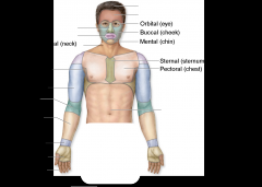
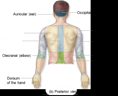
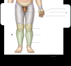
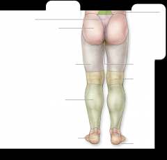
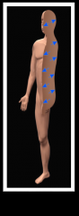
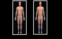
Identify the regions |
|
|
Identify the regions |
|
|
Identify the regions
|
|
|
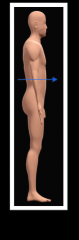
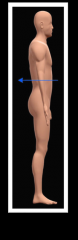
Describe anatomical position and explain its significance |
Anatomical position is also known as the universal reference point. As a result, the body is erect,face/eyes forward,upper limbs at side palms forward, lower limbs parallel, feet/toes forward. |
|
|
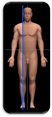
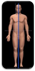
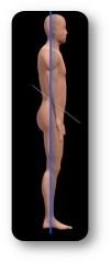
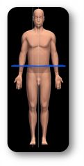
Describe the anatomical planes of selection |
1. Sagittal- any vertical plane cutting the body into left and right 2.Median/Midsagittal- a vertical plane that passes through the midline of the body divided into left and right. 3. Frontal/Coronal-divides the body into anterior and posterior portions 4. Transverse/Horizontal-divides body into sueprior and inferior portions. |
|
|
Compare and contrast bilateral and unilateral |
Bilateral occurs on both sides of the midline. However, unilateral occurs on only one side of the midline. |
|
|
Compare and contrast ipsilateral and contralateral |
Ipsilateral occurs same side of the body. Contralateral opposite sides of the body |
|
|
Define the terms that describe the location of a body structure relative to the front or back of body |
Anterior-towards the front of the body Posterior-towards the back of the body |
|
|
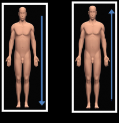
Define the terms that describe the location of a body structure relative to the head |
Superior-towards the head Inferior-towards the feet |
|
|
Define the terms that describe the location of a body structure relative to the body surface |
Superficial- Closer to the surface(on top) Deep-further from the surface (inside) |
|
|
Define the terms that describe the location of a body structure relative to the beginning of a limb |
Proximal-closer to th beginning of limb distal-further from the beginning of limb |
|
|
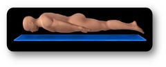
prone |
|
|
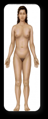
anterior |
|
|
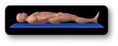
supine |
|
|
Anatomical position |
|
|
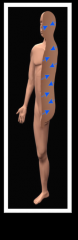
Posterior |
|
|
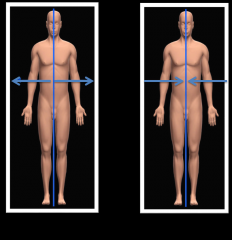
Sagittal |
|
|
Midsagittal/Median |
|
|
Frontal/Coronal |
|
|
Transverse/horizontal |
|
|
inferior and superior |
|
|
Lateral and medial |
|
|
deep |
|
|
superficial |
|
|
Proximal and distal |
|
|
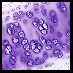
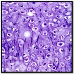
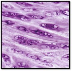
Hyaline cartilage |
|
|
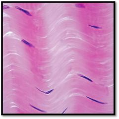
Elastic Cartilage |
|
|
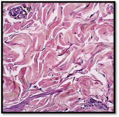
Fibrocartilage |
|
|
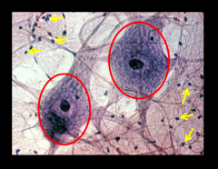
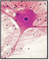
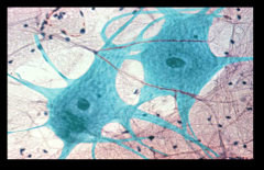
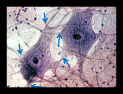
Red: Cell body Yellow: Neuroglial Cell |
|
|
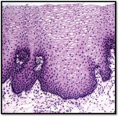
Stratified squamous (non-keratinized) |
|
|
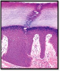
Stratified squamous (keratinized) |
|
|
identify |
|
|
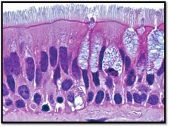
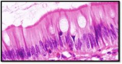
Pseudostratified columnar (ciliated) |
|
|
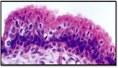
Transitional |
|
|
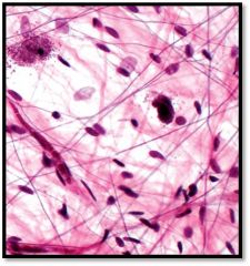
Dense regular CT |
|
|
Dense irregular CT |
|
|
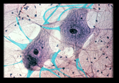
Neuron |
|
|
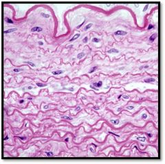
Dense Elastic CT |
|
|
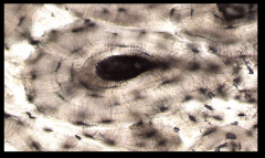
Compact bone Identify osteon & central canal |
|
|
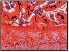
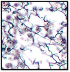
Spongy bone |
|
|
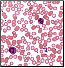
Blood identify:erothrocyte, leukocyte and platelets |
|
|
simple squamous |
|
|
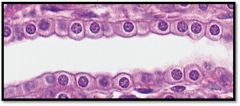
simple cubiodal |
|
|
simple columnar |
|
|
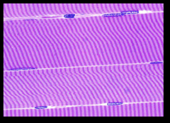
skeletal muscle |
|
|
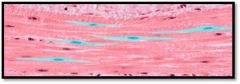
smooth muscle |
|
|
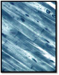
cardiac muscle |
|
|
dendrites |
|
|
areolar loose CT |
|
|
dendrites |
|
|
Recticular loose CT |
|
|
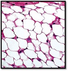
Adipose loose CT |
|
|
Epithelia form the ____ layer of the body |
surface |
|
|
Epithelia line ____ ______ and _______ |
body cavities and hollow organs |
|
|
Epthelia constitute (make up) most ______? |
Gland tissue |
|
|
What is the functional unit of the nervous tissue? |
neurons |
|
|
What are the supporting cells of the nervous tissue? |
Neuroglial cells |
|
|
The basal is surface of an epithelium is fixed to an underlying connective tissue. What structurally separates the two different tissue types? |
layers |
|
|
Epithelia lack blood vessels. What is the term that means lack of blood vessels? |
Avascularity |
|
|
If a cell is keratinized is the surface layer of cells alive or dead? |
dead |
|
|
What structure do all the three types of cartilage have in common? |
Chondrocytes |
|
|
Name the formed elements of blood |
Erothycyte, leukocyte and platelets |
|
|
The watery ground substance of blood consists of dissolved _____? |
proteins |
|
|
Compare cartilage and bone |
cartilage is semisolid matrix and it contains hyaline, fibrocartilage and elastic. bone is solid matrix and it contains spongy and compact. |
|
|
3 types of muscle |
Skeleton,cardiac and smooth muscle |
|
|
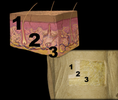
1. Epidermis 2. Dermis 3.Subcutaneous (hypodermis) |
|
|
What are the two general cell types of nervous tissue? |
Neuron & Neuroglial cell |
|
|
The functions of a neuron |
to receive,transmit and process nerve impulses |
|
|
A ___________ is where information is transferred between nerve cells (or to an effector cell, or to a sensory receptor cell) |
Synapse |
|
|
An oligodendrocyte is a neuroglial cell found in the _______ |
CNS |
|
|
While a Schwann cell is a neuroglial cell found in the ________ |
PNS |
|
|
Oligodendrocytes functions |
1. Myelinated and insultates CNS axon 2. Allow faster action potential propagation allong axons in thed CNS 3.Large cells with bulbous body and slender 4.Cytoplasmic extension |
|
|
Schwann Cells |
1. Myelinates and insulates pns axons 2. Allows for faster action potentionally propagation and an axon in the PNS 3. Cell grows around axon ,cytoplasm is squeezed out and multiple layers of cell membrane wrap the axon. |
|
|
Functions of integument |
1. Protection (physical injury,chemicals, toxics and microbes) 2. Prevents water loss 3.Temperature regulation 4.Sensory perception 5.Excretory organ-sweat 6.Formation of Vitamin D |
|
|
_______ + ________= skin |
Epidermis and dermis |