Reading...
![]()
Play button
![]()
Play button
![]()
Use LEFT and RIGHT arrow keys to navigate between flashcards;
Use UP and DOWN arrow keys to flip the card;
H to show hint;
A reads text to speech;
197 Cards in this Set
- Front
- Back
|
What do Mb and Hb carry?
|
Oxygen
|
|
|
Where is Mb located?
|
Myocytes
Transport of O2 through cytosol to mitochondria |
|
|
Where is Hb located?
|
Erythrocytes
Transport of O2 from lungs to peripheral tissues Transport of CO2 and H+ from tissues back to lungs |
|
|
Why does oxygen have low solubility in water?
|
Oxygen is nonpolar and has little opportunities for reacting with water
|
|
|
What is the oxygen carrying capacity of plasma?
|
5ml O2/liter plasma
|
|
|
What is the oxygen carrying capacity of whole blood?
|
250ml O2/liter plasma
|
|
|
What is the concentration of Hb in RBCs?
|
340 g Hb/L of cytosol
|
|
|
What is a simple protein?
|
Polypeptide only
|
|
|
What is a conjugated protein?
|
Polypeptide + cofactor
|
|
|
What is a cofactor?
|
aka: Prosthetic Group
NON-PEPTIDE group that is essential to a protein's function |
|
|
What is a prosthetic group?
|
aka: cofactor
NON-PEPTIDE group that is essential to a protein's function |
|
|
What type of protein are Mb and Hb?
|
conjugated proteins --> polypeptide + cofactor (heme)
|
|
|
What is the prosthetic group in Mb and Hb?
|
Heme
|
|
|
What is the apoprotein (polypeptide) and prosthetic group for Mb?
|
apoprotein: apoMb
prosthetic: (1) Heme |
|
|
What is the apoprotein (polypeptide) and prosthetic group for Hb?
|
apoprotein: (2) alpha, (2) beta
prosthetic: (4) heme |
|
|
In terms of the prosthetic group in Hb and Mb, what differs?
|
In Mb, only (1) heme is present.
Hb has (4). |
|
|
Within each subunit of Hb, what is present?
|
(1) polypeptide chain and (1) heme unit
Polypeptide chain can be alpha or beta |
|
|
How many subunits are needed for a functional unit?
|
(4) are needed-->(4) polypeptides, each with a heme unit
|
|
|
What is leghemoglobin?
|
Oxygen binding protein structure found in legumes in response to infection from parasites
|
|
|
How do you relate the Hb subunits to Mb?
|
The (4) Hb subunits each structurally resemble the Mb as a whole.
|
|
|
Within biology, what are the two types of metal bonding?
|
ionic
covalent |
|
|
Describe ionic metal bonding:
|
*Electrons NOT shared
*ion-ion or ion-dipole interaction *Transient Complexes *Examples: Na+, K+, Mg 2+, Ca 2+ (alkali metals and alkaline earth metals) |
|
|
Are ionic metal bonds long term or short term?
|
Transient
|
|
|
Describe covalent metal bonding:
|
*Electrons shared
*Coordinate covalent bond: both electrons from other (nonmetal) atom *Long lived *Examples: iron, zinc, copper (transitional metals) |
|
|
What is a coordinate covalent bond?
|
both electrons from other (nonmetal) atom
|
|
|
Are covalent bonds short or long lived?
|
Generally long lived (stable) complexes
|
|
|
Describe Iron within the heme complex:
|
Oxidation State: ferrous
Symbol: Fe(II) ionic equivalent: Fe 2+ Usual coordination #: 6 |
|
|
Describe Iron within the hemin complex:
|
Oxidation State: Ferric
Symbol: Fe (III) ionic equivalent: Fe 3+ Usual coordination #: 6 |
|
|
What is the coordination number?
|
Number of atoms nearest the metal atom.
|
|
|
What is the oxidation reaction of iron?
|
heme (FeII)->hemin(FeIII)+1e-
|
|
|
What is the reduction reaction of iron?
|
hemin(FeIII)+1e- -->heme (FeII)
|
|
|
What is the organic portion of heme?
|
protophyrin
|
|
|
How large is heme?
|
>70 atoms
|
|
|
Describe the bond structure of heme and the results of it:
|
*alternating double bonds
*aromatic *planar (flat) *pi electrons: intensely colored *non-polar except for (2) COO- |
|
|
Is heme completely non-polar?
|
No, (2) COO- are present
|
|
|
Within heme, there are unpaired electrons of Nitrogen. Are they available for interaction?
|
No, they are delocalized in the ring system.
|
|
|
Iron is attached to ________ in heme.
|
(4) nitrogens
Iron is tightly bound at center of (4) Nitrogens Covalent bonds ->Both electrons come from N |
|
|
When iron is within heme, it is bonded to ____ atoms. It is capable of bonding to ___ more atoms.
|
4, 2
|
|
|
When iron is attached to heme, what state is it in?
|
Ferrous - Fe(II)
|
|
|
Spatially within heme, where are the two additional bonding positions on iron?
|
The heme molecule is planar, the two additional bonding positions are perpendicular to this plate.
|
|
|
Within heme, what are the non-polar interactions?
|
Folded peptide has a pocket complementary in size and shape to heme
|
|
|
Within heme, what are the coordinate covalent bond interactions?
|
Coordinate covalent bond exists between a histidine side chain and iron
5th coordinate position (proximal histidine) |
|
|
At what coordinate covalent position does iron bond with the main polypeptide chain of heme?
|
5th
|
|
|
Within heme, what his does iron bond with?
|
Proximal his
Mb - #93?? Hb - #8?? |
|
|
Within heme, what ion pairing occurs?
|
heme COO- groups paired with (2) cationic side chains:
*arginine (R) - 45 *histidine (H) - 97 |
|
|
Where is the oxygen binding site on heme?
|
On the iron atom, 6th coordination position
|
|
|
How is the Fe-O bond formed?
|
Fe-O bond formed via an electron pair on oxygen
|
|
|
How is the distal his chain involved in oxygen binding?
|
distal his binds to O2, stabilizing Mb-O2 binding
|
|
|
Describe oxidation in heme:
|
irreversible
e is transferred hemin unable to bind O2 (not an O2 carrier) When heme is bound to Mb and Hb, oxidation occurs very slowly due to inhibition by apoprotein |
|
|
What is the rxn of oxidation of heme?
|
heme-O2 → hemin + O2–*
*O2- is called superoxide, it is a reactive oxygen species |
|
|
What is metHb?
|
Methemoglobin
Hb or Mb with hemin instead of heme |
|
|
How does metHb occur and how is it taken care of?
|
metHb naturally forms via oxidation very slowly
Heme is ferric instead of ferrous Unable to carry oxygen It is normally reversed via enzymes & reductants (metHb->Hb) |
|
|
What is methemoglobinia and how is it caused?
|
Clinically significant build up of metHb
Causes: *Food additives (nitrites) *drugs (sulfonamides, some local anesthetics) *deficiency in reversing enzymes Treatable with reducing agents such as Vitamin C |
|
|
How is methemoglobinia treated?
|
Treatable with reducing agents such as Vitamin C
|
|
|
Within Mb, how much is alpha helix?
|
75%
|
|
|
How many alpha helix does Mb have and how are they arranged?
|
8
arranged in 2 layers |
|
|
For Mb, where are the polar and non-polar side chains located?
|
Polar - Exterior
Non-polar - Embedded |
|
|
What is important regarding heme and the folded structure of Mb?
|
The folded polypeptide has a pocket for heme
When heme is bonded to the folded Mb protein, it is less oxidizable |
|
|
What about Hb over Mb gives it unique characteristics?
|
The tetrameric structure and lack of quaternary
|
|
|
When Hb and Mb go from oxygenated to deoxygenated state, how much O2 is released?
|
Hb - 4 O2
Mb - 1 O2 |
|
|
How do you define fractional saturation of oxygen?
|
Y= pO2/[pO2+P50]
P50 - pO2 where 50% is saturated |
|
|
What is the descriptive word used to describe the shape of the graph of pO2 v. saturation in Hb and Mb?
|
Hb - Sigmoidal
Mb - Hyperbolic |
|
|
For Mb, what is the p50?
|
1 torr
similar to Hb |
|
|
What is the significance of p50?
|
Defines O2 affinity of an O2 binder
|
|
|
Does a higher p50 value correspond to higher or lower O2 affinity?
|
lower O2 affinity
|
|
|
Describe the affinity curve of Mb vs Hb:
|
Mb has a hyperbolic shape
Hb has a sigmoidal shape Implications: Hb curve is steeper in middle Hb more sensitive to changes in pO2 in middle |
|
|
What is the saturation of blood as it leaves healthy lungs?
|
.98
Same in tissue arteries |
|
|
What is the saturation of blood as it leaves the tissues?
|
.62
|
|
|
During exercise what is the saturation of blood as it leaves the tissue?
|
.26
|
|
|
What is the relationship between changes in pO2 and changes in saturation?
|
small changes in pO2 doubles change in saturation
|
|
|
What is cooperative binding in hemoglobin?
|
Binding of oxygen to hemoglobin increases affinity of molecule to bond the next molecule of oxygen
Likewise for release of Oxygen |
|
|
Define allosterism in regards to hemoglobin:
|
change in one site of a structure leads to changes elsewhere in the molecule
*Increased affinity as more Oxygen binds |
|
|
What is degree of cooperativity?
|
Can range from 1-># of binding sites/molecule
|
|
|
How are the subunits in Hb joined?
|
Via:
ionic (salt links) H-bonds nonpolars |
|
|
Why is Hb a dimer of dimers?
|
dimer 1 - alpha1beta1
dimer 2 - alpha2beta2 |
|
|
When O2 binds, how does the Hb molecule shift?
|
The dimers shift relative to each other, not within though
Relative to one dimer, the other dimer: rotates 15 degrees counterclockwise and translates .8 angstrums |
|
|
What happens to the interactions between dimers as oxygen binds?
|
several intersubunit salt bridges break
|
|
|
What happens during the iron movement due to O2 bonding?
|
*in deoxyHb, iron is out of plane of heme, towards proximal his (dome shaped)
*O2 binds, drawing iron into heme plain *proximal his is pulled along *parts of the polypeptide chain attached to proximal his move as well |
|
|
As oxygen binds to hemoglobin, shifts occur and bonds break? How many and of what kind break?
|
8 intersubunit ion pairs break
|
|
|
Describe O2 binding energy and how it changes as more oxygen binds:
|
Some O2 binding energy is used in polypeptide movement/breakage
First O2 requires more breakages-->weaker bond Later O2 requires less breakages-->stronger bond |
|
|
What two forms does Hb exist in?
|
Tense and Relaxed
Tense (T): low O2 affinity many intersubunit interactions Relaxed: high O2 affinity few intersubunit interactions |
|
|
How does the affinity of Hb for O2 change?
|
Changes with metabolic needs
|
|
|
What does BPG directly do?
|
BPG lowers O2 affinity by binding to deoxyHb, not oxyHb
|
|
|
What state is Hb when BPG binds?
|
Binds only to tense state
|
|
|
Can Hb bind BPG and O2?
|
No, only one of a kind
|
|
|
What type of molecule is BPG classified as?
|
Allosteric effector
binds at a seperate site than O2 (allosteric site) Acts as a regulator of O2 affinity |
|
|
At physiological pH, what is the charge of BPG?
|
4-5 negative charges
|
|
|
How and where is BPG manufactured?
|
synthesized and degraded in RBCs by side reaction of glycolysis
|
|
|
How many BPG binding sites are available per Hb?
|
Only 1 is available per HbT
None are available in relaxed state |
|
|
Is the BPG binding site cationic or anionic?
|
Cationic
|
|
|
What subunits create the BPG binding site?
|
(2) beta chains
(or gamma in fetal) |
|
|
Why is Hbr have little affinity for BPG?
|
The conformational changes result in the BPG being covered up
|
|
|
Within Hb, what are the most important characteristics of the BPG site?
|
Arrangement of cationic groups:
N-terminal of alpha-NH3+ grou (3) basic side chains from each beta chain |
|
|
When oxygen binds, what happens to the bonded BPG?
|
The ion pairing breaks
|
|
|
Name some ways fetal Hb is different than adult Hb:
|
BPG site differs
P50 of fetal Hb is lower than adult No matter the pO2, transfer to fetal Hb occurs |
|
|
What are the isoforms of Hb in adults and fetus?
|
HbA: alpha2beta2
HbF: alpha2gamma2 |
|
|
Does HbF has lower or higher affinity for BPG, and chemically why?
|
Lower
Due to fewer cationic groups at BPG site his 143 of each betachain replaced by ser in each gamma chain |
|
|
Does Hb and Mb's affinity for Oxygen change with pH?
|
Hb - YES
Mb - NO |
|
|
Describe the bohr effect:
|
lowering pH increases P50, promoting O2 release, while raising pH decreases P50
|
|
|
Why does the bohr effect take place?
|
Increase in [H+] is due to increase in metabolism --> increase in Oxygen needs of muscles and increased Oxygen unloading
|
|
|
How is some H+ removed from the tissue?
|
carried by deoxyHb
|
|
|
What are the (3) antagonists for O2 binding?
|
BPG
H+ CO2 |
|
|
For various groups, what is the affinity (pKa) based upon?
|
local environment
|
|
|
is H+ affinity higher for HbT or HbR, and why?
|
HbT
pKa values of a few basic groups are higher on HbT than on HbR |
|
|
Does CO2 react preferentially with HbT or HbR?
|
HbT is more likely to react
|
|
|
What groups does CO2 react with on Heme?
|
amino groups
|
|
|
Where does CO bind to Hb?
|
On the Oxgyen binding sites
|
|
|
What are the relative affinities of Oxygen and CO with hemoglobin?
|
Hb=200xs
Free Heme=25,000xs |
|
|
Within free heme, the affinity for CO is much higher than in physiological form.
Why is this? |
Steric hindrance by distal his, preventing optimal CO binding
|
|
|
How much CO does it take to become fatal?
|
.1% CO for ~1hr ties up ~50% of the Hb
|
|
|
The dissociation of CO from Heme is catalyzed by?
|
light
|
|
|
Comparing T vs. R forms:
Binds? Salt Bridges? Temperature? |
T:
*binds H+, CO2 & BPG *contains 8 salt links that break on oxygenation *favored by ↑ temperature R: *binds O2, CO *fewer intersubunit interactions |
|
|
What are the signs and symptoms of Sickle Cell Disease?
|
*susceptibility to infection
*recurring pain *chronic fatigue *swollen lymph nodes *cardiac enlargement *kidney damage *jaundice *anemia *misshapen erythrocytes |
|
|
What are the codons for HbA and HbS?
|
A: GAG
S: GTG |
|
|
What are the sticky patches on Heme?
|
Differing side chains on Beta chain:
HbA has a surface glu HbS has a surface val |
|
|
What are complementary patches on Heme?
|
deoxyHb (on both HbA and HbS) have surface sites complementary to the val on each Betachain
(val is substituted for the correct one) |
|
|
What has both sticky patches and complementary patches?
|
deoxyHbS
|
|
|
How does the aggregation occur in Sicke Cell Disease?
|
deoxyHbS has both sites and so they aggregate together
binding causes precipitation as fibers this occurs where increase in [deoxyHbS] |
|
|
Aggregation of deoxyHbS is based upon:
|
aggregation rate is extremely
concentration-dependent: proportional[deoxyHbS]10 e.g., 7%↑ [deoxyHbS] produces rate doubling |
|
|
Where does deoxyHbS increase?
|
deoxyHbS increases at regions where there is a decrease in [O2]
|
|
|
Is precipitation of HbS self regulating?
|
No, it is amplifying, vicious cycle
|
|
|
What is one positive thing of the heterozygous sickle cell trait?
|
Protection against malaria
|
|
|
What is the heterozygous sickle cell trait and characteristics?
|
1/2 - HbA
1/2 - HbS Protection against Malaria Minimal SCD symptoms |
|
|
What is the treatment for Sickle Cell Disease?
|
Marrow transplant - RBC progenitor cells replaced
|
|
|
What is the functional relationship between oxygen binding by myoglobin and oxygen concentration?
|
Y=pO2/(pO2+p50)
|
|
|
What is the HbS sticky patch?
|
a surface anionic side chain (glu) on b chains of HbA is replaced by a nonpolar side chain (val )
On beta chain: glu (anionic)-> val (nonpolar) |
|
|
What is the HbS complementary site?
|
also on b chains ala 70 & leu 88
|
|
|
What two sites interact on HbS?
|
occurs via intermolecular binding of surface val of one. HbS to a complementary site on another HbS
|
|
|
What is the relationship between O2 consumed and CO2 expired?
|
For each O2 consumed,
.8 CO2 is produced |
|
|
How is most O2 in blood carried?
|
Bound to protein Hb
|
|
|
How is most CO2 transported?
|
12% is bounded to Hb in form of carbamino groups
Majoraty is soluble in H2O |
|
|
Compare the solubility of O2 and CO2 in water:
|
CO2 is 20xs more soluble in water than O2
|
|
|
Does O2 react with water?
|
No
|
|
|
Does CO2 react with water?
|
Yes
CO2 (gas) <--> CO2 (liq) + H2O <--Carbonic Anhydrase--> H2CO3 <--> HCO3- |
|
|
What is the enzyme involved in CO2 transport?
|
carbonic anhydrase
|
|
|
Where is carbonic anyhdrase located?
|
RBC cytosol
NOT in plasma |
|
|
For every HCO3- produced, how many H+ are produced?
|
1 for 1
|
|
|
When attempting to measure CO2 levels, which ones are hard to measure seperate?
|
CO2liq & H2CO3
Together, they are called disolved CO2 |
|
|
What is dissolved CO2 composed of?
|
CO2liq and H2CO3
|
|
|
What is the simplified equilibrium equation for CO2 dissolving?
|
CO2gas <--> disCO2 <--> H+ + HCO3–
|
|
|
What is the H-H equation for CO2 dissolving?
|
pH = pKa + log([HCO3– ]/[disCO2])
|
|
|
What is the ratio of basic:acidic form in CO2?
|
19-20
|
|
|
What form of dissolved CO2 is the highest?
|
HCO3-
|
|
|
What are the percentages of the dissolved CO2?
|
HCO3- - 84%
carbaminoHb - 12% disCO2 - 4% |
|
|
Can dissolved CO2 levels change w/o detremental effects on health?
|
Yes, pH (dissolved H+) must stay constant though
|
|
|
What are some harmful effects of pH change?
|
malfunction of enzymes, receptors, neurons
|
|
|
How much of H+ is carried bound by buffers?
|
60% is carried bound to buffers
|
|
|
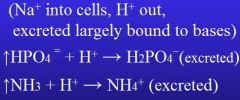
Isohydric carriage of CO2:
|
CO2 is carried with minimal change of pH
pKa shifts of Hb side chains upon O2 dissociation (deoxy-Hb/ HbT formation) enable Hb to bind & carry more H+ |
|
|
If pH rises, what is likely to happen to excitable cells and why?
|
hyperexcitability likely
due to effects on membrane proteins |
|
|
If pH lowers, what is likely to happen to excitable cells and why?
|
hypoexcitability likely
due to effects on membrane proteins |
|
|
What are some important buffers in blood and their pKas?
|
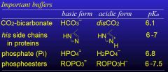
***buffers
|
|
|
What are the important buffers for intracellular and extracellular regions?
|
Intracellular:
Protein (Hb in RBC) CO2-bicarb Pi Phosphesters Extracellular: CO2-bicarb (in plasma, protein also) |
|
|
What are (3) key things of buffers?
|
Act Instantaneously
Only minimize pH changes Are overwhelmed with continuous base or acid production |
|
|
Does ventilation adjust pH quickly or slowly?
|
Quickly, usually within seconds
|
|
|
What are lungs capable of excreting?
|
They only DIRECTLY extrect CO2
Fixed "non-volatile" acids must be extreted by the kidneys |
|
|
Why are the lungs considered open system for buffering?
|
The rate of excretion can be varied
|
|
|
What basis are used for excretion of H+ by the kidneys?
|
***kidney
|
|
|
What are the characteristics of renal compensation to pH?
|
slow (hrs->days)
permanent |
|
|
Respiratory Acidosis:
|
Hypoventilation
Cause: Lung disorders Compensation: Increased renal acid excretion Note: [HCO3-] & pCO2 still high |
|
|
Respiratory Alkalosis:
|
Hyperventilation
Cause: anxiety Compensation: decrease renal acid excretion |
|
|
Metabolic Acidosis:
|
Causes: diabetes mellitus, renal failure
Compensation: increase excretion of CO2 from lungs |
|
|
Metabolic Alkalosis:
|
Cause: vomiting, antacids
Compensation: Decrease excretion of CO2 by lungs |
|
|
What are genes made of?
|
DNA
|
|
|
A complete copy of all DNA is in which cells?
|
All Cells
|
|
|
What does DNA determine?
|
cellular structure and function.
|
|
|
What is the diameter and length of a single round of DNA double helix?
|
Diameter - 20 angstrums
Length - 34 angstrums |
|
|
What is the backbone of DNA?
|
Deoxyribose Phosphate
|
|
|
What is the sugar in DNA?
|
Beta-D-2-deoxyribose
|
|
|
On the DNA sugar, where are the hydroxal groups?
|
1',3',5'
|
|
|
Why is it deoxy instead of oxyneucleic acid?
|
2' position has a hydrogen instead of a hydroxal
|
|
|
What connects the sugars within DNA?
|
Phosphate
|
|
|
What are the purine bases?
|
Adenine
Guanine |
|
|
What are the Pyrimidine bases?
|
Thymine
Cytosine |
|
|
What is a deoxynucleoside?
|
A deoxynucleoside is a deoxyribose with a base (purine or pyrimidine) attached at the 1' carbon by an N-glycoside bond
1' hydroxyl group has been replaced by a hydrogen |
|
|
What is a deoxynucleotide?
|
Deoxynucleoside that is phosphorylated at the 5' hydroxyl
There are deoxynucleoside mono-, di-, and tri- phosphates |
|
|
What type of deoxynucleoside phosphates are there?
|
Mono, Di-, Tri phosphates
dNTP's (where N is base [T,A,G,C]) dNTP's are relatively unstable |
|
|
DNA is a polymer of:
|
deoxynucleotide monophosphates
|
|
|
What is the backbone a repeating unit of?
|
Repeating identical units of deoxyribose and phosphate connected by a diester linkage
|
|
|
What are the ends of DNA called?
|
3' (bottom)
5' (top) called 3' and 5' hydroxals of the deoxyriboses |
|
|
How are complementary bases paired?
|
Via Hydrogen bonds
|
|
|
How are the phosphates within DNA arranged?
|
They are arranged on the outside of the helix to maximize their distance from each other
also helps reduce hydrolysis of the phosphate ester bond |
|
|
What does cooperativity do for the DNA molecule?
|
Cooperativity makes small changes in structure energetically unfavorable
|
|
|
How many degrees does each base pair rotate the helix?
|
36 degrees
|
|
|
What type of replication is DNA?
|
Semiconsurvative
|
|
|
What end is the dNMP added?
|
3' end via the 5'->3' polymerase
|
|
|
Addition of dMNP to DNA requires how much energy?
|
zero, addition of dMNP to DNA is energetically neutral
Hydrolysis of pyrophosphate drives the reaction |
|
|
What are the polymerization energetics?
|
1
dATP+H20<->dADP+Pi (-7.3kcal/mole) 2 dATP+DNA<->DNA-dAMP+PPi (0) 3 PPi+H2O<->2Pi |
|
|
What is the DNA polymerase III?
|
Both strands of parental DNA bind to alpha subunits of DNA polymerase III
Moving in 3'->5' direction on both templates, the polymerase places the next bases on the active sites Fast (2000/sec) Processive (thousands at a time) Big (~900 kD) |
|
|
Can a DNA polymerase III start a chain?
|
NO
Requires enzyme primase |
|
|
What is sequence of starting a chain?
|
Enzyme primase uses the template to synthesize a primer (5 base RNA using NTPs, not dNTPs)
DNA Polymerase III uses dNTPs to add DNA to RNA primer |
|
|
What is DNA polymerase I?
|
Removes RNA primer and replaces it with DNA
Does not attach it to the original DNA segment though...still has gap requiring DNA ligase |
|
|
What does the DNA ligase do?
|
Connects DNA fragments
|
|
|
The lagging strand contains short segments called:
|
Okazaki's fragments
|
|
|
The leading and lagging strands have new DNA replicated in what direction?
|
The DNA is built in a 5'->3' only
==> Both are in 5'-3' direction |
|
|
What does helicase do?
|
Unwinds the double helix DNA
|
|
|
What does the primase do?
|
synthesizes RNA primers
|
|
|
What does ssb do?
|
stabilizes DNA single strand regions
|