![]()
![]()
![]()
Use LEFT and RIGHT arrow keys to navigate between flashcards;
Use UP and DOWN arrow keys to flip the card;
H to show hint;
A reads text to speech;
88 Cards in this Set
- Front
- Back
|
Arteries |
Carry BLOOD AWAY from the heart. |
|
|
Capillaries |
Blood vessels that EXCHANGE gases, nutrients and waste products between blood and tissues and return TOWARD the heart. |
|
|
Veins |
Return blood TOWARD the heart |
|
|
Anastomosis |
The site where two or more arteries converge to supply the same tissue, such as the Circle of Willis. |
|
|
What is the structure of the artery walls? |
1. Tunica intima - a simple squamous epithelium forming smooth surface in the lumen. 2. Tunica media - circularly arranged smooth muscle cells. 3. Tunica externa - the outer protective connective tissue layer, and larger veins have longitudinal muscle. |
|
|
What is the structure of the vein walls? |
1. Tunica intima - a simple squamous epithelium forming smooth surface in the lumen. 2. Tunica media - circularly arranged smooth muscle cells, thinner in veins. 3. Tunica externa - the outer protective connective tissue layer, and larger veins have longitudinal muscle. |
|
|
What are the structural differences between veins and artery walls? Why? |
1. A/V have similar 3 layers, but relative thicknesses are different. 2. Veins have valves, have larger lumen and smooth muscle. 3. Arteries contain elastic lamellae Why? Their structure reflects their function! |
|
|
What is the difference between elastic arteries, muscular arteries and arterioles? |
The elastic arteries have greater elastic laminae for recoil and conduction, while the muscular arteries are for distributing. |
|
|
What four arteries supply the brain? |
Common carotids - L/R Vertebral arteries - L/R |
|
|
What is the circle of Willis and what is its function? |
The circle of Willis is an anastomosis of arteries around the sella turcica. If one artery becomes blocked, then the other arteries can still supply the same tissue. |
|
|
What are two major carotid branches? |
1. Anterior and middle cerebral arteries that feed the brain. 2. Ophthalmic artery |
|
|
What are the three main branches of the thoracic aorta? |
Right subclavian A Internal thoracic A Anterior intercostal |
|
|
What are the three unpaired arteries that supply the GI and spleen? |
1. Celiac artery 2. Superior mesenteric artery 3. Inferior mesenteric artery |
|
|
What are the three paired arteries that branch from the abdominal aorta? |
Suprarenal arteries Renal arteries gonadal arteries |
|
|
Why do small and med veins contain valves? |
Because they have a low blood pressure, there needs to be valves, infolds of the tunica intima, to prevent backflow of blood. |
|
|
What is the structure and function of the lymphatic system? |
The lymphatic system has the lymph fluid transported in lymphatic vessels, lymphatic organs - thymus lymph nodes tonsils and spleen. Its purpose is to return excess ISF to the circulation, transport lipids and as part of the immune system. |
|
|
What is key role of lymphatic capillaries over blood vessels? |
They are more permeable that blood capillaries. |
|
|
Where are lymphatic capillaries not located? |
1. CNS 2. Bone and bone marrow 3. Cartilage 4. Epidermis 5. Inner ear or eye orbit |
|
|
What is the similarity between veins and lymphatic capillaires? |
They both have valves and travel toward the heart. |
|
|
Why is the lymphatic system important in relation to the cardiovascular system/ |
The lymphatic system and cardiovascular system are combined as one.
|
|
|
What are three main properties of cardiac muscle cells? |
Branched cells Interconnected Joined at intercalated discs 99% contractile |
|
|
What is the pacemaker of the heart? |
SA Node |
|
|
What prevents AP from travelling directly from atria to ventricles? Why is this important? |
There is non-conductive tissue between them that prevents APs from stimulating ventricles at the same time as the atria. It allows the atria to contract first, then ventricles. |
|
|
Why is it important that the atria and ventricles do not contract at the same time? |
|
|
|
What is excitation-contraction coupling? |
The link between AP and muscle contraction - done by influx of Ca2+ ions during a plateau phase, induced by a larger influx of Ca2+ from the sarcoplasmic recticulum. |
|
|
Autorhythmic cells |
|
|
|
Cardiac Output |
HR x SV = the amount of blood pumped by each ventricle each contraction per minute. |
|
|
End diastolic volume |
The volume of blood in a ventricle at end of diastole |
|
|
End systolic volume |
The volume of blood in each ventricle after systole. |
|
|
Venous return |
The volume of blood that returns to the heart per minute, Increasing VR, increases EDV, cardiac muscle stretch and a stronger contraction. |
|
|
How does the Frank-Starling law affect stroke volume? |
Increasing the venous return causes the EDV to increase and stretching the cardiac muscle. Stretching the cardiac muscle cells produces a more optimum overlap between thick and thin filaments, leading to a stronger contraction. |
|
|
Describe each of the factors that AID venous return? |
1. Cardiac suction 2. Skeletal muscle pump 3. Venous valves 4. Sympathetic nervous system |
|
|
Explain the baroreceptor reflex? |
The baroreceptor reflex occurs when it detects a fall in blood pressure by the DROP in RECEPTOR POTENTIALS. This causes a DECREASED rate of firing in AFFERENT NERVES and is sent to the CARDIOVASCULAR CENTRE. This stimulates a sympathetic response to increase cardiac nerve activity, vasoconstrictor nerve activity and decrease the parasympathetic nerve activity. Then there is an increase in HR, SV and venous vasoconstriction, therefore a higher CO and TPR. After some time, the blood pressure is now increased to normal. |
|
|
Draw a flow chart of the changes that occur in the cardiovascularsystem in response to an increase in blood pressure and a decrease in bloodpressure. |
See notes :) |
|
|
What are baroreceptors and where are they located? |
Baroreceptors are the mechanoreceptors that respond to stretch and are located within the carotid sinuses and aortic arch. These monitor the blood flow to the brain systemic circulation. |
|
|
Listand describe the organs of the urinary system. |
LECTURE
|
|
|
Explainthe functions of the urinary system. |
LECTURE |
|
|
Describethe anatomy of the kidneys. |
LECTURE |
|
|
Describethe anatomy and components of a nephron. |
LECTURE |
|
|
Describe the anatomy and function of the ureters, urinarybladder and urethras. |
LECUTRE |
|
|
Nameand (if possible) identify the blood vessels that supply the organs of theurinary tract. |
LECTURE |
|
|
Listthe changes in the urinary system in relation to age. |
LECTURE |
|
|
Describethe effect of sympathetic and parasympathetic innervation to the urinarysystem. |
LECTUR
|
|
|
What is the purpose of the kidneys? |
They filter waste products from the bloodstream and convert the filtrate into urine. |
|
|
What is the purpose of the ureters? |
The ureters transport urine from the kidneys to the bladder via peristalsis. |
|
|
What is the purpose of the urinary bladder? |
Storage of urine |
|
|
What is the purpose of the urethra? |
To transport urine from the bladder to outside of the body. |
|
|
Where do the kidneys sit? |
Roughly the bottom of your chest to just past your ribcage. |
|
|
What are the four main functions of the kidneys? |
1. Regulation of blood volume and pressure 2. Regulation of the erythrocyte production via erythropoietin. 3. Regulation of the blood's inorganic ion balance - Na+, potassium and phosphate ions. 4. Acid base balance - through rate changes of H+ ion uptake and ammonium secretion. |
|
|
How are the kidneys held in position? |
They are NOT attached by ligaments, instead held by adipose capsule. If adipose tissue melts, then they call become mobile. |
|
|
What are the protective layers around the kidney? |
1. Renal fascia - dense irregular tissue 2. Adipose capsule - fat 3. Renal capsule - dense connective tissue |
|
|
Is the kidney an intraperitoneal or retroperitoneal organ? |
Retroperitoneal organ because it sits behind the parietal peritoneum. |
|
|
In the kidney anterior view, what order are the three vessels? |
Vein, artery, ureter |
|
|
Where does the kidney blood supply flow from and to before the renal A/V? |
In the renal artery from the abdominal aorta, then out the renal vein to the inferior vena cava. |
|
|
What is the artery flow to the kidney? |
Renal - segmental - interlobar - arcuate - interlobular |
|
|
What are the layers of the ureter? |
1. Mucosa - stretchy epithelium 2. Muscularis - 2 muscle layers: inner longitudinal and outer circular in peristalic waves to propel urine. 3. Adventina - areolar connective tissue |
|
|
Is the urinary bladder inside or outside the peritoneum? |
Retroperitoneal |
|
|
Where is the base of the bladder in females? |
Inferior to the uterus and anterior to the vagina. |
|
|
What is the trigone? |
It is the triangular area of the bladder between the two posterior urethral openings and the anterior urethral openings.
|
|
|
What are the four tunics that form the wall of the bladder? |
Mucosa, submucosa, musclaris and adventitia |
|
|
What is the function of the trigone? |
The trigone acts as a funnel for urine into the urethra as the bladder wall contracts. |
|
|
What are the parasympathetic axons? |
Pelvic splanchnic nerves that come from the spinal cord; their purpose is to stimulate urination. |
|
|
The sympathetic axons are... |
segments of the spinal cord as inhibit micturition. |
|
|
What are the functions of the nephron? |
Filtration, reabsorption or secretion |
|
|
What are kidney functions? |
To regulate the extracellular fluid, maintain acid-base balance, for waste disposal and hormone production. |
|
|
How do the kidneys regulate the ECF? |
|
|
|
What hormones do the kidneys produce? |
Erythropoietin, renin and vitamin D activation |
|
|
What is a nephron? |
A functional unit of the kidneys that contain the renal corpuscle and tubule. |
|
|
What sections does the fluid progress through in the nephron?
|
First, the renal corpuscle, then through the proximal convoluted tubule, then around the nephron loop and out the distal convoluted tubule. |
|
|
What is the renal corpuscle? |
The direct site of blood filtration by fluid pressurized from the capillaries into Bowman's capsule. |
|
|
What is the proximal convoluted tubule? |
The primary site of filtrate reabsorption (60-70%), such as glucose, salts and water. |
|
|
What is the distal convoluted tubule?
|
The last minute HORMONALLY controlled reabsorption of water and Na+ |
|
|
What is glomerular filtration? |
The non-discriminant filtration of a protein-free plasma from the glomerulus into the Bowman's capsule. |
|
|
What is tubular reabsorption? |
The selective movement of filtered substances from the tubular lumen into the peritubular capillaries, roughly 20% glomerulus plasma.
|
|
|
What is tubular secretion? |
The selective movement of non-filtered substances from the peritubular capillaries into the tubular lumen, roughly 80% of glomerulus plasma. |
|
|
Where does the peritubular capillary attach? |
From the glomerulus to the venous system, carrying non-filtered plasma. |
|
|
How do we make urine? |
By the three stages of glomerular filtration, tubular reabsorption and tubular secretion. |
|
|
What is the filtration glomerular hydrostatic pressure? |
It is the hydrostatic pressure and osmotic pressure, about 50mmHg.
|
|
|
What forces make up the hydrostatic pressure? |
Glomerular hydrostatic pressure and the capsular hydrostatic pressure |
|
|
What is the net filtration pressure?? |
The forces favouring filtration minus the forces opposing filtration |
|
|
Draw the diagram that shows the factors controlling glomerular filtration, with labels. |
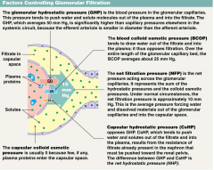
|
|
|
What is the GFR? |
The glomerular filtration rate = the amount of filtrate the kidneys produce each minute, averages around 125 ml/min. |
|
|
What will change the GFR? |
ANYTHING that changes the net filtration pressure. |
|
|
How do we control GFR? |
By adjusting the glomerular hydrostatic pressure: 1. Arteriolar vasoconstriction decreases the GFR 2. Arteriolar vasodilation increases the GFR. |
|
|
What is reabsorption? |
The selective movement of substances into the blood where out of 180 L/day of plasma filtered, there is 178.5 L/day reabsorbed. |
|
|
Where are the three locations for reabsorption of filtrate? |
Proximal convoluted tubule, loop of Henle and the distal convoluted tubule and collecting duct. |
|
|
What are the five barriers that must be crossed during transepithelial transport? |
The luminal membrane the cytosol the basolateral cell membrane the interstital fluid the capillary wall |
|
|
What is transepithelial transport? |
The five barriers that a substance must cross to travel from the filtrate to the plasma. |