![]()
![]()
![]()
Use LEFT and RIGHT arrow keys to navigate between flashcards;
Use UP and DOWN arrow keys to flip the card;
H to show hint;
A reads text to speech;
165 Cards in this Set
- Front
- Back
|
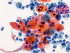
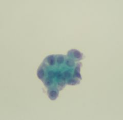
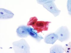
Carcinoma Epidermóide queratinizante |
|
|
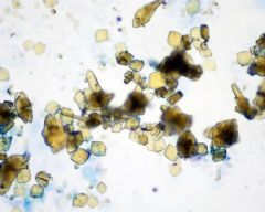
Birefringent uric acid crystals in urine |
|
|
Candida spp. in voided urine |
|
|
cels uroteliais reativas |
|
|
celulas basais do urotélio |
|
|
celulas normais do urotelio |
|
|
citologia normal com negatividade para neoplasia |
|
|
Citologia normal |
|
|
CMV-infected cells with large eosinophilic intranuclear inclusion surrounded by clear halo |
|
|
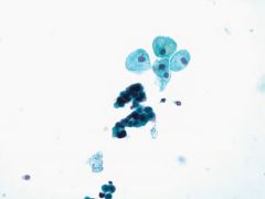
Columnar cells in voided urine |
|
|
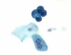
Corpora amylacea-voided urine. Corpora amylacea are laminated structures, staining blue-green here; they occur more with advancing age and are derived from degenerate cells |
|
|
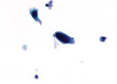
Degenerated atypical urothelial cells showing slight anisonucleosis and bland chromatin in bladder catheterized specimen |
|
|
Endometrial cells—voided urine |
|
|
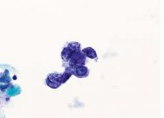
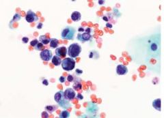
few cells exhibit classic features of high-grade urothelial carcinoma- HGUC |
|
|
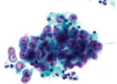
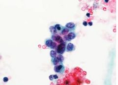
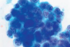
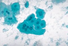
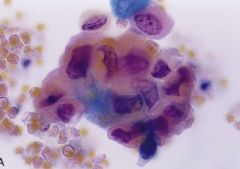
Group of malignant cells from high-grade urothelial carcinoma of bladder. The nuclei display an irregular chromatin pattern and contain nucleoli. The cytoplasm is focally eosinophilic |
|
|
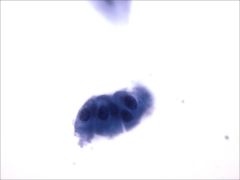
HGUC |
|
|
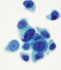
High-grade urothelial carcinoma (HGUC) displays coarse chromatin and nuclear membrane irregularity |
|
|
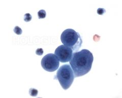
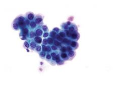
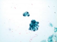
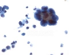
High-grade urothelial carcinoma (HGUC) present as a cohesive group of malignant cells |
|
|
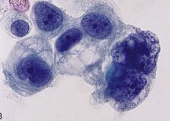
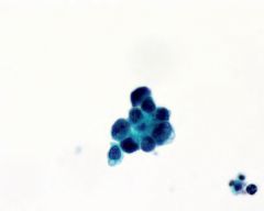
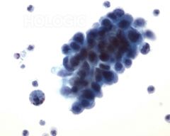
High-grade urothelial carcinoma (UC). A cluster of malignant urothelial cells, some with prominent nucleoli |
|
|
High-grade urothelial carcinoma (UC). Numerous isolated malignant cells have enlarged nuclei with coarsely textured chromatin and an increased nuclear-to-cytoplasmic ratio |
|
|
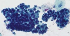
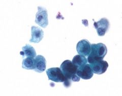
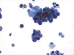
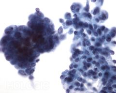
High-grade urothelial carcinoma Cluster of malignant cells exhibiting many diagnostic features including pleomorphism |
|
|
High-grade urothelial carcinoma Cluster of malignant cells with hyperchromatic nuclei. |
|
|
High-grade urothelial carcinoma coarse chromatin pattern |
|
|
High-grade urothelial carcinoma marked nuclear hyperchromasia |
|
|
High-grade urothelial carcinoma of bladder. Large pleomorphic tumor cells with irregular nuclei and a coarsely granular chromatin pattern |
|
|
High-grade urothelial carcinoma of bladder. The nuclei are very large and contain prominent nucleoli |
|
|
High-grade urothelial carcinoma with squamous differentiation |
|
|
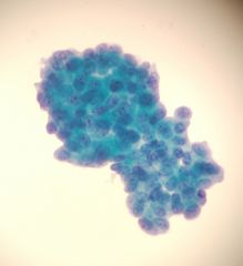
High-grade urothelial carcinoma. Considerable nuclear pleomorphism and hyperchromasia are seen. The nucleocytoplasmic ratio is high |
|
|
High-grade urothelial carcinoma—voided urine. |
|
|
Histiocyte-containing intracytoplasmic laminated inclusions consistent with malakoplakia |
|
|
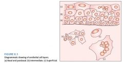
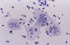
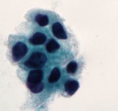
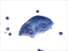
Large superficial umbrella and smaller pyramidal or basal cells. Some of the superficial cells are multinucleated and contain nucleoli |
|
|
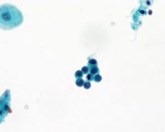
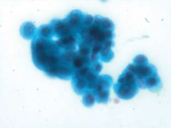
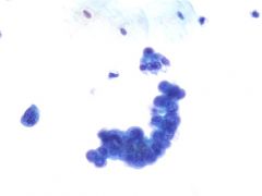
low-grade urothelial lesion (catheterized specimen). Benign cell clusters have a smooth outline (“collared” groups). |
|
|
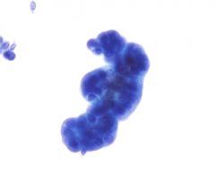
low-grade urothelial lesion Low-grade lesions tend to exfoliate as clusters with irregular edges |
|
|
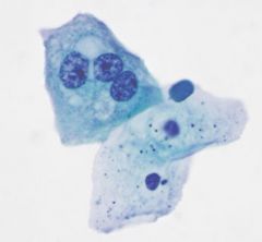
Multinucleated superficial urothelial cell. |
|
|
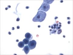
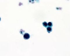
Normal urothelial cells. A papillary cluster of urothelial cells with rounded edge is often seen in patients with proven ureteric calculi |
|
|
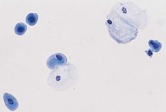
Normal urothelial cells. The large cells are from the superficial and the small cells from the basal layers |
|
|
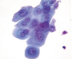
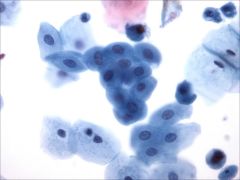
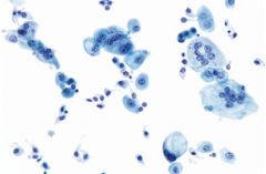
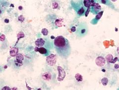
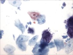
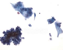
Normal voided urine. Most benign voided urine samples show a mixture of urothelial cells and squamous cells In voided urine, most of the urothelial cells are of intermediate type, with an oval or pyramidal shape |
|
|
Polyoma virus infected cell. The cell in the centre of the field has an eccentrically placed nuclei, the chromatin of the cell appears glassy |
|
|
Polyoma virus infected cells |
|
|
Polyoma virus |
|
|
Polyomavirus infection. Some enlarged, round nuclei are virtually replaced by a glassy, homogeneous inclusion. |
|
|
Polyomavirus-infected cells with large intranuclear homogeneous inclusions |
|
|
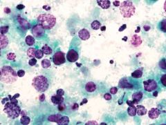
Reactive urothelial cells (catheterized urine). Coarsely vacuolated cytoplasm is characteristic of benign, reactive changes and uncommon in malignancy. |
|
|
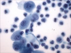
Reactive urothelial cells |
|
|
reativas uroteliais |
|
|
Renal tubular epithelial cells. The cells are of a size similar to that of immature urothelial cells and have uniform, round nuclei |
|
|
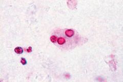
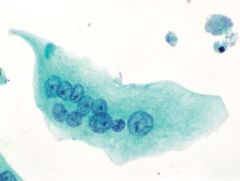
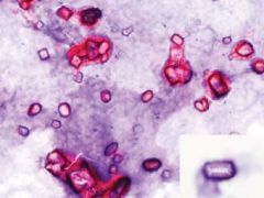
Schistosoma haematobium. Ova of the Schistosoma haematobium. Voided urine |
|
|
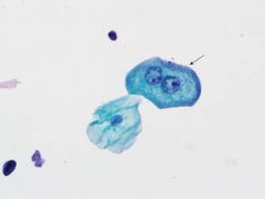
Single abnormal urothelial cells with enlarged hyperchromatic nuclei in urothelial carcinoma in situ of bladder |
|
|
Aspeto das umbrella cells em urotélio |
|
|
|
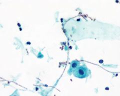
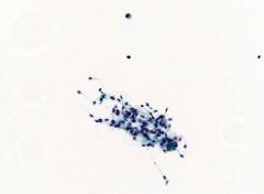
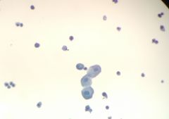
Spermatozoa-voided urine |
|
|
Superficial umbrella cells, with a multinucleated cell in an umbrella configuration |
|
|
Superficial urothelial (arrow) and superficial squamous cell. Superficial urothelial cells (umbrella cells) are often bi- or multinucleated and have abundant cytoplasm |
|
|
Triple phosphate crystals.typical coffin lid appearance |
|
|
Umbrella cell com duas celulas pavimentosas |
|
|
Umbrella cells. These are the largest urothelial cells and cover the surface of the urothelium. Normal columnar urothelial cells are also present |
|
|
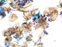
Uric acid crystals—voided urine. Uric acid crystals showing large variation in appearance |
|
|
uroteliais reativas |
|
|
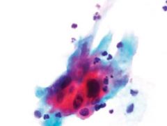
Urothelial carcinoma (UC) variants. Some of the malignant cells show squamous differentiation, manifested by cytoplasmic orangeophilia |
|
|
Urothelial carcinoma (UC) variants. Some UCs have foci of adenocarcinoma |
|
|
Urothelial cells in a papillary group from a patient with calculus disease. Note smooth borders that suggest a benign process |
|
|
Urothelial cells with prominently vacuolated cytoplasm |
|
|
Voided urine in a case of high-grade urothelial carcinoma of bladder. The loose cluster of cells shows large, moderately irregular nuclei with a high nucleocytoplasmic ratio |
|
|
Agrupamento de baixo grau |
|
|
Alto grau com elevada hipercromasia formação papilas pleomorfismo e escasso citoplasma |
|
|
ALTO GRAU |
|
|
Alto grau |
|
|
Baixo grau |
|
|
Baixo grau |
|
|
Baixo grau |
|
|
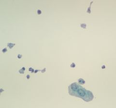
Basais reativas com inflamação |
|
|
Basais reativas |
|
|
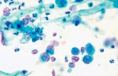
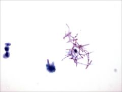
Cândida hifas e esporos |
|
|
Cel umbrella |
|
|
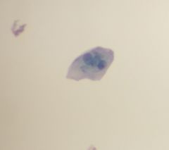
Citologia suspeita |
|
|
Citologia suspeita |
|
|
Esporos e hifas |
|
|
Baixo grau |
|
|
Baixo grau |
|
|
Baixo grau |
|
|
Suspeito |
|
|
Baixo grau |
|
|
Suspeito |
|
|
Baixo grau |
|
|
Cocos e esporos de candida |
|
|
Baixo grau |
|
|
Baixo grau |
|
|
Baixo grau |
|
|
Suspeito |
|
|
Lesão baixo grau |
|
|
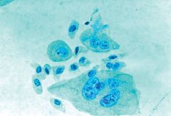
Pavimentosa à esquerda e uroteliais á direita |
|
|
Proteína com PMN |
|
|
Suspeito |
|
|
Suspeita célula isolada |
|
|
Citologia suspeita |
|
|
Citologia suspeita |
|
|
Umbrella cell |
|
|
Umbrella com PMN e uroteliais degeneradas |
|
|
Uroteliais a de baixo um pouco reativa |
|
|
Uroteliais com aspeto colunar |
|
|
Uroteliais intermédias |
|
|
Uroteliais normais |
|
|
Uroteliais reativas |
|
|
Urotelial umbrella |
|
|
Urotelial reativa núcleos aumentados |
|
|
Adenocarcinoma
|
|

|
Alto grau em urina
|
|
|
Alto grau urina
|
|
|
Bacterias
|
|
|
Candida
|
|
|
Carcinoma epidermoide queratinizante
|
|
|
Células uroteliais de aspecto colunar
|
|

|
Células uroteliais num fundo inflamatório
|
|
|
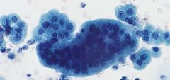
Cluster urotelial benigno- contornos nucleares normais núcleo central cromatina granular aumento nuclear e celular uniforme
|
|
|
Grupo de células uroteliais com aumento nuclear e cromatina grumosa – sugestivo de Lesão de Baixo Grau
|
|
|
Lesao de alto grau em urina
|
|
|
Lesão de alto grau urina
|
|
|
Lesão de alto grau
|
|
|
Lesão de alto grauu
|
|
|
Lesao de baixo grau - Células isoladas e em pequenos grupos
|
|
|
Umbrella cell
|
|
|
Urina normal- Células pavimentosas, uroteliais benignas e PMN
|
|
|
Urina normal- celulas uroteliais reativas
|
|

|
Urina normal- uroteliais em agrupamento
|
|
|
Urina por cateterização cristais
|
|

|
Uroteliais
|
|
|
pavimentosas e uroteliais
|
|
|
Umbrela cell dta e e urotelial esq devido ao aspeto citoplasma e ligeiro reforço que o citoplasma faz á periferia
|
|
|
Umbrella cell- multiplos nucleos com nucleolos proeminentes
|
|
|
candida com esporos e hifas e inflamção com PMN
|
|
|
pavimentosas quetainizadas com cocos e esporos candida
|
|
|
Urina- agrupamento uroteliais basais, com uroteliais intermedias
|
|
|
cocos á dta com hifas á esq e PMN (inflamação)
|
|
|
Urina- PMN (inflamção) com urrotelial e pavimentosa
|
|
|
Urina- uroteliais basais
|
|
|
Urina- uroteliais devido ao formato cuboidal e citoplasma espumoso
|
|
|
uroteliais dta e urotelial esq devido ao citoplasma e forma cuboidal
|
|
|
Urina - lesão de baixo grau
|
|
|
Lesao de baixo grau urina
|
|
|
Urina - metaplasia uroteliais
|
|
|
Urina- carci queratinizante
|
|
|
Urina lesao baixo grau
|
|
|
Lesão de alto grau
|
|
|
alto grau Cells of high grade urothelial carcinoma are pleomorphic and nuclei may contain a prominent, irregular nucleolus
|
|
|
alto grau Cells of high grade urothelial carcinoma have high N-C ratios
|
|
|
alto grau Cells of high grade urothelial carcinoma may be clustered
|
|
|
alto grau Chromatin in nuclei of high grade carcinoma is coarse and hyperchromatic
|
|

|
alto grau Chromatin in nuclei of high grade carcinoma is coarse, hyperchromatic and often irregularly dispersed
|
|
|
alto grau Nuclei may be pointed in high grade urothelial carcinoma
|
|
|
alto grau Nuclei of high grade urothelial carcinoma are often eccentric with convoluted, thickened membranes
|
|
|
alto grau Nuclei of high grade urothelial carcinoma may have multiple irregular nucleoli
|
|
|
alto grau urina
|
|
|
alto grau Urothelial tumors may be papillary or non-papillary
|
|
|
alto grau
|
|
|
baixo grau cells in the clusters of low grade urothelial carcinoma have high N-C ratios
|
|
|
baixo grau Cells of low grade urothelial carcinoma may be in small groups with some single cells
|
|
|
baixo grau Chromatin in low grade urothelial carcinoma is usually granular and evenly distributed
|
|
|
baixo grau clusters in low grade urothelial carcinoma may or may not be papillary
|
|
|
baixo grau Compare the normal urothelial on the right to the crowded cluster on the left.
|
|
|
baixo grau loose cluster of cells from low grade urothelial carcinoma can be compared to reactive urothelial cells
|
|
|
baixo grau Nuclei in low grade urothelial carcinoma sometimes bulge out of the cytoplasm
|
|
|
baixo grau Nuclei of low grade urothelial carcinoma are irregular and may appear to have notches or grooves
|
|
|
baixo grau Nucleoli are usually indistinct or absent in low grade urothelial carcinoma
|
|
|
benigno clusters of cells
|
|
|
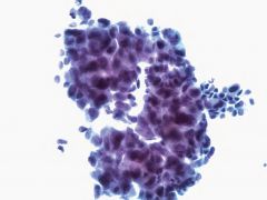
Lesão de alto grau. High grade urothelial carcinomas often are abundantly cellular
|
|
|
uroteliais reativas nuclear enlargement, hyperchromasia, prominent nucleoli and occasiona
|