Reading...
![]()
Play button
![]()
Play button
![]()
Use LEFT and RIGHT arrow keys to navigate between flashcards;
Use UP and DOWN arrow keys to flip the card;
H to show hint;
A reads text to speech;
103 Cards in this Set
- Front
- Back
|
What is a safe amount of blood to take from an animal?
|
Blood is about 10% body weight in kg. It is safe to take 1% of that
|
|
|
What are the techniques for filling tubes?
|
clean venipuncture - no tissue
largest vein possible CBC and biochemical profile needs 5-6 mL of blood microtainer |
|
|
What are possible causes for causes hemolysis from blood collecting?
|
forcing blood into tube
using smaller than a 20g needle vacuum tubes |
|
|
Why is important to fill to the line on the tube
|
to keep the ratio of blood to anticoagulant in balance
inadequate filing of purple top tube will lead to too much EDTA |
|
|
What is the order of draw for filling tubes?
|
Blue
Red Green Purple Gray |
|
|
What does the blue tube top have and what is it used for?
|
citrate
panel for individual factor testing |
|
|
What does the red tube top have and what is it used for?
|
serum tube
Biochem profile |
|
|
What does the green tube top have and what is it used for?
|
haparin
plasma profile |
|
|
What does the purple tube top have and what is it used for?
|
EDTA
chelates Ca used for CBC, PCV, blood film and cell differentiation |
|
|
What does the gray tube top have and what is it used for?
|
sodium fluoride
glucose measurement and lactate measurement inhibits glcolysis |
|
|
How many times should you invert a tube for proper mixing?
|
8 -10 times
|
|
|
Which tube is used for the CBC?
|
Purple top (EDTA)
|
|
|
Which of the following tubes is most commonly used for the biochemical profile?
|
Red top (clot tube no additive)
|
|
|
Which of the type is used for plasma biochemical profile?
|
Green top heparin
Also use heparin for blood gas |
|
|
What are proper sample handling procedures for the CBC?
|
analyze in an hour
make a film and refrigerate tube |
|
|
What is the protocol for blood for serum biochemical profile?
|
use a fasted sample
allow clot for 30 minutes centrifuge remove serum refrigerate harvested serum until analyzed freeze if you can't analyze in 24 hours |
|
|
What increases mean cell hemoglobin?
|
Hemolysis
Lipemia Icterus Extreme leukocytosis Heinz bodies oxyglobin |
|
|
What are the possible reasons for having decreased mean cell hemoglobin concentration?
|
Severe iron deficiency
Regenerative anemia with increased reticulocytes |
|
|
How do you calculated reticulocytes?
|
Percentage of reticulocytes times RBC count = absolute retic
|
|
|
Why is it unlikely for horses and cows to usual produce reticulocytes in the blood?
|
They RBCs mature in the bone marrow
|
|
|
How do you interpret 0 - 10,000 ul of reticulocytes in dogs?
|
Non-regenerative anemia
|
|
|
How do you interpret 10,000 - 60,000 ul of reticulocytes in dogs?
|
Poorly regenerative anemia
|
|
|
How do you interpret 60,000 - 200,000 ul of reticulocytes in dogs?
|
mild to moderate regeneration
|
|
|
How do you interpret greater than 200,000 ul of reticulocytes in dogs?
|
maximum regeneration
|
|
|
How long does it take reticulocytes to mature in dogs?
|
24 - 48 hours from release to maturation
|
|
|
If you see aggregate retics and punctate retics, which are counted in a reticulocyte count?
|
Aggregate retics
|
|
|
When do you see aggregated reticulocytes? Punctated?
|
About 12 hours
10-12 days |
|
|
What is an example of what an abnormal leukogram can indicate?
|
The presence of inflammation
See numeric data and morphologic abnormalities |
|
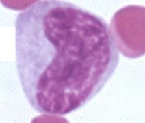
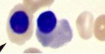
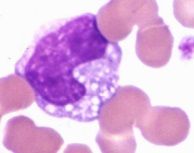
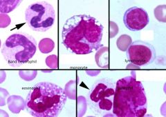
What is this?
|
A monocyte
|
|

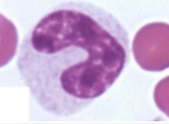
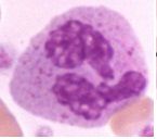
What is this?
|
Mature Segmented neutrophil
|
|
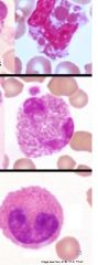
What is this?
|
Metamyelocyte
|
|
What is this?
|
Band Neutrophil
|
|
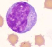
What is this?
|
Lymphocyte
|
|
What is this?
|
Nucleated RBC
|
|
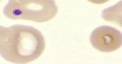
What is this?
|
Howell-Jolly Body
nuclear remnants in circulated RBCs |
|
What is this?
|
Spherocyte
|
|
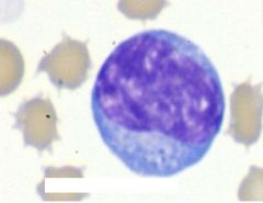
What is this?
|
Reactive Lymphocyte
|
|
What is this?
|
Mature Monocyte
|
|
What is this?
|
Basophil
|
|
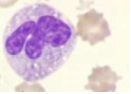
What are these?
|
Eosinophils
|
|
|
What are the common blood leukocytes?
|
Neutrophils - phagocytic and killing cells
Band neutrophils - immature neutrophils released when inflammation is present Metamyelocytes - less common but seen during profound inflammation Important in horses |
|
|
What are examples of lymphocyte subpopulations?
|
B - cells = hummoral immunity
T - cells = CMI Large granular lymphocytes - NK cells Reactive lymphocytes Plasma cells |
|
|
What are the functions of monocytes?
|
Phagocytosis
Antigen presentation to T lymphocytes Iron storage and recycling Cytokine production |
|
|
What are the functions of eosinophils?
|
Modulate inflammatory response
Phagocytosis Defense agains helminth parasites Seen in allergic disease - mast cells |
|
|
What are basophils?
|
Contain heparin
Histamine And numerous proteins Increased concentration of basophils is associated with parasitic infections |
|
|
What is packed cell volume?
|
The percentage of whole blood composed of erythrocytes
Measured after centrifugation |
|
|
What are the three layers of the microhematocrit tube after it has been spun?
|
Plasma
Buffy coat RBCs |
|
|
What is composed of the buffy coat?
|
Leukocytes
nRBCs Platelets Microfilaria |
|
|
What does plasma color indicate?
|
Small animals should have clear and colorless plasma layer
Yellow pigmentation is suggestive of icterus Large animals plasma maybe pale yellow White/pink and opague = lipemia either do to chylomicrons with postprandial collection or high cholesterol (abnormal lipid metabolism) Red = free hb due to hemolysis If PCV isn't decrease it is likely in vitro |
|
|
What is red plasma associated with?
|
Intravascular hemolysis if PCV is decreased
|
|
|
What is yellow plasma associated with?
|
Icterus in large and small animals; maybe normal in large animals
|
|
|
What does a blood film help you do?
|
Get a differential cell count and calculated absolute concentrations
Assess morphology |
|
|
How do you prepare a blood film?
|
Push technique with 2 clean slides
Stabilize bottom slide Hold push slide at a 45 or 60 degree angle Push smoothly and quickly Dry rapidly |
|
|
What are the three zones of a blood film?
|
Body
Counting area Feather edge |
|
|
What can be seen in the body zone of a blood film?
|
Rouleaux
Agglutination Heartworm do not look at cell morphology here |
|
|
What can be seen in the counting area zone of a blood film?
|
Cell morphology
Differential count Platelet estimation |
|
|
What can be seen in the feathered edge area of a blood film?
|
Platelet clumps
Hemoparasites Heartworm Big cells Do not look at cell morphology here |
|
|
Why must one invert the purple top tube before making a blood film or before machine analysis?
|
Because the rocker mixes inadequately
|
|
|
What is a torocyte?
|
an artifact of drying
|
|
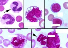
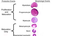
Fill in these WBCs
|
|
|
Identify these WBCs
|
|
|
|
What do you expect with less than 500 semented neutrophils per microliter?
|
Increased susceptibility to bacterial infections
|
|
|
What do you see with inflammation?
|
Increased neutrophil production
Lps and inflammatory mediators G-CSF is key in stimulation of granulopoiesis, maturation of precursors and mobilization from bone marrow |
|
|
What is a left shift?
|
Increased concentration of immature neutrophils (bands usually)
Occurs with normal neutrophil, neutrophilia or neutropenia If with neutropenia = more sever inflammatory response If bands are greater than mature neutrophils = degererate left shift See increases of G-CSF |
|
|
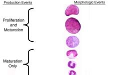
What is orderly maturation?
|
Concentration of each cell increases with the degree of maturity
|
|
|
What is disorderly maturation?
|
Usually a neoplastic process
Very severe consumption |
|
|
What is leukemia?
|
Neoplastic cells in blood or bone marrow- associated hematologic abnormalities
Concentration of neoplastic cells variable non detectable so greater than 500,000 per microliter |
|
|
What is a lymphoproliferative disorder?
|
Neoplasm if lymphocytes and plasma cells
more common in vet med |
|
|
What is myeloproliferative disorder?
|
Neoplasm arising from bone marrow stem cells
|
|
|
What are some examples of lymphoproliferative disorders?
|
Lymphoma/sarcoma - confined to tissues and seen in older animals
Lymphocytic leukemia - neoplasia in marrow and or blood (acute and chronic) seen in younger animals Multiple myeloma - plasma cells present in bone marrow, rapidly fatal and seen in older dogs |
|
|
What are the 5 main types of Myeloid Leukemias?
|
1) Red cell leukemia - acute erythroid leukemia a) myeloblast and rubriblasts b) erythroid predominance
-polycythemia vera - chronic mature cells found 2) Netrophils = granulocytic leukemia 3) Monocytes = monocytic leukemia 4) Neutrophils and Monocytes = Megakaryocytic leukemia = +/- osteosclerosis and |
|
|
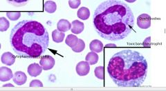
What are characteristics of toxic changes for neutrophils?
|
Usually indicates inflammation
See accelerated production Persistence of ribosomes and RER See diffuse increased basophilia of cytoplasm Presence of Dohle bodies (aggregates) Cytoplastic vacuolation - intense localized/systemic infection; sterile inflammation and drug toxicity Seen in acute pancreatitis |
|
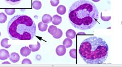
Identify these Neutrophils
|
|
|
|
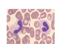
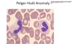
What is neutrophil hypersegmentation?
|
Usually an unimportant finding
Neutrophils greater than 5 lobes Usually a result of normal aging or endogenous/exogenous glucocorticoids |
|
What is wrong with this Neutrophil
|
|
|
|
What is the leukocyte response to inflammation?
|
Consumption of neutrophils in inflammatory response = increase production and early release
Total neutrophil is dependent on balance between consumption and production There are species differences and lesional differences |
|
|
What would we see in an excitement and physiologic response like with epinephrine?
|
Increased blood flow through flow through microcirculation
2-4 fold increase neutrophil concentration Most cats/healthy animals see lymphocytosis and neutrophilia Return within normal in one hour |
|
|
When would we see a stress response in animals?
|
Illness, pain, metabolic disturbances
Iatrogenic Corticosteroid producing tumors Netrophilia and lymphopenia Monocytosis +/- Eosinopenia |
|
|
What would you see in a neutrophilia due to inflammation?
|
Left shift and neutrophil greater than 2 times upper limit
|
|
|
When would you see in a neutrophilia due to excitement?
|
Lymphocytosis, +/- lymphopenia +/- monocytosis, no left shift
|
|
|
What would you see in a neutrophilia due to stress?
|
No left shift, lymphopenia +/- monocytosis
|
|
|
Where do we see lymphocytosis?
|
Excitement response
Lymphoproliferative disease Antigenic stimulation Ehrlichiosis |
|
|
When do we see neutropenia?
|
Consumption
Immune-mediated destruction Lack of production by bone marrow Reversible - viral injury, chemo Irreversible - FeLV |
|
|
When do you see lymphopenia?
|
Steroid response
Acute viral infections Immunodefeciency (uncommon) SCID in arabian foals, basset hounds |
|
|
When do you see monocytosis?
|
Inflammation
Stress response |
|
|
When do you see Eosinophilia?
|
Parasitism (with long tissue contact)
Hypersensitivity Lesions producing eosinophil chemoattractants Ex) heartworm, hookworm, dermatitis, asthma |
|
|
When do we see Basophilia?
|
Usually accompanies eosinophilia
|
|
|
What does it mean when you have macrocytic anemia?
|
There is regeneration
|
|
|
What does it mean when there is microcytic anemia?
|
Iron deficiency anemia
|
|
|
Where do you examine RBC and WBC morphology on a blood film?
|
In the counting area (between the feathered edge and the body)
|
|
|
What does it mean when we nucleated RBCs?
|
Ribosomes and mitochondria are still present because it is still young cell.
If you see it in the blood you know that the bone marrow is pumping out immature red cells |
|
|
What are causes for anemia?
|
Bone marrow
Blood loss Blood destruction |
|
|
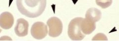
What is Hypochromasia?
|
increased central palor, decreased color in RBCs
Less hemoglobin - iron deficiency anemia |
|
|
What are Spherocyte? how are the important clincally?
|
they are the most diagnostic RBC cell change in a dog most likely due to immune hemolyic anemia
|
|
|
What are important Erythrocyte shapes?
|
Spiculated- projections seen with electrolyte imbalances, kidney disease, rattlesnake envenomation (echinocytes)
-Acanthocyte - few unevely distributed projection due to lipid changes in RBC membrane (hepadic lipidosis (cats) hemangiosarcoma (dogs) -Keratocyte - breaking open blisters seen with Schistocytes - fragmented RBCs - due to iron deficiency anemia, DIC, vascular tumors Sphereocytes - ball like seen with immune mediated hemolytic anemia |
|
|
What are Eccentrocytes and when do you see them?
|
Shift of hemoglobin to once side of the cell resulting in a clear zone outlined by membrane
Caused by oxidative damage such as may be seen with ingestion of onion in dogs. Often seen in conjunction with Heinz body formation |
|
|
What are stomatocytes?
|
Mouth like clear area in the center of the RBC
A few are usually present and are insignigicant Hereditary stomatocytosis reported in alaskan malamutes, miniature schnauzers and Drentse partrijhound |
|
|
What is heinz body anemia?
|
Oxidatively denatured hemoglobin
Causes met hemoglobin which can't carry oxygen , toxic to liver cells Hemoglobin that is denature causes the Red cell to be stiff and unable to change shape and it is more likely to be phagocytizes. Due to decreased receptors = more likely to see an immune mediated hemolytic anemia |
|
|
In what diseases are we likely to see heinz body formation?
|
Acetaminophen in cats
Propylene glycol in cats Ilness in cats Onion in all species Garlic powder in all species Cephalosporins in dogs Zinc toxicosis in penny ingestion Phenothiazine - horses Wilted red maple leaves - horses Kale - cattle Sheep- copper toxicosis |
|
|
What are basophilic stippling of RBCs?
|
Abnormal aggregation of ribosomes
Appear as small basophilic granules Normal in ruminants May see with very regenerative anemia in cats and dogs If you see significant amount in small animals consider lead poisoning |
|
|
What are Howell-Jolly Bodies?
|
Nucleated RBCs
Normally seen with regenerative anemia Non- functioning spleen or splenectomy increased corticosteroids Consider lead posioning |
|
|
What does it mean if you seen Rubriblasts and/or prorubricytes in peripheral blood?
|
Either the bone marrow is destroyed due to trauma or there is a red cell leukemia
|
|
|
What do parasites that affect RBCs do?
|
Cell loses hemoglobin making organism easier to see in cytoplasm
|