Reading...
![]()
Play button
![]()
Play button
![]()
Use LEFT and RIGHT arrow keys to navigate between flashcards;
Use UP and DOWN arrow keys to flip the card;
H to show hint;
A reads text to speech;
15 Cards in this Set
- Front
- Back
|
Be able to identify the mediastinum
know the bumps and lumps of the heart (borders) know the 5 basic densities know the MDPLOTS KNOW THE POSITION SIZE CONTOUR STUFF |
this is everything you need to know from the first radiology lecture
|
|
|
|
|
|
on a lateral view, what is the most posterior structure?
|
mitral valve
|
|
|
|
|
|
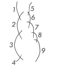
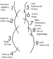
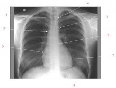
Brachiocephalic vein
Superior vena cava Right Atrium Left Subclavian artery Aortic Knob Left atrial appendage Left ventricle Air bubble |
|
|
Kerley B lines are found in radiographs of what problem?
|
CHF
|
|
|
you are looking at an xray of a patient and you notice Increased heart size -- cardiothoracic ratio >0.50. You see a large hila with indistinct margins and pleural effusion. What does this patient have?
|
CHF
|
|
|
batwing and butterfly pattern are seen in what?
|
CHF
|
|
|
what causes mitral stenosis (normally)
|
rheumatoid disease
|
|
|
you see deposits of hemosiderin and calcification in the lungs...what might be going on?
|
mitral stenosis
|
|
|
A “quiet” condition clinically. Dyspnea, cough, leg swelling, chest pain, palpitations
May follow infective endocarditis May result from rupture of cordae tendineae or papillary muscle this describes |
mitral regurgitation
|
|
|
pt presents with breathlessnes that gets worse with physical activity. The pt also reports coughing at night when lying down in bed. What do they have?
|
Aortic stenosis
|
|
|
if you have an enlarged left ventricle and engorged SVC you likely have
|
aortic insufficiency
|
|
|
to see pericardial effusion what is required?
|
>200cc fluid volume
|
|
|
patient has Pleural effusion and pericardial effusion that has developed after MI. What do they have?
|
Dressler's Syndrome
|