Reading...
![]()
Play button
![]()
Play button
![]()
Use LEFT and RIGHT arrow keys to navigate between flashcards;
Use UP and DOWN arrow keys to flip the card;
H to show hint;
A reads text to speech;
9 Cards in this Set
- Front
- Back
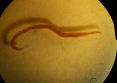
family of both Ancyclostoma duodenale (female pictured) and Ancyclostoma caninum
common names? are they part of the order that means "burseate nematodes"? |
Family Ancyclostomatidae. aka hookworm family. "turn people into wasted people"
Ancyclostoma duodenale is the "old world human hookworm" Ancyclostoma caninum is the canine hookworm yes. |
|
Ancyclostomata duodenale host and location on host? (female pictured)
geography? |
human, intestine (male pictured)
tropical and subtropical countries |
|
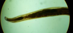
Ancyclostoma duodenale description (pictured: female)
|
stocky body;
Transverse straitations, pair of cervical papillae and located at the esophagus. Funnel shaped buccal capsule large and thick wall. Two large ventral cutting plates, dorsal plate small and teeth in the depths of the capsule |
|
|
Ancyclostoma duodenale female details
|
Tail ends in spine, vulva behind the middle of body, two threadlike ovaries and uteri and oviducts are intercalary wound about the intestine. Uteri join to form vagina. Ovijector and terminal parts of uteri are filled with elliptical, hyaline, thin-shelled eggs
|
|

Ancyclostoma duodenale male details
|
Copulatory bursa has small dorsal lobe and two large lateral ones. Dorsal Ray Has seven digitations: external dorsal ray, three lateral rays arising from bases posterolateral, mediolateral and extnolateral and ventral rays lateroventral and ventroventral.
Brownish needle like spicules serve to transfer seminal fluid form the male to female...associated with gabernaculum in the dorsal wall of the cloaca |
|
|
Ancyclostoma duodenale life cycle
|
make 10-30k eggs/day.
Eggs pass in feces onto the soil, two molts of juvenile, they lose rhabditiform esophagus and become strongyliform. Free-living juveniles have long narrow buccal cavity, strongyliform third stage has unnotched pointed tail. Third stage infective juveniles burrow through skin, enter lymphatic or veins and are then carried by the blodstream tot he heart and to lungs, break out of capillaries into alveoli, crawl up bronchi and to trachea to the pharynx and swallowed...intestine juveniles go through third molt and molt a fourth time |
|
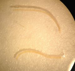
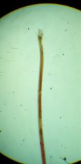
Ancyclostoma caninum description (pictured: adult male and female)
|
Larger than duodenale
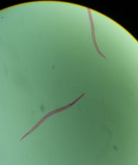
each of the two dental plates has 3 teeth (pictured: filariform juveniles). |
|
|
Ancyclostoma duodenale (and other A.'s) causes this disease
how contracted? |
Ancylostomiasis aka miner's anaemia, Egyptian chlorosis, brickmaker's anaemia, tunnel disease.
Primary problem anaemia. contracted when walking barefoot. |
|
|
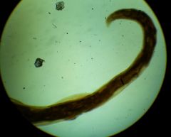
Ancyclostoma caninum Life cycle
|
eggs -- feces --> hatch in feces/soil/litter -->
1st-stage rhabditiform juveniles (pictured) -> molt to 2nd stage -> molt to infective 3rd stage strongyliform infective juvenile -> burrow through hair follicles or are swallowed -> if swallowed, they go directly to small intestine -> if entry by skin, juveniles migrate blood->->lungs-> trachea-> swallowed Some juveniles make way to mammary glands of bitches and pass to offspring via milk |