Reading...
![]()
Play button
![]()
Play button
![]()
Use LEFT and RIGHT arrow keys to navigate between flashcards;
Use UP and DOWN arrow keys to flip the card;
H to show hint;
A reads text to speech;
52 Cards in this Set
- Front
- Back
|
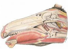
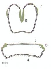
What are the cartilages of the equine nostrils?
|
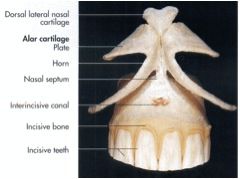
Dorsal and ventral lateral cartilages
Alae or wings Lateral wing for each nostril – skin, muscle, fibrous CT Medial cartilage of both nostrils – cartilaginous base: Lamina of alar cartilage – dorsal Cornu of alar cartilage – ventral |
|
|
In the equine nose, what is the alar fold a continuation of?
|

Mucous membrane fold continuous rostrally with a fold of skin attached to the lamina of the alar cartilage and caudally with the ventral nasal concha
|
|
|
Where are the true and false nostril of the equine nose?
|
True - ventral to alar fold
False - dorsal to alar fold (nasal diverticulum (2) |
|
|
What is the nasal diverticulum?
Why is it clinically relevant? |

Blind pocket located dorsal to the alar fold
Clinical relevance - to be aware of with passing a nasogastric tube. |
|
|
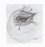
Where is the nasolacrimal duct orifice in the equine nostril?
|

Nasolacrimal duct orifice (3)
External opening of the nasolacrimal duct Located on floor of vestibule |
|
|
What are the nose and nostril cartilages of the ox? How does the nose help in identification?
|
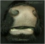
Dorsal and ventral lateral cartilages
Planum nasolabiale – ox The planum nasolabialis is glandular and should be moist. You can take nose prints for identification. :) |
|
|
What are the features of the small ruminant nose and nostrils?
|
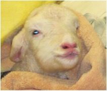
Dorsal and ventral lateral cartilages
Planum nasale – small ruminant Philtrum – small ruminant |
|

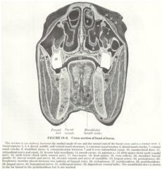
What are a, a', a", a'''?
|
a Nasal septum
a’ Membranous part a” Cartilaginous part a’’’ Osseous part – perpendicular plate of ethmoid bone and vomer bone |
|
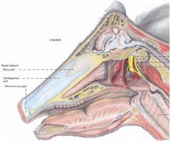
What are a, a', a', a'"?
|

a Nasal septum
a’ Membranous part a” Cartilaginous part a’’’ Osseous part – perpendicular plate of ethmoid bone and vomer bone |
|
|
Is the equine choana vertical or horizontal? What nasal areas is it located between?
|
horizontal
Located between the nasal cavity and the nasopharynx |
|
|
Is the ruminant choana horizontal, vertical or oblique?
|
Oblique
|
|
|
What is the fold of the dorsal nasal concha of the equine?
|
Straight fold – courses from the dorsal nasal concha to the nostril
|
|
|
Which equine nasal meatus communicates with the maxillary sinus?
|
The middle nasal meatus
|
|
|
Which nasal meatus of the equine do you want to pass a nasograstric tube through? Why?
Which meatus can you get hung up in? |
The ventral nasal meatus.
Because it leads to the choana. The dorsal nasal meatus can be a dead end for your nasogastric tube. |
|
Which equine nasal meatus opens up as you move caudally?
|
The ventral nasal meatus
|
|
|
What fold is in the dorsal nasal concha of the bovine?
|
The straight fold
|
|
|
What is the largest nasal meatus in the bovine?
|
The ventral meatus
|
|
|
Which bovine nasal meatus opens into the nasomaxillary opening (maxillary sinus)?
|
The middle nasal meatus
|
|
|
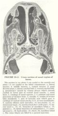
What divides the middle nasal meatus in the ruminant? What does it divide into?
|
Divided into dorsal and ventral passages caudally by the middle nasal concha
|
|
|
What are the interesting feature of the ruminant cheek?
|
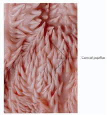
Conical buccal papillae. They are mechanical, not sensory.
|
|
|
T/F ruminant canines and incisors are movable.
|
True - this is thought to be a way to protect the dental pad from getting hit in the same spot every time.
|
|
|
What subsets of sublingual salivary gland do horses have?
|
Polystomatic only, so there is no sublingual salivary duct.
|
|
|
Should performing surgery on the parotid salivary duct in the horse make you nervous?
|
Yes, because the duct is surrounded by a lot of vasculature
|
|
|
What subgroups of sublingual salivary gland do the bovine have?
|
Monostomatic portion is located ventral to the rostral half of the polystomatic portion
Drains at sublingual caruncle Polystomatic portion drains directly into floor of mouth on the sublingual fold |
|
|
what salivary glands drain at the sublingual caruncle in the bovine?
|
Mandibular and monostomatic sublingual salivary glands.
|
|

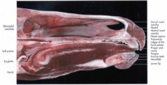
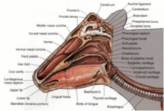
Name these equine oral cavity structures!
|
A. Sublingual caruncle
B. Lingual frenulum C. Fungiform papillae D. Filiform papillae E. Vallate papillae F. Foliate papillae G. Lingual tonsil H. Palatine tonsil I. Stump of palatoglossal arch |
|
|
Is the horse palatine tonsil located in the wall of the pharynx?
|

No, they are located laterally to the tongue.
|
|

Name these ruminant oral cavity structures!
|
A. Sublingual caruncle
B. Lingual frenulum C. Fungiform papillae D. Filiform papillae E. Conical and lenticular papillae F. Vallate papillae G. Region of palatoglossal arch H. Lingual tonsil |
|
|
What are the boundaries to the intrapharyngeal ostium of the equine pharynx?
|
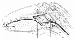
Located between the nasopharynx and laryngopharynx
Formed by the palatopharyngeal arch (4) and free border of soft palate (3) |
|
|
why is the pharyngeal recess of the equine nasopharynx clinically important?
|
It is another blind pouch that the nasogastric tube can get stuck in. Important to pass the tube with the curve pointing down.
|
|
Can you name these equine tonsils/openings?
|

1. Oropharynx
2. Nasopharynx 3. Palatine tonsil 4. Lingual tonsil 5. Tonsil of the soft palate 6. Pharyngeal tonsil 7. Tubal tonsil (located around the auditory tubes) |
|
|
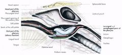
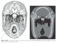
What are the dorsal and ventral boundaries of the guttural pouch?
|

Dorsal - base of cranium and atlas
Ventral - pharynx and beginning of esophagus |
|
|
How many guttural pouches are there? Do they communicate?
|

There are R and L guttural pouches that do not communicate. Each side has medial and lateral compartments (1 and 2)
|
|
|
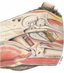
What is the medial compartment of the guttural pouch in contact with?
|

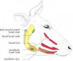
The medial compartment is in contact with the internal carotid artery, ventral cerebral vein (from occipital vein), glossopharyngeal n. (CN IX), vagus n. (CN X), accessory n. (CN XI), hypoglossal n. (CN XII), cranial cervical ganglion, and sympathetic trunk. Ventrally, the medial compartment is in contact with the medial retropharyngeal lnn.
|
|
|
What does the lateral compartment of the guttural pouch have coursing along its surface?
|
The lateral compartment has the facial nerve coursing along its lateral surface along with the maxillary artery and vein.
|
|

What can you ID here?
|
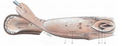
1. Medial compartment of guttural pouch
1’. Lateral compartment of guttural pouch 2. Stylohyoid bone 5. Medial retropharyngeal lnn. 18. Septum between guttural pouches |
|
|
How does air get into the guttural pouch?
|

Through the pharyngeal opening of the auditory tube that opens during expiration and swallowing
|
|
|
What equine structure does this describe:
it is a mucosal fold attached to the lateral wall of pharynx and medial wall of auditory tube, located ventrally? |
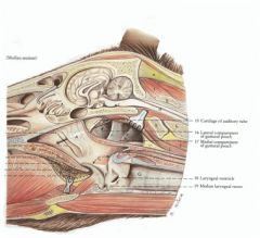
Plica salpingopharyngea
|
|
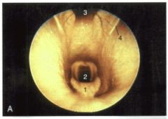
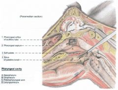
What are 1 through 4?
|

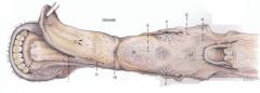
1 - epiglottis
2 - opening into trachea 3 - pharyngeal recess 4 - external auditory openings |
|
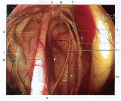
Here we are in the guttural pouch. What can we see?
|
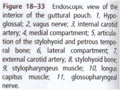
see key
|
|
|
What are some important structures to avoid during surgical drainage of the guttural pouch?
|
Parotid duct
Linguofacial v. Common carotid a. External carotid a. Hypoglossal n. Glossopharyngeal n. |
|
|
What forms the pharyngeal tonsil caudally in the ruminant?
|

The pharyngeal septum
|
|
|
What might you need to reflect in the ruminant to see the external auditory opening?
|
The pharyngeal septum
|
|
|
What is the name of the structure that the palatine tonsil surrounds in the ox?
|
Tonsillar sinus
|
|

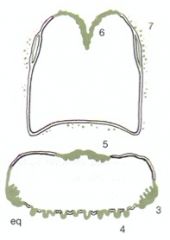
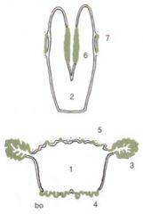
How are the ruminant tonsils different between small ruminants and ox (shown)?
|
1. Oropharynx
2. Nasopharynx 3. Palatine tonsil 4. Lingual tonsil 5. Tonsil of the soft palate 6. Pharyngeal tonsil 7. Tubal tonsil |
|
|
What muscle raises the soft palate to close the choana during swallowing in the large animals?
|

levator veli palatini m.
|
|

Name b, c, d, e
|

Tensor veli palatini m. (B)
Levator veli palatini m. (C) Pterygopharyngeus m. (D) Palatopharyngeus m. (E) |
|
|
what is the only muscle that dilates the pharynx in the large animals (as in the small animals)
|
stylopharyngeus m.
|
|
|
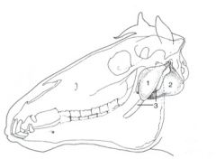
These yellow areas are palpable, what are they?
|
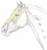
Facial crest
Ventral and caudal borders of the mandible Angle of mandible Nasoincisive notch Infraorbital foramen Zygomatic arch Zygomatic process of frontal bone Supraorbital foramen Nuchal crest |
|
|
What are the palpable features of the ruminant head?
|
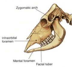
Facial tuberosity
Poll / intercornual protuberance Temporal line Zygomatic arch Infraorbital foramen |
|

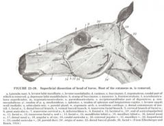
ID CNs 15, 16, 17 here
|
15. Accessory n. (CN XI)
16. Vagus n. (CN X) 17. Sympathetic trunk and cranial cervical ganglion |
|
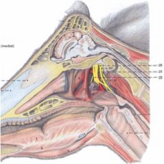
What are the CNs called 20, 21, 22?
|

20. Vagus n. (CN X)
21. Pharyngeal br. 22. Cranial cervical ganglion |