Reading...
![]()
Play button
![]()
Play button
![]()
Use LEFT and RIGHT arrow keys to navigate between flashcards;
Use UP and DOWN arrow keys to flip the card;
H to show hint;
A reads text to speech;
93 Cards in this Set
- Front
- Back
- 3rd side (hint)
|
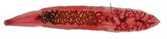
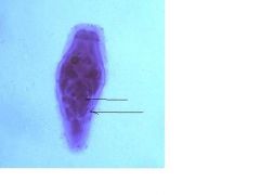
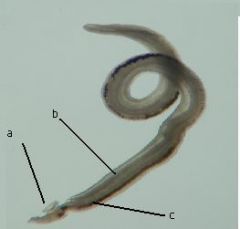
Clonorchis sinensis
Human Liver Fluke Adult |
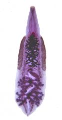
What species? What life stage?
|
Common name is Human Liver Fluke
|
|
|
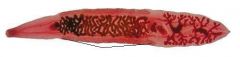
Incomplete digestive sys
found in bile ducts of host Single anterior sucker hermaphrodite |

What observable feature would a diagnostician use in making this identification? List enough features to distinguish it from all similar structures
|
Can you see a digestive system or suckers present
|
|
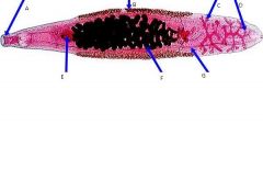
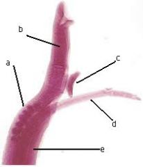
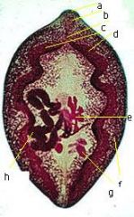
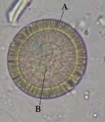
Identify structures at ends of pointers
|
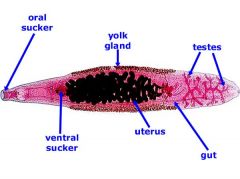
A. Anterior Sucker
B. Vitellaria (yolk glands) C/D Testes E. Ventral Sucker F. Uterus w/eggs G. Intestinal Caecum |
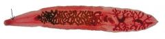
Start at the anterior end
|
|
|
Pharnyx
Muscular throat - pulls in food, breaks it up and pushes it into esophagus referred to as the masticating orgain |

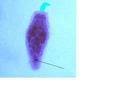
Identify structure at end of pointer, what is it used for?
|
food goes through this organ
|
|
|
Esophogus
tube that moves food to intestinal caecum |
Name structure at the end of pointer and its purpose
|
a tube
|
|
|
The single ovary.
produces ova which is released into ovaduct |
Identify structure at end of pointer and its purpose
|
There is only one in flukes
|
|
|
The mouth.
purpose is opening into the digestive system |

What is this structure and what is its purpose
|
The opening itself
|
|
|
Flame cells and their collecting ducts
Vitelline glands and their ducts |
What is located in this region (visible and may not be visible)
|
There are two different systems that occupy this space.
|
|
|
Describe the male reproductive system of a fluke from testes to genital pore
|
Sperm starts in testes, and is collected into vas efferens. The two tubes become the vas deferens which turns into the seminal vesicle. This is a temp storage site for sperm. The seminal vesicle joins and ejaculatory duct.The prostate complex is in this area. Some may have a cirrus and cirrus pouch which ends in the genital atrium
|
|
|
|
Flukes:
What is the purpose of the vitellaria? |
produce vitelline cells that are carried by ducts to oviduct. These are the yolk glands that produce yolk cells.
|
|
|
|
In Flukes:
What is the purpose of Mehlis' gland? |
Secretes enzymes that cause shell granules in the yolk cells to coalesce and solidify to form the egg shell.
|
|
|
|
In Flukes:
What is Laurer's canal? |
vestigial vagina
it is attached to the duct of the seminal receptabcle |
|
|
|
In Flukes:
What is the purpose of the excretory system and name the 4 main structures |
maintains osmolarity.
flame cells - functional portion of system. collects water and pushes through ducts. collecting ducts - funnels water to bladder excretory badder - sac-like tubule excretory pore - spincter muscle that periodically dilates and releases water. |
|
|
|
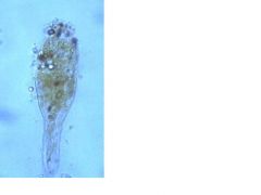
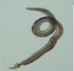
Miracidium of a fluke
1st larval stage |

What type of organism is this, and which life stage?
|
free swimming
|
|
|
Miracidium of a fluke.
1st larval stage. |
What type of organism is this, and what life stage?
|
free swimming
|
|
|
In flukes:
what is the first stage after egg? |
Miracidium
is the first larval stage |
|
|
|
In flukes:
where must an egg be deposited in order to continue life cycle? |
in water
|
|
|
|
What does a miracidium do?
|
It is free swimming, non feeding. finds and enter first intermediate host (certain freshwater snails)
|
|
|
|
How does a miracidium swim?
|
by using the cilia located on its epidermal plates
|
|
|
|
How does a miracidium enter its first intermediate host?
|
once it finds a snail, it swims along mucus layer and finds a crevice. It attaches via apical papilla forming a suction cup and holds on while releasing digestive fluid from the apical gland. The penetration glands release histolytic enzymes which dissolve tissues. Using the apical papilla to wiggle in it burrows through body wall and adjacent tissues where it forms a sporocyst
|
|
|
|
Apical papilla
|
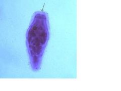
What is the structure at the end of the pointer?
|
used for attachment
|
|
|
Epidermal plates with cilia
|

What is covering this organism?
|
look at the thick brushlike border
|
|
|
developing embryos
|
Identify the structures at the end of the pointers
|
what is the purpose of this organism
|
|
|
Germinal cells
|
Identify structure at end of pointer
|
hmmm hard to tell what they are yet?
|
|
|
eyespots
|

If you could see two back to back C shaped objects in this view. What would they be?
|
|
|
|
What is missing from the miracidium
|
digestive tract
reproductive system suckers |
|
|
|
What is lifespan of a miracidium
|
less than 24 hours
(average is 12 hours) |
|
|
|
What is diagnostic importance of the miracidium
|
none
|
|
|
|
how many developing embryos may be present in a miracidium?
|
up to 6
|
|
|
|
how does the miacidium make developing embryos?
|
through polyembryonation
|
|
|
|
Sporocycst. The second larval stage of a fluke. it contains rediae.
|
What is the stage of the life cycle and what does it contain?
|
|
|
|
What may you find in a sporocyst. (identifiable structures)?
|
Body wall, brood chamber, birth port, developing embryos and germinal cells
|
|
|
|
What is the longevity of a sporocyst?
|
approx 4 weeks
|
|
|
|
How does a sporocyst eat?
|
Through body wall. No digestive system
|
|
|
|
What is the function of a sporocyst?
|
produce rediae.
|
|
|
|
What is the difference between a sporocyst and a daughter sporocyst?
|
Daughter sporocysts contain cercaria, instead of rediae
|
|
|
|
What life stage is the redia?
|
The 3rd larval stage, 2nd generation
|
|
|
|
where are redia found?
|
produced inside of snail in the sporocyst by poly embryony. Stays in body cavity and surrounding tissue
|
|
|
|
What is the function of the redia?
|
produces next generation (cercariae)
|
|
|
|
Main visible difference between redia and sporocyst?
|
redia have digestive tract, while the sporocyst does not.
|
|
|
|
What is the diagnostic importance of the redia?
|
none
|
|
|
|
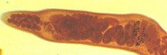
Redia of the flukes. It is the 3rd larval stage.
|
What is this type organism and what life stage?
|
fluke
|
|
|
A. Cercariae
B. Mouth C. Pharnyx D. Collar E. Tail |

Identify the lettered structures
|
|
|
|
A. Body - part that evolves into body of juvenile
B. Tail - used for swimming C. Oral Sucker - attaches to next host D. Penetration glands - used for burrowing into next host E. primordium Region: primordium - will develop into other organs |
Identify structures and function
|
|
|
|
What life stage causes swimmer's itch?
|
cercariae
|
|
|
|
how does cercariae find host?
|
photoreceptors to find general location, then chemoreceptors will find next host
|
|
|
|
Where are cercariae found?
|
produced inside snails. leaves snail becomes free-swimming in aquatic environment, find and enter next host.
|
|
|
|
What is the 2nd intermediate host for C. sinensis?
|
certain freshwater fish
|
|
|
|
What is the final host of the cercariae of the S. Mansoni?
|
human
|
|
|
|
What are the larval stages of the fluke?
|
Egg -> miracidium -> sporocyst -> redia -> cercariae - > metacercariae -> juvenile -> adult
|
|
|
|
what is a metacercariae?
|
An encysted juvenile fluke. Not always present in life cycle of fluke (i.e. blood flukes lack encystment)
|
|
|
|
What is the function of the metacercaria?
|
transmissable stage, that must be orally ingested by next host
|
|
|
|
Where are metacercariae found?
|
in 2nd int host (fishes/crustaceans)
|
|
|
|
how is the metacercariae made?
|
cyst wall secreted by cercaria, hardens and forms cyst wall.
|
|
|
|
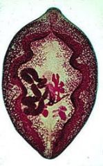
metacercaria of fluke
|
What is this life stage?
|
|
|
|
A. Oral sucker
B. Cyst wall C. Ventral sucker |
Identify structures
|
|
|
|
metacercaria
a. cyst wall b. oral sucker c. excretory bladder d. ventral sucker |
Identify structures and life stage
|
|
|
|
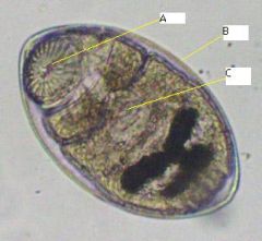
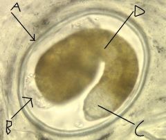
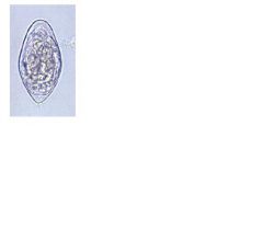
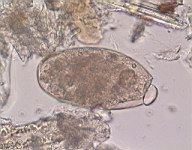
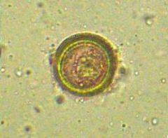
Clonorchis sinensis
small size rimmed operculum (small spine is sometimes present) |
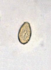
Identify species and give diagnostic features
27-35 um long 12-20 um wide |
fluke or tape?
|
|
|
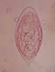
Clonorchis sinensis
rimmed operculum small size small spine is sometimes present |
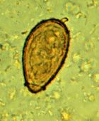
Identify species and give diagnostic features
<50 um |
|
|
|
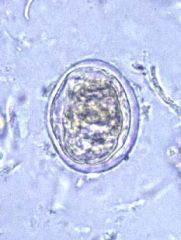
Clonorchis sinensis
small size rimmed operculum small spine is sometimes present |
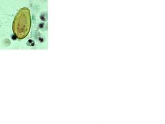
Identify species and give diagnostic features
<50 um |
|
|
|
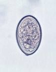
Schistosoma mansoni
No operculum Large posterolateral spine |
Name species and give diagnostic features
>100 um |
|
|
|
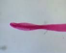
Schistosoma hematobium
No operculum terminal spine on large eggs |
Identify species and give diagnostic features
|
|
|
|
Schistosoma hematobium
Terminal spine on large eggs |
Identify species and diagnostic feature
|
|
|
|
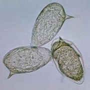
Schistosoma japonica
small posterolateral spine on medium sized egg |
Name species and give diagnostic features
65-105 um long 50-90 um wide |
|
|
|
Schistosoma japonica
small posterolateral spine on medium egg |
Name species and give diagnostic features
Moderate sized |
|
|
|
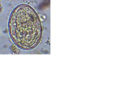
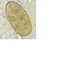
Fasciolopsis buski
non-rimmed operculum on large egg |
Name species and diagnostic features
>100 um |
|
|
|
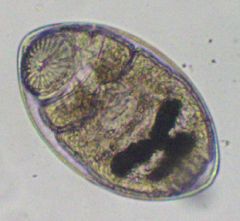
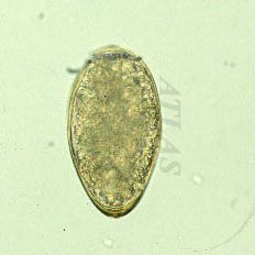
Paragonimus westermani
rimmed operculum medium egg |
Name species and give diagnostic features
70-120 um long 45-65 um wide |
|
|
|
Paragonimus westermani
rimmed operculum on med egg found in human sputum or feces |
Name species and diagnostic features.
Where might this egg be found? |
|
|
|
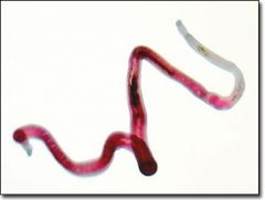
Schistosoma mansoni
male |
Name species and gender
|
|
|
|
Schistosoma mansoni
a. Ventral sucker b. Gynocophoric canal c. Testes |
Identify species and structures
|
|
|
|
a. testes
b. esophagus / esophageal glands c. ventral sucker d. female schistosoma mansoni e. gynecophoric canal |
Identify structures
|
|
|
|
Schistosoma mansoni
female |
Name species and gender
|
|
|
|
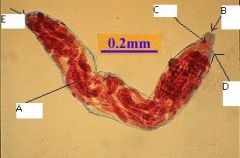
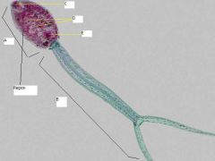
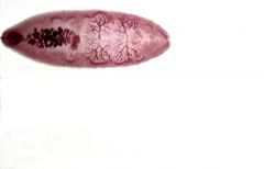
Paragonimus westermani
|
Identify species
8 to 16 mm long 4 to 8 mm wide |
|
|
|
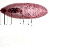
a oral sucker
b pharynx c esophagus d intestinal ceaca e ovary f vitellaria g testes h uterus |
Identify structures
|
|
|
|
Fasciolopsis buski
|
Name species
2 - 8 cm long 8 - 20 mm wide |
|
|
|
a oral sucker
b ventral sucker c seminal vesicle d uterus e. ovary f. mehlis' gland g testis h vitellaria |
Identify structures
|
|
|
|
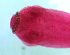
The Scolex inculding the acetabula, rostellum and hooks
and the neck Taenia pisiformis adult |
What is visible in this picture?
|
|
|
|
a. hooks
b. rostellum c. suckers d. neck |

Identify structures
|
|
|
|
2 testis
3 uterus 4 genital atrium 5 vas deferens 6 vagina 7 ovaries 8 vitallarium |
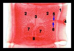
Identify structures (except 1)
|
|
|
Identify species and structures
|

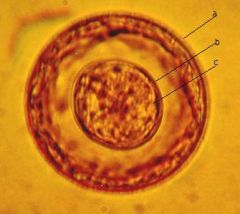
Hymenolipis diminuta
a. outer true shell b. embryophore (inner shell) c. oncosphere |
|
|
Identify species and structures
|

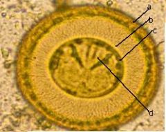
Hymenolipis diminuta
a. true outer shell b. embryophore (innershell) c. oncosphere d. hooks |
|
|
Identify species and structures
|

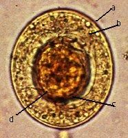
Hymenolipis nana
a outer shell b polar filaments c embryophore w/polar knobs d oncosphere |
|
|
Identify species and structures
|

Taenia saginata or solium
a. thick striated embryophore b larva w/ hooks |
|
|
|
Taenia saginata or solium
Diagnostics: larva w/hooks = tapeworm thick striated embryophore = taenia honeycomb surface |
Identify and give diagnostic features
|
|
|
|
Diphyllobothrium latum
|
Identify species
|
|
|
|
Diphyllobothrium latum
non-rimmed operculum on medium egg |
Identify species and give diagnostic features
58-75 um long 40-50 um wide |
|
|
|
cyclophyllidea
|

name order
|
|
|
|
hyatids
Echinococcus granulosus or Echinococcus multilocularis |
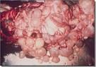
Identify structures and possible causative agent
|
|
|
|
How many uterine branches in the gravid proglottid of Taenia saginata?
|
15 - 30 pairs
|
|
|
|
How many uterine branches in the gravid proglottid of Taenia solium
|
7 - 13
|
|
|
|
What is the diagnostic characteristic of the H. nana proglottid?
|
proglottid less than 1 mm w/saclike uterus
|
|
|
|
Difference between H.nana and H. diminuta proglottids?
|
H. nana is <1 mm
H. diminuta is 1.5 - 4 mm |
|
|
|
Which tape has a rosette shaped uterus?
|
Diphyllobothrium latum
|
|