Reading...
![]()
Play button
![]()
Play button
![]()
Use LEFT and RIGHT arrow keys to navigate between flashcards;
Use UP and DOWN arrow keys to flip the card;
H to show hint;
A reads text to speech;
32 Cards in this Set
- Front
- Back
- 3rd side (hint)
|
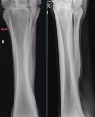
Standard views
|
Lateral
DP DMPLO DLPMO |
|
|
|
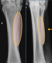
Wolff's law
- - - Periostitis - - |
Wolff's law (adaptive)
- modelling of bone according to the stresses placed on it - coritcal modelling is an adaptive response to increased load during exercise -cyclic loading of the MCIII/MTIII can lead to microfractures Periostitis (pathologic) - direct trauma . inflammation of periosteum and/or subperiosteal hematoma . new bone typically not deteced for 14 days . quiescent in 6 to 12 wks -produced in repsonse to microfractures (bucked shins) . heat, pain, swelling, variable degrees of lameness |
|
|
|
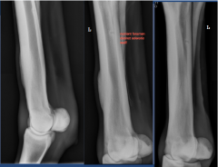
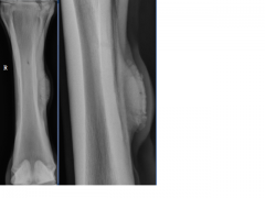
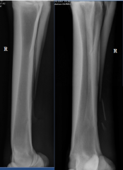
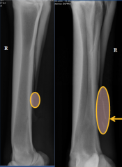
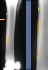
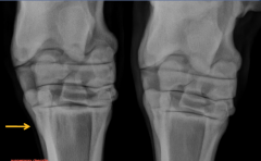
Cortical stress fractures
radiographic findings: |
focal dorsal cortex thickening (along periosteum and endosteal cortex)
lucent lines (stress fractures) reaction wll quiescent in 6 to 12 wks (can require surgical intervention) |
|
|
|
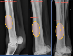
lateral view, DMPLO, DLPMO
bone production and stress fracture |
|
|
|
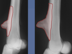
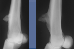
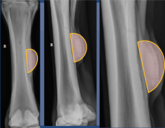
Chronic traumatic periostitis
exuberant bone production on dorsal margin |
|
|
|
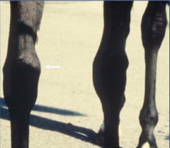
Periostitis - "Splints"
|
Secondary to damage of the interosseous ligament
usually young horses recently started in work involved proximal 1/2 of MCIII/MTIII and II and IV (II and III in forelimb, III and IV in hindlimb) Radiographically: |
new bone production between splint bone and cannon bone
periosteal reaction on the splint bone mild soft tissue swelling - heat and pain - can be incidental |
|
|
Periostitis MCII
thickening, enlargement of MCII fusing MCIII to MCII exuberant bone production should be similar to MCIV |
|
|
|
Periostitis
fibrous tissue focal soft tissue swelling injury of interosseous ligament |
|
|
|
Fractures
MC/MT II and IV |
most common on the distal third
fractures of distal third are most common in horses >5 years of age proximal fractures are often complicated by infection -proximity to tarsometatarsal joint heal with bone prodcution - laterally into suspensory ligament may have concurrent suspensory desmitis |
|
|
|
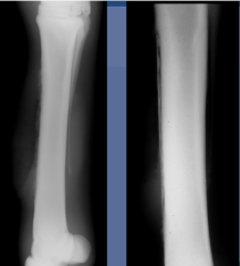
traumatic MC IV fracture
segmental displacement of distal body |
|
|
|

Chronic MT IV fracture
healing - smooth bone production, blurred indistinc margin, bridging ossesus callus if medial, suspensory ligament problems |
|
|
|
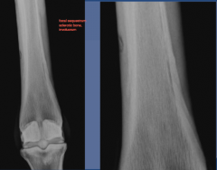
Sequestrum
|
radiopaque piece of bone that has lost its blood supply
MC/MTIII are prone to sequestrum formation and infection following soft tissue injury -thick dorsal cortex -minimal soft tissue coverage - poor vascular blood supply (sclerotic, necrotic, any bone, more common in long bones, little blood supply from nutrient foramen, need blood supply from soft tissue, thickened dorsal cortex) radiographic findings: |
sclerotic bone fragment (sequestrum)
involucrum: margin of sclerotic bone that borders the sequestrum cloaca: opening into the involucrum may take 7 to 14 d following injury to see - periosteal reaction proximal and distal to sequestrum -soft tissue swelling associated with infection process |
|
|
dorsal margin of cortex, MCIII
seperating from parent bone -sequestrum |
2 wks later
severe soft tissue, irregular sclerotic bone, seperation from parent bone |
|
|
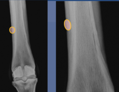
Sequestrum
-focal sclerotic bone involucrum |
|
|
|
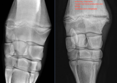
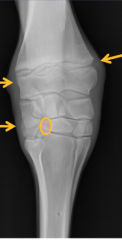
Desmitis - origin of suspensory
|
suspensory ligament originates from the proximal palmar/plantar aspect of the cannon bone
tearing of attachment results in periostitis radiographic findings: |
increased opacity (sclerosis) on proximal aspect of MC/MT III
- crescent shaped lucency surrounded by sclerosis avulsion fragment may be seen |
|

|

suspensory desmitis
square shaped radiolucency, crescent shaped fairly uncommon |
|
|

|
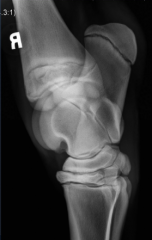
angular limb deformity
valgus - laterally displaced varus - medially displaced |
|
|
|
physitis
|
young horsese (4-12 mos)
also 2 yr old in training (growing, high plane of nutrition, aseptic) disruption of endochondral ossification variable lameness common locations: radiographic findings: |
common locations:
distal radius distal metacarpus/metatarsus radiographic findings: irregular and asymmetrical widening of the physis flaring and periosteal proliferation of the metaphysis and epiphysis soft tissue swelling can be associated with angular limb deformity |
|

|
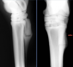
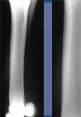
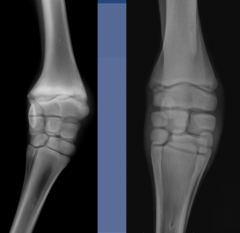
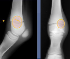
normal vs physitis
medial aspect of limb, more common not smooth margin of physis widening irregularity, bone production and flaring epiphysis and metaphysis |
|
|
|
Angular limb deformity
- - - |
Angular limb deformity
-congenital (in utero) . . . . -developmental . . -acquired . . . |
Angular limb deformity
-congenital (in utero) . positioning . exposure to toxin or infection . nutrition . skeletal maturity -developmental .developmental orthopedic disease . nutrition, exercise, overloading, trauma -acquired . trauma . fracture . infection |
|
|
angular limb deformity:
level of maximum deviation |
-diaphysis
-distal radial physis - distal radial epiphysis - epiphyseal growth imbalance - incomplete ossification of carpal bones - flaccidity or damage to periarticular structures |
|
|

|
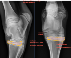
angular limb deformity
-used to define level of maximum deviation . irregular physis . wedge shaped epiphysis . delayed development lateral styloid process . cuboidal bone disease |
13 degree angle of rotation
angle of maximum deviation at distal radial epiphysis note narrowed lateral aspect of third carpal bone |
|
|
incomplete ossification of carpal bones and tarsal bones
|
seen primarly in young foals
- premature, dysmature, twin - incomplete ossficiation, not enough nutrients radiographically: -small rounded bones/lack normal cuboidal shape - enlargement of carpal joint spaces - bones lack normal angular shape - granular appearing bones - collaps and malformation of bones - angular limb deformity not formed enough to support weight of animal, stress to bones, long term djd |
|
|

|
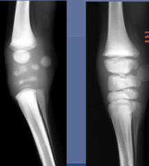
incomplete ossification of carpal/tarsal bones
- small round, not completely formed - widening of jt space - minimal ossification |

|
|

|
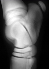
incomplete ossification
|
|
|

|
Tarsal Bone Collapse
|
incomplete ossification of central and third tarsal bones secondary to skeletal immaturity
radiographically - dorsal or lateral collapse of the central or third tarsal bones - dorsal fragmentation - sclerosis - angular limb deformity (tarsus valgus) - may develop degenerative joint disease of the distal intertarsal and tarsometatarsal joints |
|
|
Septic Arthritis
|
most common in foals
- failure of passive transfer and delayed closure of gut - pneumonia - oomphilitis - enteritis/colitis in adult horses it is usually associated with direct trauma to the joint or associated with an injection radiographic bone changes may be apparent in 7 to 10 days after the onset of clinical signs |
radiographic findings:
soft tissue swelling bone lysis irregular outline to subchondral bone periosteal bone proliferation (aggressive) abscence of changes does not rule out septic arthritis Classification: P type - begins in physis, involves bone on both sides E type - begins in epiphysis S type - begins in synovium T type - begins in tarsus C type - begins in carpal bones |
|

|
Early S-type septic arthritis
|
|
|

|
P type septic arthritis
|
distal third metacarpal bone physis
bone lysis, sclerosis, and periosteal bone production of the epiphysis and metaphysis sequestrum |
|

|
P type septic arthritis
distal radial metaphysis, physis, epiphysis bone lysis and bone sclerosis soft tissue swelling subcutaneous emphysema |
|
|

|
C type septic arthritis
carpal bones soft tissue swellin bone lysis and sclerosis |
|
|

|
septic arthritis and osteomyeltitis
|
|