![]()
![]()
![]()
Use LEFT and RIGHT arrow keys to navigate between flashcards;
Use UP and DOWN arrow keys to flip the card;
H to show hint;
A reads text to speech;
13 Cards in this Set
- Front
- Back
|
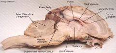
Picture of sheep brain |
|
|
|
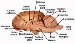
Picture of sheep brain 2 |
|
|
|
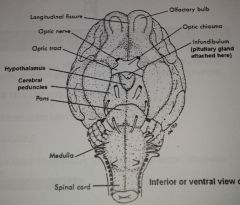
Diagram of sheep brain, inferior view |
|
|
|
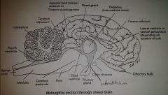
Diagram of sheep brain, midsagittal view |
|
|
|
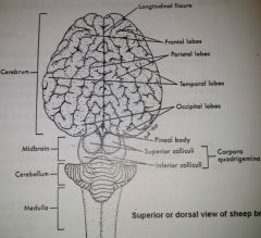
Diagram of sheep brain, dorsal view |
|
|
|
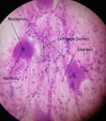
Neuron cells (400x) |
|
|
|
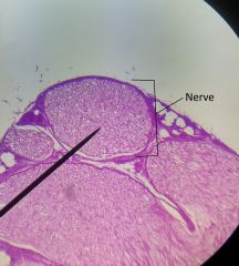
Nerve and blood vessels slide (closeup of nerve) |
|
|
|
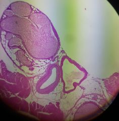
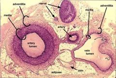
Nerve and blood vessels slide |
|
|
|
more nerves and blood vessels |
|
|
|
artery vein nerve diagram |
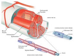
note that some axons are myelinated and others are unmyelinated |
|
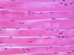
review: what type of tissue is on the slide |
skeletal muscle |
|
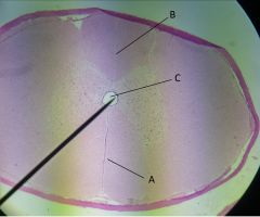
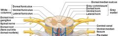
Identify the indicated structures |
A - Ventral median fissure (i think) B - Dorsal median sulcus C - central canal Note: generally it is possible to tell the difference between the fissure and sulcus, because the Dorsal Median Sulcus is longer |
|
|
Diagram showing cross section of the spinal cord |
|