![]()
![]()
![]()
Use LEFT and RIGHT arrow keys to navigate between flashcards;
Use UP and DOWN arrow keys to flip the card;
H to show hint;
A reads text to speech;
45 Cards in this Set
- Front
- Back
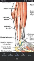
What are the muscles of the Lateral compartment of the leg? |
Fibularis longus & Fibularis brevis |
|
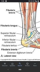
Superior fibular retinaculum |
A thickening of crural fascia found on the lateral side of the ankle posterior to the lateral malleolus. |
|

Inferior fibular retinaculum |
On the lateral side of the ankle, anterior to the lateral malleolus. |
|
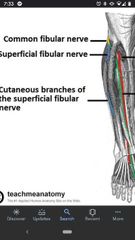
Superficial fibular nerve |
A branch off the common fibular nerve. Innervates the lateral compartment of the leg. Primary cutaneous nerve of the dorsum of the foot. |
|
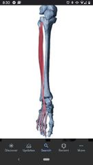
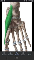
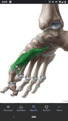
Fibularis longus |
Proximal Att: head of fibula & lateral surface of fibula. Distal Att: base of first metatarsal and medial cuneiform bone. Actions: everts & plantarflexes the foot. Innervation: Superficial fibular nerve |
|
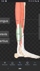
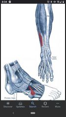
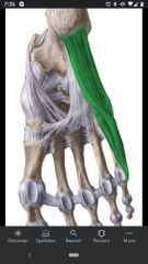
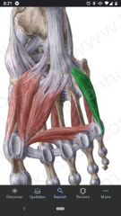
Fibularis brevis |
Proximal Att: inferior part of lateral surface of fibula. Distal Att: tuberosity of 5th metatarsal bone. Actions: evert & plantarflexes the foot. Innervation: superficial fibular nerve |
|
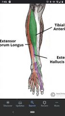
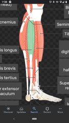
What are the muscles of the anterior compartment of the leg? |
Fibularis tertius, extensor digitorum longus, extensor hallucis longus, & tibialis anterior. |
|
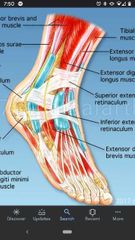
Superior/inferior extensor retinaculum |
On the anterior surface of the ankle, transverse thickenings of crural fascia that hold tendons in place. Inferior retinaculum is Y-shaped and is attached to calcaneus. The superior extends across tendons superior to ankle joint. |
|
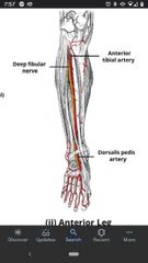
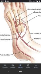
Anterior tibial artery |
Deep to the extensor hallucis longus tendon at the level of superior extensor retinaculum. Proximally between the extensor digitorum longus & anterior tibialis muscles. Branch off the popliteal artery. |
|

Deep fibular nerve |
Branch off the common fibular nerve. Runs along side the anterior tibial artery. Innervates the anterior compartment of the leg. |
|
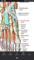
Dorsalis pedis artery |
Continuation of the anterior tibial nerve, deep to the inferior extensor retinaculum. |
|
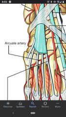
Arcuate artery |
A branch off the dorsalis pedis artery that crosses the proximal ends of the metatarsal bones. |
|
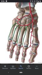
Dorsal metatarsal arteries |
3 branches off the arcuate artery. |
|
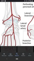
Lateral tarsal artery |
Arises from the dorsalis pedis artery near the ankle joint and passes deep to the extensor digitorum brevis & extensor hallucis brevis muscles. |
|
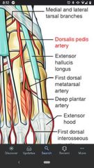
Deep plantar artery |
Arises from the dorsalis pedis artery near the origin of the arcuate artery. Anastomosis with the plantar arch on the sole of the foot. |
|
Dorsal digital branches of the deep fibular nerve |
Between the great toe and the second toe. |
|
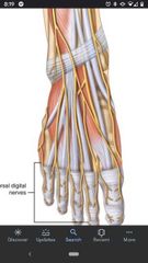
Deep dorsal branches of superficial fibular nerve |
Cutaneous branches between toes 2-5. |
|
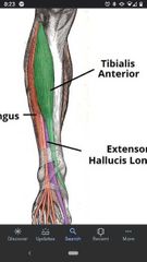
Tibialis anterior muscle |
Proximal Att: superior half of the lateral surface of the tibia Distal Att: base of first metatarsal and medial and inferior surfaces of medial cuneiform. Actions: dorsiflexes and inverts the foot Innervation: deep fibular nerve |
|

Extensor hallucis longus |
Proximal Att: middle part of anterior surface of fibula and interosseous membrane. Distal Att: dorsal aspect of base of distal phalanx of great toe. Actions: extends great toe & dorsiflexes the foot. Innervation: deep fibular nerve |
|
Extensor digitorum longus |
Proximal Att: tibia & superior 3/4ths of anterior surface of interosseous membrane. Distal Att: extensor expansions of distal phalanges of lateral 4 digits. Actions: extends lateral 4 digits & dorsiflexes the foot. Innervation: deep fibular nerve |
|
Fibularis tertius |
Proximal Att: inferior third of anterior surface of fibula & interosseous membrane. Distal Att: dorsum of base of 5th metatarsal. Actions: dorsiflexes and everts the foot. Innervation: deep fibular nerve |
|
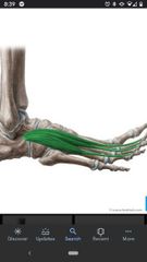
Extensor digitorum brevis |
Proximal Att: calcaneus Distal Att: extensor expansions of digits 2-5. Actions: extends digits Innervation: deep fibular nerve |
|
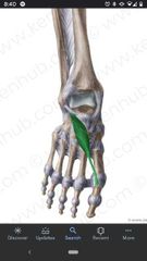
Extensor hallucis brevis |
Proximal Att: calcaneus. Distal Att: extensor expansion of digit 1. Actions: extends great toe. Innervation: deep fibular nerve |
|
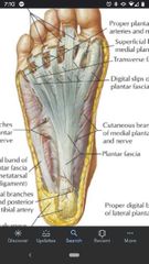
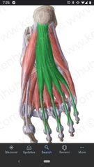
Plantar aponeurosis |
Supports the longitudinal arch and divides distally to into 5 bands, one for each toe. |
|
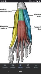
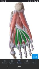
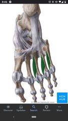
First layer of sole muscles |
Flexor Digitorum brevis, abductor hallucis, abductor digiti minimi |
|
Common/Proper plantar digital nerves |
Branches of the medial & lateral plantar nerves. |
|
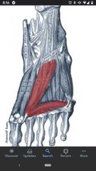
Flexor digitorum brevis |
Proximal Att: calcaneal tuberosity & plantar aponeurosis. Distal Att: middle phalanges of digits 2-5. Actions: flexes toes 2-5. Innervation: medial plantar nerve
|
|
Abductor hallucis |
Proximal Att: medial process or calcaneal tuberosity, flexor retinaculum & plantar aponeurosis Distal Att: medial side of base of proximal phalanx of 1st digit. Actions: abducts and flexes 1st digit Innervation: medial plantar nerve |
|
Abductor digiti minimi |
Proximal Att: medial & lateral side of calcaneal tuberosity & plantar aponeurosis. Distal Att: lateral side of base of proximal phalanx of 5th digit. Actions: abducts & flexes the 5th digit Innervation: lateral plantar nerve |
|
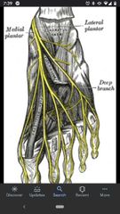
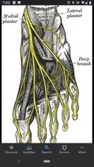
Medial/lateral plantar nerves |
Divisions of the tibial nerve. Medial innervates toes 1-4 & lateral innervates toes 4 & 5. |
|
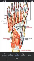
Medial/lateral plantar arteries |
Divisions of the posterior tibial artery. Run along side the medial & lateral plantar nerves. |
|
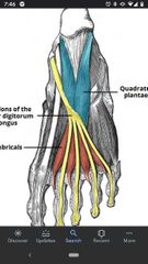
Muscles of the 2nd layer of the sole |
Quadratus plantae, flexor digitorum longus tendon, lumbricals |
|
Quadratus Plantae |
Proximal Att: medial & lateral margin of calcaneus Distal Att: posterolateral margin of tendon of flexor digitorum muscle. Actions: flexes digits 2-5. Innervation: lateral plantar nerve |
|
Lumbricals |
Proximal Att: tendons of flexor digitorum longus muscle. Distal Att: medial aspect of extensor expansion of digits 2-5. Actions: flexes proximal phalanges & extends middle and distal phalanges of digits 2-4. Innervation: medial plantar nerve & lateral plantar nerve. |
|
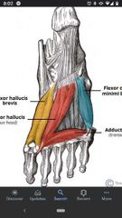
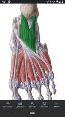
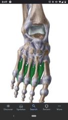
Muscles of 3rd layer of the sole |
Flexor hallucis brevis, tendon of the flexor hallucis longus, adductor hallucis, flexor digiti minimi brevis |
|
Flexor hallucis brevis (medial & lateral heads) |
Proximal Att: plantar surfaces of cuboid and lateral cuneiforms. Distal Att: both sides of proximal phalanx of first digit. Actions: flexes proximal phalanx of 1st digit. Innervation: medial plantar nerve |
|
Adductor hallucis |
Proximal Att: base of metatarsals 2-4 (oblique head), plantar ligaments of MTP (Transverse head). Distal Att: lateral side of base of proximal phalanx of 1st digit Actions: adducts great toe. Innervation: deep branch of lateral plantar nerve. |
|
Flexor digiti minimi |
Proximal Att: base of 5th metatarsal. Distal Att: base of proximal phalanx of 5th digit. Actions: flexes proximal phalanx of 5th digit Innervation: superficial branch of lateral plantar nerve |
|
4th layer of Sole muscles |
Plantar interosseous, dorsal interosseous, fibularis longus tendon, tibalis posterior tendon. |
|

Plantar arch |
At the base of metatarsal bones, formed by the lateral plantar artery and anastomosis with the deep plantar artery. |
|
Deep plantar artery |
A branch of the dorsalis pedis artery. The medial end of the plantar arch. |
|
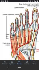
Plantar metatarsal arteries |
Arise from the plantar arch. |
|

Dorsalis pedis artery |
Continuous with anterior tibial artery distally. |
|
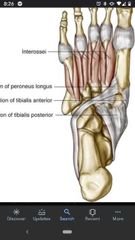
Plantar interossei |
Proximal Att: plantar surface of metatarsals 3-5. Distal Att: medial sides of bases of phalanges of digits 3-5. Actions: adducts digits 3-5 & flexes MTP joints. Innervation: lateral plantar nerve |
|
Dorsal interossei |
Proximal Att: adjacent sides of metatarsals 1-5. Distal Att: medial side of proximal phalanx of second digit (1st), lateral sides of proximal phalanx of digits 2-4. Actions: abducts digits 2-4 & flexes MTP joints Innervation: lateral plantar nerve |