![]()
![]()
![]()
Use LEFT and RIGHT arrow keys to navigate between flashcards;
Use UP and DOWN arrow keys to flip the card;
H to show hint;
A reads text to speech;
31 Cards in this Set
- Front
- Back
|
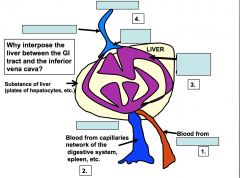
What is the largest endocrine organ? What is covered by? It divides the parenchyma (bulk of the liver=hepatocytes) into lobules Glycogen storage is one of the functions of the organ above. What else stores glycogen? Where is this organ found and why? |
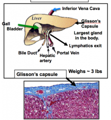
Liver Covered by the Glisson's capsule (CT) -We have very little CT in liver perenchyma in humans (thicker in animals ex. pigs) Glycogen is also stored in cardiac and smooth muscle It is found between the small intestine and heart because all of the toxins can be taken out of the liver before it enters the heart |
|
|
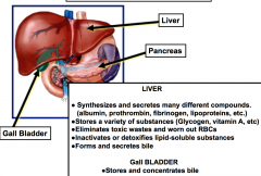
Functions of the liver Synthesizes many compounds like prothrombin, fibrinogen, lipoprotein and? What does it store? What does it eliminate? What does it inactivate or detoxify? What does it form and secrete? |
|
|
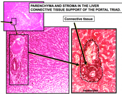
The liver is divided into 6 sided unit structures called? The little space around the above is? Each point in the unit structure is called? And is composed of what 4 things? In the unit structure are what cells that are linearly arranged and are in "plates" (polygonal)? |
Classic lobule Little space is the connective tissue Portal Triad there is an artery, vein, duct, and lymphatic vessel embedded in connective tissue Hepatocytes |
|
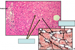
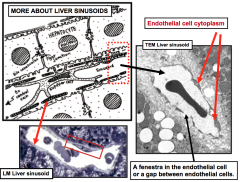
Between hepatocytes arranged in plates are discontinuous capillaries with endothelial cells are called? What supports the discontinuous capillaries of the above? |
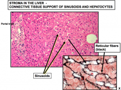
Sinusoids Reticular walls (collagen type III) support walls of discontinous capillaries (White is the lumen of the sinusoids) |
|
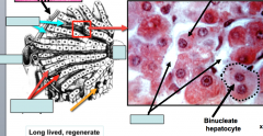
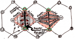
In the portal triad: There is a hepatic artery that brings __ into the liver There is a portal vein that comes from the __ and brings __ into the liver There is also a bile duct and a lymphatic vessel Blood goes from the __ through the __ into the __ Hepatocytes are stacked together and make bile which is transported from the __ towards the __ Hepatocytes (polygonal) can be nucleate or binucleate (4n DNA instead of 2N due to no cytokinesis) |
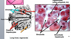
The Hepaticartery– brings oxygenated blood (25% of mix) into the liver Portalveincomes from GI tract -brings poorlyoxygenated blood (75% of mix) into the liver -(aswell as toxins from the intestine, breakdownproducts of blood cells from the spleen, and endocrine secretions from thepancreas and enteroendicrinecells of the gi tract) Blood goes down through portal triad through sinusoid to the central vein Hepatocytesmake bile and are transported from all the hepatocytes towards bile duct -Opposite the direction of the flow of blood! Hepatocytes can be nucleate or binucleate (4n amount of dna instead of 2n) no cytokinesis |
|
|
Hepatocyte Structure: Has a central nucleus, extensive rough ER, and smooth ER that deal with toxic molecules and glycogen metabolism On 4 sides of hepatocytes are? On the other 2 sides of the hepatocytes are? Which sides are the extensive microvilli brush border found? What are found in between hepatocytes and what do they do? What other organelle does the liver have? |
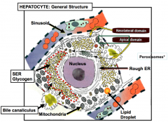
On 4sides are other hepatocytes Onthe other 2 sides are sinusoid Havean extensive microvilli brush border on the sidesthat face the sinusoid Bile canaliculi are between hepatocytes and transfers bile Peroxisome: make catalyse which breakdown hydrogen peroxide |
|
|
Liver hepatocytes are filled with? What is one of it's major functions? What organelle does this occur in? |
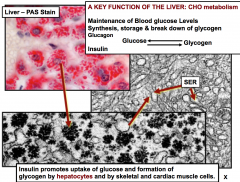
Filled with glycogen molecules Mainfunction is carbohydrate metabolism -Maintain blood glucose levels by synthesizing or breaking down glycogen -Insulin promotes uptake of glucose and formation of glycogen -Also stores glycogen -specifically happens in the smooth endoplasmic reticulum |
|
|
Besides carbohydrate metabolism, the smooth ER is involved in? How does it do this? -Other roles of SER include synthesis of lipoproteins, bile salts and VLDL (very low density lipoproteins) |
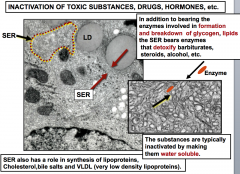
Involved in inactivation of toxic substances, drugs, hormones -detoxifies barbiturates, steroids and alcohol It inactivates them by making them water soluble (can pee them out) |
|
|
Role of Rough ER in hepatocytes Synthesizes? This includes __ which contributes to colloid pressure of plasma and transports non soluble substances Also includes __ and __ for clotting And __ which transports iron in plasma to bone marrow/spleen/liver |
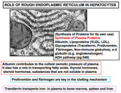
Plasma protein |
|
|
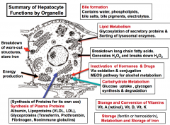
Summary of Hepatocyte Function: 1. Bile formation 2. Lipid metabolism 3. Breakdown of fatty acids and hydrogen peroxide 4. Inactivation of hormones and drugs (toxins) 5. Carbohydrate metabolism 6. Storage of vitamins 7. Storage of Iron from RBCs 8. Synthesis of plasma protein 9. Energy production |
|
|
|
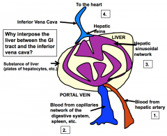
Cream= where hepatcotyes are (parenchyma) Bloodis coming from hepatic artery(oxygenated) Bloodcomes in from portal vein (poorly oxygenated) Goesthrough purple channels sinusoids Makesits way to hepatic vein, inferior cava, and to the heart |
|
|
Besides the classic lobule, there are 2 other ways to organize the liver: One is the Portal Lobule It is used to describe? It is a boundary that joins __ and the center is the __ This triangular region is the part of the classic lobule that secretes __ that drains into the __ of the __ |
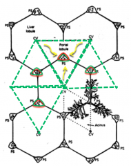
PortalLobule: -Used to describe the exocrine function of the liver such as bile secretion and flow -Boundary that joins three central veins and the center is the portal triad •The triangular region is that partof the classic lobule that secretes bile that drains into the bile duct in theportal triad |
|
|
Besides the classic lobule, there are 2 other ways to organize the liver: The other is the Hepatic (portal) Acinus *This is the smallest functional unit to describe the liver parenchyma -It is also used to describe another exocrine function of the liver The Hepatic (portal) Acinus is diamond shaped. What is found at the ends of its long axis? What is found at the ends of its short axis? |
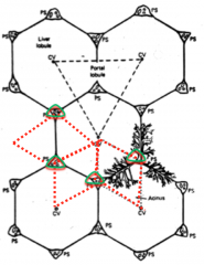
Hepatic (portal) Acinus Atthe end of it's long axis are central veins At the ends of the short axis areportal triads |
|
|
Hepatic (portal) Acinus Is divided in to 3 zones: Zone 1 is in the __ of the hepatic portal acinus and is the region closest to? Zone 3 is the __ of the hepatic portal acinus and is the region closest to? Zone 2 is the region in between Blood comes in from which zone? What zone sees the most amount of oxygen? What zone sees the most amount of toxin? What zone sees the least amount of toxin? If you have a patient whose bile duct is clogged, which zone would be most effected -Sinusoids go all the way through every zone in the hepatic acinus Oxygen content, metabolic activity, and distribution of hepatic enzymes varies across the zones. Damage that occurs from exposure of toxic materials are explained using these different zones. Because of this hepatocytes are capable of? |
Hepatic (portal) Acinus Zone1 (w/ thick black line in middle): Region closest to portal triads Zone 3: 2 regions at the ends of the hepatic acinus, closest to the central veins Zone 2: region in between Mix of oxygenated andpoorly oxygenated Blood comes in from Zone 1 -Blood then passes through sinusoids to zone 2, zone 3 and out the central vein. Zone 1 sees highest amount of oxygen Zone 1 also sees the most toxins Zone 3 sees the least amount of oxygen -Zone 1. Hepatocytes can regenerate and proliferate |
|
|
Macrophages roam in the sinusoid space are called? |
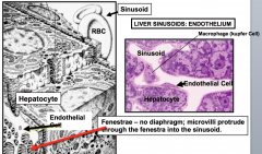
Macrophages = Kupffer cells |
|
|
- |
|
|
Hepatocytes also have mitochondria and rough ER What is between the hepatocytes and wall of endothelial cells? Microvilli can go through here |
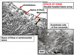
Spaceof Disse |
|
|
In the Space of Disse, what cells store Vitamin A and lipids? Why is it bad to have when you have liver disease? |
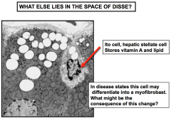
Ito Cell -Bad to have when you have liver disease because they can differentiate into a fibroblast cell type and are responsible for causing fibrosis in liver (thickening/scarring of CT) |
|
|
Hepatocytes dump bile into __ which eventually reach? What is bile involved in? It also is involved in secretion of lipids, cholesterol, bile salts, bilirubin, iron, and copper -Bile canaliculi is lined by cholangiocytes -Bile going from the hepatocytes into the bile canaliculi is an active process |
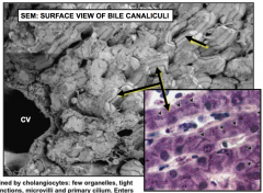
Hepatocytes dump bile into bile canaliculi (ductile network) which eventually reach the bile duct Bile is involved in the absorption of fat |
|
What is this structure? What does it do? What are 2 distinct characteristics of this structure? |
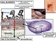
Gallbladder -Smallpouch that holds bile *1. mucosalfolds are tortuous *2. hugesmooth muscle (muscularis) Has a mucosa (epithelium-folds and lamina propria) -Adventitia has loose connective tissue -has simplesquamous epithelial lining |
|
|
What concentrates the bile in the gallbladder? These cells are attached by? What is pumped out into the basal lateral region in the intercellular spaces? Where does it go from there? |
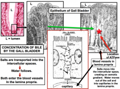
Epithelialcells in the mucosal folds of the gallbladder concentrate bile Tight junctions Saltisbeing pumped out into basal lateral regionin the intercellular spaces andwater follows -large number of sodium pumps present Both go out of the cell and into the capillaries of the laminapropria -therebyconcentrating the bile within the cell |
|
|
What arises from overlying epithelium? Which one is associated with ducts (secretory region which enters a duct system and end up in small intestine to perform their function)? Which one is not associated with ducts? They make their hormones and pump them into surrounding vasculatures This is also called the? |
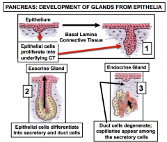
Exocrineand endocrine glands -exocrineglands in the pancreas -endocrine glands are not associated with ducts -endocrineportion of the pancreas are the Islets oflangerhans |
|
|
The Pancreas is covered by? What 3 things does it synthesize? |
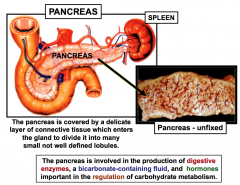
Pancreas is coveredby CT which enters the gland to divide it into lobules 1. digestive enzymes 2. bicarbonate-containing fluid which neutralizes the acidic chyme from the stomach 3. hormones for the regulation of carbohydrate metabolism |
|
|
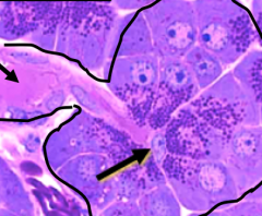
In the exocrine portion of a pancreatic lobule, there are groups of cells that form an __ which make? This group of cells are polar meaning, the nuclei is found? And the the zymogen granules (which contain enzymes and are bright pink) are found? Part of the intralobular duct is in the secretory cells (acinus) and is lined by? What is the function of the above? Digestive enzymes are dumped into the intercalated duct goes to the __ and then to the __ that goes into the __ |

Exocrinepancreas: group of cells form acinus which make digestive enzymes Have nuclei at the bottom and apex filled with zymogen granules Epithelial cells called centroacinar cells Centroacinar cells make the bicarbonatesolution that neutralizes pancreatic fluid going from the stomach to the smallintestine Interlobular duct (outside the acinus) and then into the main pancreatic duct which goes into the duodenum |
|
|
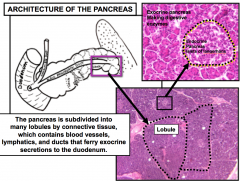
The pancreas is subdivided by CT which has blood vessels/lymphatics/ducts that carry exocrine secretions to the duodenum Parotid vs Pancreas glands: both serous secreting glands -How can you tell them apart? |
Pancreas has Islets of Langerhans = Endocrine pancreas |
|
|
Exocrine cells (acinus cells) are regulated by? Centroacinar cells are regulated by? |
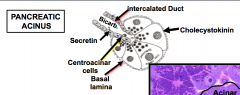
Exocrine cells are regulated by cholecystokinin Centroacinar cells are regulated by secretin |
|
What is the arrow pointing to? |
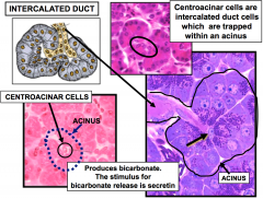
Centroacinar cell |
|
|
Exocrinepancreatic cells are making digestive enzymes and bicarbonate fluid that entersintercalated duct (intralobular duct) It enters bigger ducts thatlie between the lobules (interlobular ducts) then go into the main pancreaticduct and into the duodenum which are characterized by? |
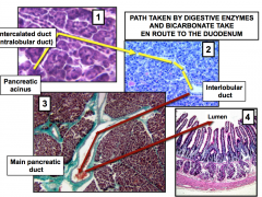
Characterized by brunner's glands |
|
What is this structure? It contains: 1. Beta cells that secrete __ which causes? 2. Alpha cells secrete __ which causes? 3. D cells secrete __ which causes? 4. PP cells secrete __ which causes? |
Islets of langerhans are highly vascular with capillaries where hormones are secreted 1. Betacells –> Insulin –Causes decrease of blood glucose and coverts it to glycogen (liver, skeletal muscle & fat cells) 2. Alphacells –> glucagon -Causes increase of blood glucose by glycogenbreakdown (liver, release of glucose) 3. D-cells –> Somatostatin -Causes inhibition of insulin & glucagon secretion (inhibits alpha and beta cells) 4. PPcells –> pancreatic polypeptide -Causes inhibition of enzyme & bicarbonate release |
|
|
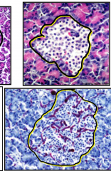
Numerous __ capillaries lie within the islets __ capillaries occuramong the pancreatic acini. |
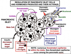
Numerous fenestrated capillaries lie within the islets Non-fenestrated capillaries occuramong the pancreatic acini |
|
|
What is a marker of pancreatic cancer? It secretes growth factors, hemokines, cytokines, etc. It changes the extracellular matrix and becomes like a fibroblast -it is able to travel through the extra cellular matrix |
Stellate Cell |