![]()
![]()
![]()
Use LEFT and RIGHT arrow keys to navigate between flashcards;
Use UP and DOWN arrow keys to flip the card;
H to show hint;
A reads text to speech;
54 Cards in this Set
- Front
- Back
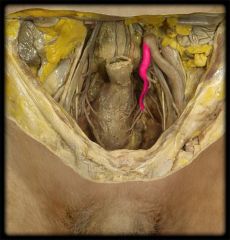
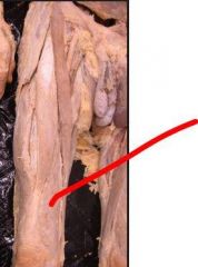
What is the structure highlighted in pink? At what level does it bifurcate? |
Common iliac arteries. Bifurcates at level of L4. |
|
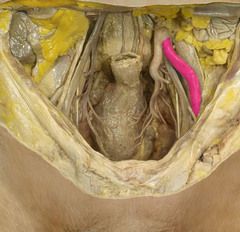
What is the structure highlighted in pink? What structure does it change into as it passes deep to the inguinal ligament? |
External iliac artery Changes into the femoral artery after it passes deep to the inguinal ligament. |
|
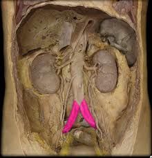
Identify the vascular structure highlighted in pink. What region of the body does it supply? |
Internal iliac artery. Supplies the pubic region of the body. |
|

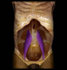
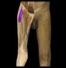
Identify the highlighted structure. What is its origin(s) and insertion(s)? |
Psoas major Origin: Transverse processes of all lumbar vertebrae and the sides of T12-L5 Insertion: Lesser trochanter of the femur |
|

Identify the highlighted structure. What is its origin(s) and insertion(s)? |
Iliacus Origin: Iliac crest/fossa, anterior sacro-iliac ligaments Insertion: Tendon of psoas major and lesser trochanter |
|
What is the innervation of the highlighted structure? |
Femoral nerve |
|
What is the innervation of the highlighted structure? |
Anterior rami of spinal nerves |
|

What are the actions associated with the highlighted structure? |
Hip flexion Controls trunk deviation |
|
|
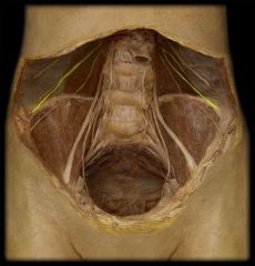
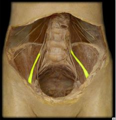
At what spinal level does the ilioinguinal nerve originate? |
L1. |
|
|
At what spinal level does the iliohypogastric nerve originate? |
L1. |
|
|
At what spinal level does the genitofemoral nerve originate? |
L1-L2. |
|
|
At what spinal level does the obturator nerve originate? |
L2-L4. |
|
|
At what spinal level does the femoral nerve originate? |
L2-L4. |
|
|
At what spinal level does the sciatic nerve originate? |
L4-S3 |
|
|
At what spinal level does the pudendal nerve originate? |
S2-S4 |
|
|
At what spinal level does the lateral cutaneous nerve of the thigh originate? |
L2-L3 |
|
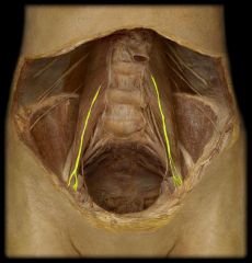
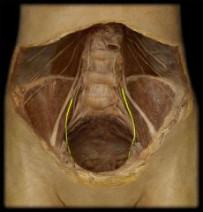
Identify the highlighted nervous structure. From what spinal level does it originate? |
Ilioinguinal Originates from L1 |
|
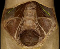
Identify the highlighted structure. From what spinal level does it originate? What is the structure associated with it? |
Genitofemoral nerve Originates from L1-L2. Psoas major is associated with it. |
|
Identify the highlighted structure. |
Subcostal nerve. |
|
Identify the highlighted structure. From what spinal level does it originate? |
Femoral nerve. Originates from L2-L4. |
|
Identify the highlighted structure. From what spinal level does it originate? |
Obturator nerve Originates from L2-L4. |
|
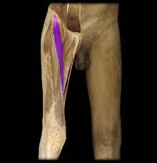
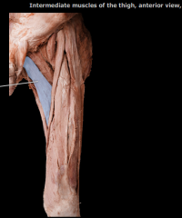
Identify the highlighted structure. What is its origin and insertion? |
Sartorius Origin: Anterior superior iliac spine Insertion: Medial surface of tibia (superior part) |
|

a) What is the highlighted structure's innervation? b) If the above nerve supply was compromised, what movements might be affected? |
a) Femoral nerve b) Flexion, abduction, and lateral rotation of hip joint; flexion of knee joint |
|
|
What is the origin and insertion of the rectus femoris? |
Origin: Anterior inferior iliac spine; ilium (superior to acetabulm) Insertion: Patella (via quad tendon); tibial tuberosity (via patellar lig) |
|
What are the actions associated with the highlighted structure? |
Extends knee joint Stabilizes and helps iliopsoas flex hip joint |
|

Identify the highlighted structure. a) What is its origin(s)? b) What is its insertion(s)? |
Vastus lateralis Originates on the lateral lip of the linea aspera and the greater trochanter Inserts onto the tibial tuberosity via the quad tendon/aponeurosis, and the patella via the patellar ligament and aponeurosis. |
|
Identify the following structure. What are its origin(s) and insertion(s)? |
Vastus medialis Originates on the intertrochanteric line and the medial lip of the linea aspera Inserts onto the tibial tuberosity via the quad tendon/aponeurosis, and the patella via the patellar ligament and aponeurosis. |
|
What are the actions associated with the highlighted structure? |
Extends the knee joint |
|

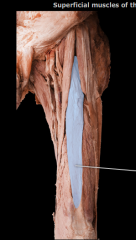
a) Identify the highlighted structure. b) What muscle lies immediately superficial to it? |
a) Vastus intermedius b) Rectus femoris lies superficial to it. |
|

Name the origin(s) and insertion(s) of the highlighted structure. |
Origin: anterior/lateral surface of the femur Insertion: tibial tuberosity via the quad tendon, and the patella via the patellar ligament. |
|

Name the innervation of the highlighted structure. |
Femoral nerve |
|

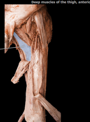
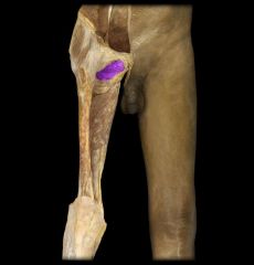
Identify the highlighted structure. Which structure is reflected to expose it? |
Adductor brevis The adductor longus is reflected to expose the entirety of the brevis. |
|
Identify the highlighted structure. |
Adductor longus. |
|

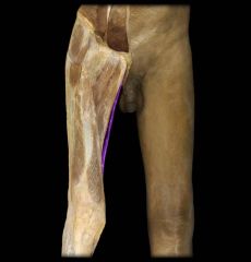
Name the origin and insertion of the highlighted muscle. |
Origin: Pubic body (just inferior to the crest) Insertion: Linea aspera (middle third) |
|
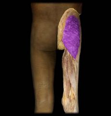
Name the origin and insertion of the highlighted muscle. |
Origin: Inferior pubic ramus, pubic body Insertion: Linea aspera (proximal part) |
|

What are the actions associated with the highlighted muscle? |
Hip adduction and to some extent, hip flexion. |
|
|
What muscle lies deep to adductor longus and brevis? What are its origins and insertions? |
Adductor magnus (adductor and hamstring part) Origin: inferior pubic ramus, ischial ramus (adductor part); ischial tuberosity (hamstring part) Insertion: linea aspera, medial epicondyle, gluteal tuberosity (adductor part); adductor tubercle (hamstring part) |
|
|
Which nerve innervates the adductor magnus? |
Adductor part: obturator nerve Hamstring part: tibial nerve |
|

Identify the highlighted structure. What is its origin(s) and insertion(s)? |
Gracilis Origin: pubic body, inferior pubic ramus Insertion: medial tibia (superior portion) |
|
List the actions associated with the highlighted structure. |
Hip adduction, knee flexion, medial rotation |
|
Identify the highlighted structure. What is its origin(s) and insertion(s) |
Obturator externus Origin: Obturator foramen margins and obturator membrane Insertion: trochanteric fossa of femur |
|

What are the actions associated with the highlighted structure? Which nerve would compromise these movements if it were damaged? |
Action: Lateral rotation of hip, holds pelvis in place Nerve: Obturator nerve |
|

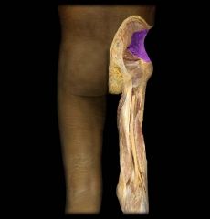
a) Identify the highlighted structure. b) What is its origin? c) What is its insertion? |
a) Gluteus maximus b) Sacrum/coccyx, ilium, sacrotuberous ligament c) Iliotibial band, gluteal tuberosity |
|
a) What is the highlighted structure's innervation? b) What actions might be compromised with damage to the above nerve? |
a) Inferior gluteal nerve b) Hip extension, lateral rotation of the hip |
|
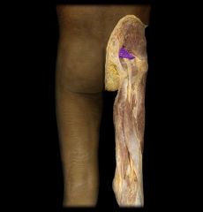
a) Identify the highlighted structure. b) What is its origin? c) What is its insertion? |
a) Gluteus medius b) Ilium (between anterior and posterior gluteal lines) c) Lateral surface of the greater trochanter |
|
a) Identify the highlighted structure. b) What is its origin? c) What is its insertion? |
a) Gluteus minimus b) Ilium (betwee anterior and posterior gluteal lines) c) Anterior surface of greater trochanter |
|

a) What is the highlighted structure's innervation? b) What actions might be compromised with damage to the above nerve? |
a) Superior gluteal nerve b) Medial rotation of the hip, abduction |
|

a) What is the highlighted structure's innervation? b) What actions might be compromised with damage to the above nerve? |
a) Superior gluteal nerve b) Medial rotation of the hip, abduction |
|
a) Identify the highlighted structure. b) What is its origin? c) What is its insertion? |
a) Tensor fascia latae b) Anterior superior iliac spine and iliac crest c) Iliotibial tract |
|

a) What is the highlighted structure's innervation? b) What actions might be compromised with damage to the above nerve? |
a) Superior gluteal nerve b) Flexion of the hip joint, may have instability as it assists gluteus maximus with stabilizing the extended hip joint. |
|

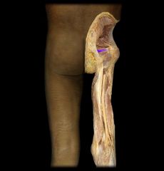
a) Identify the highlighted structure. b) What is its origin? c) What is its insertion? |
a) Obturator internus b) Obturator foramen and membrane c) Medial surface of greater trochanter |
|
a) Identify the highlighted structure. b) What is its origin? c) What is its insertion? |
a) Piriformis b) 2-4 sacral segments, greater sciatic notch, sacrotuberous ligaments c) Superior surface of the greater trochanter |
|
a) What is the highlighted structure's innervation? b) What actions might be compromised with damage to the above nerve? |
a) Nerve to obturator internus b) Lateral rotation of extended hip joint and abduction of flexed hip joint |
|

a) What is the highlighted structure's innervation? b) What actions might be compromised with damage to the above nerve? |
a) Anterior rami of spinal nerves b) Abduction of flexed hip joint and lateral rotation fo extended hip joint |