![]()
![]()
![]()
Use LEFT and RIGHT arrow keys to navigate between flashcards;
Use UP and DOWN arrow keys to flip the card;
H to show hint;
A reads text to speech;
69 Cards in this Set
- Front
- Back
|
What kind of culture must be used to run diagnostic tests? |
pure cultures |
|
|
What are the major types of media |
General (Complex) Contains necessary nutrients, vitamins, minerals, etc.. Most types of non-fastidious organisms, but not all are able to grow on this type of media. EX: tryptic soy broth, nutrient agar Enriched-contains added ingredients like blood, serum, vitamins, growth factors etc.. EX: Blood agar Differential-Contains and indicator designed to cause certain organisms to develope a different appearance from other microbes growing on same medium. EX: MacConkey agar, EMB agar Selective-Permits growth of certain organisms while preventing growth of others. EX: Mannitol Salt agar, EMB agar Selective/differential (does both) |
|
|
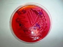
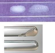
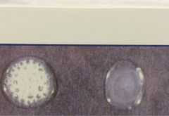
EMB (Eosin Methylene Blue) Agar |
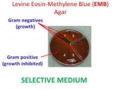
Purplish Color Selective for gram negatives Differential for lactose fermenters and fecal coliforms + result=dark purple colonies. Produces the enzyme b-galactosidase to ferment lactose to an acidic by-product. If pink colonies show it means it's a slow lactose fermenter. - result=colorless colonies. Appear same color as EMB Coliforms: green metallic sheen on colonies. Denotes vigorous lactose fermentation to an acidic by product.
|
|
|
MacConkey Agar |
Selective for gram negative Differential for lactose fermenters. + lactose fermenation=pink-lavendar colonies. Produces the enzyme B-galactosidase to ferment lactose to acidic by products. - lactose fermenation: colorless colonies. |
|
|
Starch agar |

Differential for bacteria that can hydrolyze starch; like bacillus, corynebacterium and clostridium. +starch hydrolysis: clear zone (often golden color halo) around bacterial growth AFTER adding iodine. Produces alpha-amylase oligo 1, 6-glucosidase to break down starch into smaller sugar subunits. -starch hydrolysis: dark blue or brown agar (no clear zone) after adding iodine. |
|
|
MSA (mannitol salt agar) |

Selective for staphylococcus species (7.5% salt) Differential for mannitol fermenters. Phenol red dye detects acid production by turning yellow. + mannitol fermentation- yellow agar. produces mannitol dehydrogenase to break down mannitol to acidic by products. - mannitol fermen. color of origional red agar. |
|
|
Blood Agar |
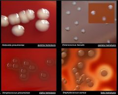
Tryptic Soy agar enriched with 5 or 10% whole sheep blood. It is widely used in the culture of medical specimens and for many types of fastidious bacteria. Differential for hemolysis reactions. Gamma Hemolysis: no change in agar appearance; absence of hemolysis Alpha hemolysis: greenish color to agar; partial destruction of RBC's. Beta hemolysis: Clear zone around colonies; total destruction of RBC's. |
|
|
Columbia CNA (Colistin-nalidixic acid) with 5% sheep blood |
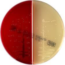
Selective for growth of gram positives, particularly staphylococci, streptococci and enterococci. Inhibits the growth of gram negatives b/c of the presence of the antibiotic colistin and nalidixic acid. Enriched with the addition of blood Differential for hemolytic reactions |
|
|
Hektoen Enteric (HE) agar |
differential for lactose fermentation and hydrogen sulfide production Selective for mainly salmonella, shigella; E. coli can also grow on it. + Lactose fermen=yellow to salmon-pink colonies. fermentation of lactose sugar produces acidic by products. -lactose fermen=blue-green colonies. +hydrogen sulfide-black colonies. (Reduces Sulfur to H2S which reacts with ferric ammonium citrate in the medium to create a black precipitate. |
|
|
Triple Sugar Iron (TSI) Agar |

Differential for glucose, sucrose and/or lactose fermentation; also hydrogen sulfide production. Helps to distinguish gram- enteric bacilli in the family Enterobacteriaceae. Contains iron. -fermentation=red slant, red butt + fermentation of only glucose=red slant, yellow butt (glucose broken down to acidic by product) + fermentation of glucose, lactose and or sucrose=yellow slant, yellow butt (2/3 sugars broken down to acidic by products) +hydrogen sulfide (H2s) blackened media; sulfur reduced to H2S (H2s reacts with ferrous sulfate in medium to form black precipitate) + gas; lifting of agar or appearance of cracks in agar |
|
|
Phenol Red Carbohydrate Fermentation Broth |
Differential for carbohydrate fermentation using pH indicator (phenol red) Can be designed to test any one specific sugar. Used to identify bacteria in general, especially useful in distinguishing gram neg. enteric bacilli but may be used for other organisms too. + fermentation: Yellow broth (the specific carbohydrate used) is broken down into acidic by-products. - fermentation: original red broth + gas production: sometimes gas is produced as a result of fermentation and is seen as bubblescollected in the Durham tube. (upside down tube) |
|
|
Citrate agar slant |
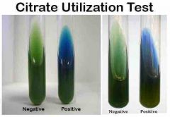
Differential for citrate utilization. If microbe can use citrate as it's only available source for carbon. Useful in identifying Gram negative enteric bacilli in the family Enterobacteriaceae
+ citrate= any amounts of blue. Organism produces citrate – permease to transport citrate (only source of carbon) into its cells to be utilized.
- citrate=green
* A heavy amount of culture is needed |
|
|
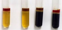
Urea Broth |
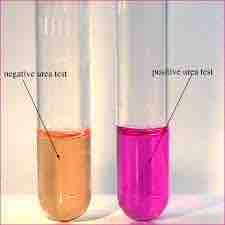
Differential for urease production. Useful in distinguishing Proteus (urease +) from salmonella or shigella (urease -).
Urease is an enzyme that breaks down urea to ammonia
Phenol red (pH indicator) changes to bright pink at alkaline pH
+ urease=dark pink (urease hydrolyzed urea to ammonia)
- urea=original salmon (orangish) or yellow color. |
|
|
Phenylalanine Agar |
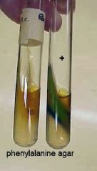
Differential for phenylalanine deaminase production. Helps to distinguish between gram negative enteric bacilli. + phenylalanine deaminase: green, when iron III chloride added. Organism has broken down phenylalanine to phenylpyruvic acid using phenylalanine deaminase. - phenylalanine deaminase: no color change (except for added chemical) |
|
|
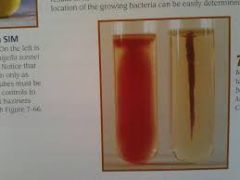
SIM Medium (semisolid) |
Differential for hydrogen sulfide production; motility and indole production. Especially useful in identifying gram negative enteric bacilli in the Family Enterobacteriaceae. 3 tests are run in ONE tube of medium--indole production, hydrogen sulfide (H2S) production and motility a. hydrogen sulfide (H2S) + H2S: blackened medium; H2S reacts with ferrous ammonium sulfate to produce the balck precipitate, ferric sulfide - H2S: :No color change b. Motility 1. Indicates if organism possesses flagella for movement. + motility: growth diffusing away from stab line indicates flagella are present. - motility: growth only along stab line c. Indole production +pink or red ring when Kovac's reagent is added; break down of amino acid, tryptophan by enzyme, tryptophanase to indole. - indole; no color chage when Kovac's added. |
|
|
Motility/ GI medium/TCC (MOT) |
Can be used as an alternate to SIM due to SIM's difficult interpretation. Differential for determining whether the organism has flagella as a means of movement. It contains an indicator called triphenyltrazolium chloride (TCC) which turns red in the presence of bacteria. This color traces the growth of the bacteria in the medium. + motility: medium displays a diffuse pattern (turbidity) of red color away from stab line or throughout the medium indicates organism possesses flagella. - motiltiy: red color only along the line of inoculation and no turbidity in the rest of the medium. |
|
|
optichin disk susceptibility Test |
paper disk impregnated with the scant amount of the antibiotic optichin. It is used to presumptively identify Strep pneumoniae which is sensitive to it, from other species of streptococci. The organism to be tested is "painted" on a Blood agar plate and an optichin disk is placed in the middle of the "Lawn" It's then incubared at 35c in a candle jar for 24 hrs. + result: (presumptive for S. pneumoniae)14+ mm zone of inhibition around a 6 mm optichin disk. - result: smaller inhibition zones indicate further testing required. Likely organism is not S. pneumoniae. |
|
|
Coagulase Test |
Distinguishes Staph. aureus from other gram positive cocci The enzyme, coagulase, causes blood plasma to clot + coagulase: clotting of plasma (solid) indicates coagulate produced - coagulase: no clotting (liquid) |
|
|
Oxidase Test |
Cytochorme c oxidase is an enzyme oxidizes tetramethyl-p-phenylenediamine (colorless) into a colored by-product. It is used to distinguish Pseudomonasand Neisseria from other organisms. + oxidase: purple or black; Cytochrome c Oxidase produced which oxidizes tetramethyl-p-phenylenediamine into a colored by product. -oxidase: no color change. |
|
|
Catalase Test |
Helpful in distinguishing Staphylococcus from Streptococcus. Catalse breaks down hydrogen peroxide (H2O2) to water and oxygen gas. + catalase: any bubbling; enzyme catalase produced by organism which broke down H2O2 to H2O and O2 -Catalase: no bubbling |
|
|
Chocolate agar |
Cooked blood agar. Will grow a wide range of organisms. Contains X & V growth factors which will allow the growth of Neisseria and Hemophilus. Used for respiratory, eye, ear, nose and throat specimens. NOT used for stool or wound specimens. |
|
|
Does refrigeration or freezing stop all bacterial growth? |
NO. refrigeration slows it down and freezing halts it but will resume once thawed. |
|
|
Staphylococcal food poisoning |
Causative agent: S. areus. Causes food intoxication Staphylococcal food poisoning is a gastrointestinal illness. It is caused by eating foods contaminated with toxins produced by Staphylococcus aureus . The most common way for food to be contaminated with Staphylococcus is through contact with food workers who carry the bacteria or through contaminated milk and cheeses. |
|
|
Food infection |
An overgrowth of bacteria that establishes themselves in GI tract |
|
|
Food intoxication |
Bacteria release toxins that make you sick |
|
|
Fecal coliform |
facultatively anaerobic, rod-shaped, gram-negative, non-sporulating bacterium. This bacteria generally originate in the intestines of warm-blooded animals. |
|
|
Salmonellosis |
Caused by salmonella; food infection. To avoid: Cook poultry, ground beef, and eggs thoroughly. Do not eat or drink foods containing raw eggs, or raw (unpasteurized) milk. |
|
|
Shigellosis |
Caused by shigellosa; food infection
A safe water supply is important for the control. and is probably the single most important factor in areas with substandard sanitation facilities Transmitted by the fecal-oral route, most infections are transmitted from person to person. Where persons who are infected may be present, the risk of transmission and infection increases with poor hand hygiene, ingestion of contaminated food or water, inadequate sanitation and toileting, overcrowding, and sexual contact. Because of its quite common person-to-person spread,it has long been associated with outbreaks in daycare centers, nursing homes, institutional settings (like prison), and cruise ships. |
|
|
E.coli; gastroenteritis (all types) |
Caused by E. coli Food infection.
Wash utensils. Use hot soapy water on knives, countertops and cutting boards before and after they come into contact with fresh produce or raw meat.Keep raw foods separate. This includes using separate cutting boards for raw meat and foods, such as vegetables and fruits. ... Wash your hands. |
|
|
botulism |
caused by colostridium; food intoxication commonly found in soil, on raw fruits and vegetables, on meat and fish, and on many other foods and surfaces.Their spores are tough, and cannot be killed with boiling water or heat without including canning pressures. Bacteria (the bacteria that grow out of germinated spores) can multiply quickly in a moist, oxygen-free environment and create a very powerful poison. One teaspoonful is enough to kill 100,000 people.Improper home canning creates the perfect environment to grow bacteria. bacteria.Because food contaminated by this bacteria may look and smell normal, you cannot tell by looking at the food whether it is poisoned by this bacteria. |
|
|
What are the different types of agar plates |
EMB MacConkey Agar MSA Starch Agar Blood Agar Columbia CNA HE Agar |
|
|
What are the different broth mediums |
TSI Agar Phenol Red Citrate Slant Urea broth PA SIM MOT/TCC |
|
|
What enzyme is useful in detecting is E. coli? |
beta-D-glucuronidase and tryptophanase (indole formation) |
|
|
what are the characteristics and diagnostic media results for E. coli? |
Gram negative rod shaped bacillus usually found in the gut of warm blooded mammals. MacConkey Agar test= + for lactose SIM= + for indole MOT/TCC= + Citrate Agar= Negative Urea broth= Negative PA= negative for deaminase production H2S=Negative |
|
|
what are the characteristics and diagnostic media results for Salmonella |
Gram negative Rod shaped aerobic bacteria possessing flagella. MacConkey Agar= Neg. lactose MOT/TCC=Pos. Citrate Agar=Positive Urea=Neg PA=Neg H2S=Positive |
|
|
what are the characteristics and diagnostic media results for Proteus mirabilis |
Gram-negative, facultatively anaerobic, rod-shaped bacterium. MacConkey Agar= Neg. lactose MOT/TCC=Pos. Citrate Agar=Negative Urea=Pos. PA=Pos. H2S=Positive |
|
|
what are the characteristics and diagnostic media results for bacillis subtilis |
cells are rod-shaped, has spores, Gram-positive bacteria that are naturally found in soil and vegetation Catalase: Positive MOT/TCC: Positive Starch Agar: Positive Phenol Red: weakly positive Oxidase: +/- Citrate: positive |
|
|
what are the characteristics and diagnostic media results for Staphylococcus areus |
gram + cocci in clusters, non motile, non spore forming, facultatively anaerobic Catalase: Positive Phenol Red: Positive glucose ferment. Coagulase Plasma: Positive Mannitol Salt Agar: Positive mannitol ferment. |
|
|
what are the characteristics and diagnostic media results for Staph. epidermis |
Gram + cocci, non motile, often appear in pairs or tetrads. May also appear as single cells or in small clusters. Facultatively anaerobic Catalase: Positive Phenol red: Positive Coagulase: Neg. Mannitol Salt agar: Neg. |
|
|
what is an epidemiologist |
scientist who fights infectious diseases. and study the causative agents of disease. ie; virus', bacteria, fungi, protozoa. |
|
|
Contact transmission |
the physical meeting of a source and a new host, often through person to person contact. ie: touching, shaking hands, kissing, direct contact w/body lesions, intimate sexual contact and mother to infant via breast milk. |
|
|
Indirect contact |
transmission through fomites. Pseudomonas are commonly transmitted this way. |
|
|
droplet spread |
when a pathogen is carried on a particle larger than 5 micrometers. ie; sneezing, coughing |
|
|
Which organism in most frequently found in human infections? |
Staphylococci. Most common infections by S. aureus are skin infection but more serious illnesses such as bacteremia, endocarditis and meningitis may be observed in the hospital environment. |
|
|
ASI Staphslide latex test |
aggultination test that detects clumping factor (coagulase) and protein A by plasma coated latex particles. Tests for Staph. aureus |
|
|
EnteroPluri Test Rapid ID |

Test offers 15 biochemical tests in 12 compartments within a self contained plastic tube. tube contains its own sterile inoculating wire. Bacterial specimen must be a pure culture of a gram negative bacillus that is oxidase negative. all negatives read as "0" and positives are given a number. |
|
|
ELISA test |
a test that detects and measures antibodies in your blood. This test can be used to determine if you have antibodies related to certain infectious conditions. Indirect: looks for antibodies of patient Direct: looks to antigen |
|
|
How much bacteria is normally found in a gram of hamburger |
1 mil/gram. That's why it has such a short shelf life. |
|
|
What were the results of the RT and Refrig. beef culture on the EMB plates? |
Growth exhibiting a metallic shiny green sheen=coliforms
Dark purple colonies=lactose fermentation Green sheen + purple colony = probably E. coli. RT beef had more growth on it. Showed presence of salmonella. |
|
|
What were the results of RT and refrig beef on HE Agar |
Blue-green colonies = negative lactose fermentation and most likely are salmonella or shigella. Colonies which are yellow to salmon pink are lactose fermenters and may be E. coli or other coliforms. Black/grey color= H2S (hydrogen sulfide) |
|
|
What were the numbers of bacteria per gram for chicken |
Room temp= 1: 100/million per gram Refrig= 1: 10 million per gram |
|
|
Urine is sterile when it leaves the kidneys? T or F |
True. As it passes through the urethra it picks up bacteria that normally inhabit the surface tissues of the lower end of the urethra near the external opening. Bacteria in urine doesn't = infection. Only if there's a large # of organisms present.
|
|
|
What is the most common bacterial offender for bladder and UTi's |
E. coli. Usually due to improper anal hygiene. Women are also more succeptible to get it due to the smaller urethra. Other pathogens to cause infection are: Proteus, pseudomonas aeruginosa, enterococci, candida albicans, Staph. aureus, Staph saprophyticus (becoming more common especially in young sexually active females) myobacterium tuberculosis, salmonella and shgella and Klebsiella. |
|
|
What are some routes of entry for Bladder infections |
Contamination by Coliforms- E.coli Catheterization-especially pseudomonas, proteus and staphylococcus. Direct contact- with infected persons. Organisms can get on fingers and then person can touch genitalia, sexual contact as well. Tight pants synthetic underwear-cotton is better multiple layers of clothing improper hygiene bubble bath-soap dries out urethral opening sex |
|
|
How can bacteria gain access to the kidneys and cause infection |
1. Hematogenous (via blood) Bacteria from other pars of the body can gain access to the kidneys via the blood and set up infection 2. Lymphatic System-they can be carried to the kidneys through lymph system 3. ureter-they can ascend the ureters up to the kidneys to establish infections |
|
|
how many bacteria in urine is considered an infection |
A culture with excess of 10,000 may indicate the beginning of an infection. 100,000 or more organisms per ml indicate a urinary tract infection. Normal urine contains 100-1,000 colonies of bacteria per ml of urine. Usually from normal skin flora around urethral opening. any bacteria present in a catheter bag should be considered an infection as the bag and the urine it collects should be sterile. |
|
|
What is a clean catch |
properly cleaning the genitalia with moist wipes and providing a "midstream catch" of urine to be cultured to check for infection |
|
|
What could cause false results when culturing a urine specimen |
normal flora contaminants in urine that may overrun pathogen Overgrowth of normal flora due to delay in culturing or specimen not refrigerated Delay may cause death of fastidious pathogens. |
|
|
How is the total number of bacteria per ml of urine found |
By the number of colonies on a streak plate X 100 (diluation) X 100 (calibrated loop) |
|
|
What is a calibrated gram stain |

fast method of determining the gram reaction and morphology, and also the number of cells per milliliter of urine. A slide with a 1-cm square grid etched in it is smeared with a designated amount of the urine specimen. A large number of cells appearing on the slide calculated by using the grid, indicates a possible infection.
|
|
|
Which mediums should be used for human specimen cultures |
1. Blood Agar-grows many types of human pathogens. 2. MacConkey or EMB (selective for gram -) 3. CNA blood agar (selective for gram+) 4. Hektoen enteric agar (select. for gram - enteric bacilli) 5.Thioglycolate Medium (promotes growth of very fastidious aerobes and anaerobes. 6.Lowenstein-Jensen medium-should be used if mycobacterium tuberculosis is suspected. |
|
|
How does an antibiotic sensitivity test benefit a patient that comes in w/an infection |
so that treatment can be started before infection has time to worsen or spread. Usually a broad spectrum antibiotic is prescribed until the results of the tests are known and the drug can be changed if necessary. |
|
|
Gonorrha |
Caused by Neisseria gonorrhea. also known as gonococci (plural), or gonococcus (singular), is a species of Gram-negative coffee bean-shaped diplococci bacteri. Spread through sexual contact. |
|
|
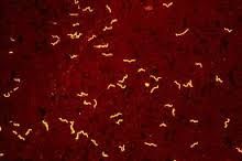
Syphilis |
caused by Treponema pallidum is a Gram-negative bacteria which is spiral in shape. It is an obligate internal parasite. Has to be gram stained with due to it's high lipid content. Spread from person to person by direct contact with a sore, known as a chancre. Chancres occur mainly on the external genitals, vagina, anus, or in the rectum. Chancres also can occur on the lips and in the mouth. Transmission occurs during vaginal, anal, or oral sex. |
|
|
what basic principles of antibody mediated immunity are utilized in an ELISA assay |
If antibodies are present in patient |
|
|
How does an ELISA indirectly detect infection by a disease causing agent |
determines antibody |
|
|
What is the function of the secondary antibody and chromogen in an ELISA |
Secondary antibody is attached to an enzyme. when chromogen is added if a patients has the antibodies the enzyme that is linked to the secondary antibody facilitates a chemical reaction that changes the color of the chromogen. A color changes indicates patient has antibodies to illness. |
|
|
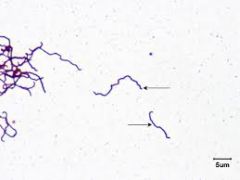
Lyme Disease |
spirochete bacterium-borrelia bergdorferi Transmitted to people through the bite of infected ticks, such as deer and black legged. Can't be xfered from person to person. They require meals of blood obtained from warm blooded animals. After it bites victim it transf. the infectious bacteria after 36-48 hrs of attachment. Symptoms include: bull's eye rash surrounding bite, flu like symptoms, headache, chills, muscle aches and fatigue. If untreated neurological difficulties and respiratory and GI problems can happen. |
|
|
Pricipitin Ring Test |
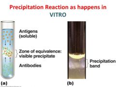
Precipitation reactions are based on the interaction of antibodies and antigens. They are based on two soluble reactants that come together to make one insoluble product, the precipitate . These reactions depend on the formation of lattices (cross-links) when antigen and antibody exist in optimal proportions. |