Reading...
![]()
Play button
![]()
Play button
![]()
Use LEFT and RIGHT arrow keys to navigate between flashcards;
Use UP and DOWN arrow keys to flip the card;
H to show hint;
A reads text to speech;
55 Cards in this Set
- Front
- Back
|
Mucle arrises from what type of tissue organ (endoderm, mesoderm, or ectoderm?)
|
Mesoderm
|
|
|
Four ways to classify muscle include
|
type: skeletal, cardiac, smooth
voluntary vs involuntary striated vs smooth contractile components/ myofilaments |
|
|
Skeletal muscle cells (one fiber) is a cylindrical _____with _______nuclei
|
Syncytium
-multiple, peripherally located |
|
|
Syncytium definition
|
a cell divides without undergoing cytokinesis
|
|
|
In skeletal muscle the fibers are_____packed, (organized/disorganized), (striated or smooth)
|
tightly packed, organized cytoplasmic protein filaments and striated
|
|
|
Connective tissues assocated with skeletal muscle include three layers
|
epimysium, perimysium, endomysium
|
|
|
Epimysium
|
Connective tissue around entire muscle
|
|
|
Perimysium
|
Connective tissue around groups of myofibers
|
|
|
Endomysium
|
Connective tissue around individuals myofibers
|
|
|
Is there an extensive or lacking capillary network in skeletal muscle
|
an extensive capillary network to meet the high energy requirement of skeletal muscle demands
|
|

Connective tissue type on skeletal muscle
|
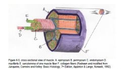
cross-sectional view of muscle: A. epimysium B. perimysium C. endomysium D. myofibrils E. sarcolemma of one muscle fiber F. collagen fibers
|
|
|
How do you distinguish the I band from the A band on skeletal muscle?
|
Dark = A band
Light = I band dArk vs lIght |
|
|
A Sarcomere is a _____unit that runs from __line to ___line
|
single contraction unit that runs from z line to z line
|
|
|
The Sarcolema of a muscle fiber can be thought of the
|
plasma membrane of the cell
|
|
|
The Sarcoplasmic reticulum of a muscle fiber can be compared to the ____ of a generic cell
|
Endoplasmic reticulum
where Ca++ is released for contraction |
|
|
Differences in the myofilament of the I and A bands
|
I band has no myosin included and the A band has both myosin and actin
|
|
|
Distinguish between the A, H, and A band
|
I band is actin, troponin, and tropomysin (stains light -more eosinophilic)
H band is thick and myosin only A nad is both actin and myosin with a darker stain |
|
|
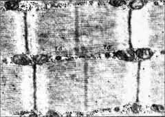
Which band changes in lenght during a contraction/ shortening of the sarcomere?
|
The H band width becomes shorter and there is no changein teh A band
|
|
|
Name the most superficial connective tissue around a microfiber?
|
Endomyosin
|
|
|
What are the three thin filaments/ muscle proteins
|
actin (2 strands of fibrous actin in 2xhelix)
tropomyosin (fibrous, located b/w strands of actin 2x helix) Troponin (regulatory components, located along actin 2x helix and attached to tropomyosin) Troponin C is associated with Ca++ |
|
|
What is the thick filaments/ muscle protein
|
myosin- has ATP-ase activity within strand and 2 heavy chain tails connected together
|
|
|
How does a muscle contraction take place?
|
acetylcholine released from a motor nerve binds to its receptors on the muscle cell,
a wave of depolarization spreads along the sarcolemma into the transverse tubules (these extend into the interior, so the depolarization wave goes deep into the myofiber), a calcium channel called DHPR is activated, opening a calcium channel on the SER called the ryanodine receptor, Ca++ is released from the sarcoplasmic reticulum, Ca++ binds to a protein called ‘troponin’ and allows myosin to interact with actin, which is necessary for energy + ADP to be formed from ATP; the energy causes the myosin head to bend, pulling actin along the myosin filament. |
|
|
What is the difference between a ‘motor endplate’ and a ‘motor unit?’
|
motor endplate is the myoneural junction: the synapse between the motor axon and
the skeletal fiber; the neurotransmitter is acetylcholine, and there is an enzyme, acetylcholinesterase, in the synaptic space that inactivates acetylcholine. The ‘motor unit’ is one alpha (lower) motor neuron in the spinal cord ventral horn and all the myofibers it innervates |
|
|
What is the neurotransmither that initiates depolarization which spreads via t-tubules
|
Acetylcholine (ACh)
|
|
|
What are the t-tubules?
|
Transverse tubules are invaginations of the cel membrane that extend b/w myofibrils
|
|
|
What are the terminal cisternae?
|
Expansions of the SER and located adjacent to T-tubules
|
|
|
Depolarization of sarcolemma is conducted down T-tubule to the triad junction where the _____ activates the_______ on the sarcoplasmic reticulum
|
DHPR- dihydropyridine receptor activating the RyR (ryanodine receptor)
|
|
|
An actin-myosin complex forms when _____binds to Calcium and the thick and thin filaments "slide" across one another as the myosin changes conformation when it______
|
Troponin C
Hydrolyzes ATP |
|
|
Muscle Fibers can be classified by fnxts and colors including ___-
|
Red fibers(Type 1= endurance)
White fibers (Type II = sprint) Intermdt fibers (type IIa) |
|
|
What is the difference between ‘red’ (type I) myofibers and ‘white’ (type II) myofibers?
|
Red fibers (type I) are slow twitch but long-endurance, look more ‘red’ because they have
a higher content of myoglobin and cytochromes, more mitochondria and have aerobic metabolism. White fibers (type IIb) are fast twitch but of short duration – they tire rapidly, have anaerobic metabolism, low content of myoglobin and cytochromes and have fewer mitochondria. There are actually fibers intermediate between these two called type IIa. |
|
|
True or False: The red:white fiber ratio in muscle can be modified
|
yes by training and use
|
|
|
Muscle Spindle is located_______and serves to _______
|
b/w endomysium and perimysium and is part of the regulation mechanism of muscle contraction
|
|
|
What is a muscle spindle?
|
stretch receptors found usually in the c.t. of muscle. They contain miniature
versions of skeletal muscle called “intrafusal fibers” (hence, regular fibers are called “extrafusal fibers”). They are innvervated by gamma motor neurons (not alpha motor neurons like those going to extrafusal fibers). They send sensory information to the spinal cord |
|
|
The sensory nerves in the muscle spindle can ____
|
sense if the muscle is straight or contracted and detect the degree of the stretch of the intrafusal fibers
|
|
|
The gamma efferent (motor) nerves in muscle spindle maintain ___
|
tension in the intrfusal fibers
|
|
|
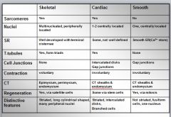
Cardiac muscle is (smooth/ striated ) and (voluntary/voluntary)
|
striated and involunatry
|
|
|
Distinguishing characteristics of cardiac vs skeletal muscle include
|
smaller, branched cells, only one centrally located nucleus per cell (some have 2) and intercalated discs (joining cells for end-end communication)
|
|
|
What are ‘satellite’ cells?
|
Spindle-shaped cells that lie within the basal lamina but outside the sarcolemma.They retain the ability to divide (remember,
the nuclei of the myofiber cannot divide). Thus after injury, especially in young animals, satellite cells can form some new myofibers |
|
|
What are the cardiac intercalated discs?
|
an electron dense junctional comples that contains gap junctions and desmosomes that transmit contractile stimuli and create the functional syncytium
|
|
|
What are the three types of junctions within the intercalted disks?
|
Fascia adherens
Desmosomes Gap Junctions |
|
|
What are the differnt functions of the three types of junctions of the intercalated disks?
|
Fascia adherens- provide intercellular adhesion, anchors actin filaments
Desmosomes- provide interacellular adhesion (prevent separation during contraction) Gap Junctions- allow for rapid communication of electric and chemcial signals |
|
|
For the cardiac muscle contraction the intrinsic conduction system is carried out by_________
|
Purkinje Fibers and low electrical reistance of gap jnxts
|
|
|
What are the purkinje fibers?
|
modified cardiac myocytes that are specializaed for orchestrated contraction
|
|
Purkinje Fibers
|
Modified cardiac myocytes specialized for the orchestrated contraction
|
|
|
Distinguishing characteristics of smooth muscle
|
non-striated
involuntary cells are spindle-shaped One nucleus per cell (elongated and oval) scant CT separated them unorganized myofilaments |
|
|
The analogous structure in smooth muscle for T-tubules is the
|
caveoli
|
|
|
The analogous structure in smooth muscle that holds filaments in register is the
|
dense bodies
|
|
|
Smooth muscle contraction also involves the myosin and actin interaction but is also regulated by different types of ___
|
surface receptors that activate diff second messenger systems. this involves phosphorylation of myosin light chains
|
|
|
How do the three types of muscles regenerate themselves?
|
skeletal muscle-uses satellite cells and connective tissue scars
Cardiac-very little regeneration (transplating stem cells) Smooth muscle (normal cell division) |
|
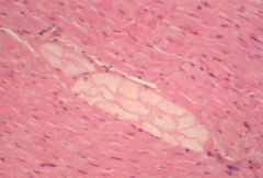
smooth muscle
|
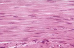
smooth muscle
|
|
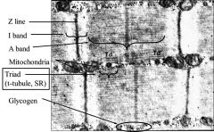
TEM skeletal muscle
|
TEM skeletal muscle
|
|
comparison chart
|

chart
|
|
|
does smooth muscle contain gap jnxts?
|
yes
|
|
|
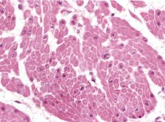
What is the morphology of smooth muscle cells?
|
They are long, spindle-like cells (you usually can’t see this on H&E because they blend
together and form a smooth mass); they have one centrally-positioned, elongated nucleus, (which may appear folded due to contraction after death). Smooth muscle is non-striated and involuntary |
|

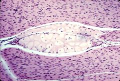
muscle spindle
|
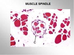
muscle spindle= stretch receptors found usually in the c.t. of muscle. They contain miniature
versions of skeletal muscle called “intrafusal fibers” (hence, regular fibers are called “extrafusal fibers”). They are innvervated by gamma motor neurons (not alpha motor neurons like those going to extrafusal fibers). They send sensory information to the spinal cord |