Reading...
![]()
Play button
![]()
Play button
![]()
Use LEFT and RIGHT arrow keys to navigate between flashcards;
Use UP and DOWN arrow keys to flip the card;
H to show hint;
A reads text to speech;
33 Cards in this Set
- Front
- Back
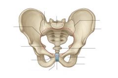
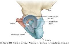
Label this diagram
|
|
|
Find these on the following diagram (or bone):
1. Ilium: - Ala - Iliac fossa - Anterior superior & inferior iliac spines - Iliac crest - Posterior superior & inferior iliac spines - Tubercle of iliac crest - Greater sciatic notch - Arcuate line - Gluteal lines posterior, anterior, inferior - Tuberosity of ilium - Auricular surface 2. Ischium: - Body of the ischium - Ramus of the ischium - Ischiopubic ramus - Obturator foramen - Ischial spine - Greater sciatic notch - Lesser sciatic notch - Ischial tuberosity 3. Pubis: - Body of pubis - Superior ramus - Pectineal line (pecten pubis ) - Inferior ramus - Pubic crest - Pubic tubercle - Pubic arch |
|
|
|
Where do the bones of the hips come together and describe this in a juvenile
|
- Ilium, ischium and pubis meet together at the acetabulum
- In the juvenile these bones are separated by a tri-radiate cartilage which appears like a Mercedes sign - The bones fuse around that ages of 20-25 years |
|
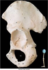
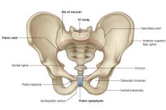
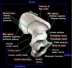
Label this diagram
|
1 acetabular notch
2 acetabulum 3 anterior gluteal line 4 aiis 5 asis 6 body of ilium 7 body of ischium 8 body of pubis 9 greater sciatic notch 10 iliac crest 11 iliopubic eminence 12 inferior gluteal line 13 inferior ramus of pubis 14 ischial spine 15 ischial tuberosity 17 lesser sciatic notch 19 obturator foramen 20 pectineal line 21 posterior gluteal line 23 auricular surface 24 pubic tubercle 25 ramus of ischium 27 superior ramus of pubis 28 tubercle |
|
|
How is the ischial tuberosity palpated?
|
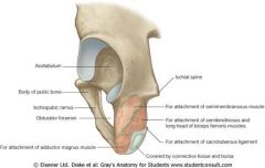
- Gluteus maximus covers the ischial tuberosity so to palpate it, the hip must be flexed similar to sitting
- The ischial tuberosity can get sore if sit down for too long → Paddler's bottom |
|
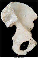
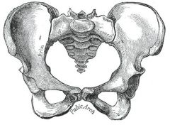
Label this diagram
|
1 aiis
2 asis 3 arcurate line 4 auricular surface 5 body of ischium 6 body of pubis 7 greater sciatic notch 8 iliac crest 9 iliac fossa 10 iliac tuberosity 11 iliopubic eminence 12 ischial spine 13 ischial tuberosity 15 lesser sciatic notch 18 pectineal line 21 pubic crest 22 tubercle |
|
|
Labels these on a model:
- Head of femur - Neck - Greater and lesser trochanters - Intertrochanteric line - Intertrochanteric crest - Trochanteric fossa - Linea aspera - Gluteal tuberosity - Pectineal line - Supracondylar lines - Intercondylar fossa - Femoral condyles - Patella surface - Epicondyles - Adductor tubercle |
|
|

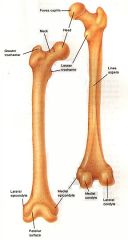
Label this diagram
|
2 Greater trochanter
3 head of femur 4 intertrochanteric line 5 lesser trochanter 6 neck of femur 9 shaft of femur |
|

Label this diagram
|
2 Greater trochanter
3 head of femur 4 intertrochanteric line 5 lesser trochanter 6 neck of femur 9 shaft of femur |
|
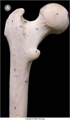
Label this diagram
|
2 gluteal tuberosity
3 greater trochanter 4 head of femur 5 intertrochanteric crest 8 lesser trochanter 9 linea aspera 11 femoral neck 12 quadrate (adductor) tubercle 13 spiral line (linea aspera) 134 trochanteric fossa |
|
|
Label this diagram
|
1 iliac crest
2 iliac tubercle 3 ASIS 4 superior ramus pubis 5 pubic tubercle 6 symphysis pubis 7 body of pubis 8 inferior ramus 9 head of femur 10 lesser trochanter Not that the joiunt is about 1/2 way along the inguinal ligament and inferior |
|

Label this diagram
|
2 gluteal tuberosity
3 greater trochanter 4 head of femur 5 intertrochanteric crest 8 lesser trochanter 9 linea aspera 11 femoral neck 12 quadrate (adductor) tubercle 13 spiral line (linea aspera) 134 trochanteric fossa |
|
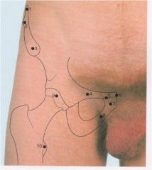
Label this diagram
|
1 iliac crest
2 iliac tubercle 3 ASIS 4 superior ramus pubis 5 pubic tubercle 6 symphysis pubis 7 body of pubis 8 inferior ramus 9 head of femur 10 lesser trochanter Note: the iliofemoral joint is about 1/2 way along the inguinal ligament and inferior |
|
|
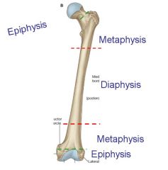
Describe the parts of the femur bone
|
|
|
|
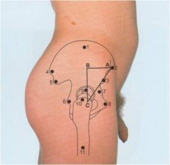
Label this diagram
What is Bryant's Triangle and what is its significance? |
1 ilium
2 ASIS 3 AIIS 4 PSIS 5 PIIS 6 ischial spine 7 iliopubic eminence 8 body of pubis 9 head of femur 10 greater trochanter 11 shaft - Bryant's triangle = ABC - This is constructed with the patient supine as a rough means of detecting disturbance of the normal anatomy of the femoral head and neck - Pathology of the femoral head or neck which displaces the greater trochanter (e.g. fracture) will tend to shorten BC |
|
Label this diagram
What is Bryant's triangle and why is it clinically significant? |
1 ilium
2 ASIS 3 AIIS 4 PSIS 5 PIIS 6 ischial spine 7 iliopubic eminence 8 body of pubis 9 head of femur 10 greater trochanter 11 shaft - Bryant's Triangle = ABC - This is constructed with the patient supine as a rough means of detecting disturbance of the normal anatomy of the femoral head and neck - The length of C is gauged on each side, and the sides compared. Pathology of the femoral head or neck which displaces the greater trochanter will tend to shorten the BC side |
|
|
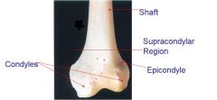
Describe the distal end of the femur
|
|
|
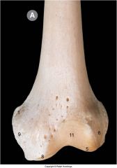
Label this diagram
|
1 adductor tubercle
5 lateral condyle 6 lateral epicondyle 8 medial condyle 9 medial epicondyle |
|
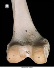
Label this diagram
|
1 adductor tubercle
4 intercondylar fossa 5 lateral condyle 6 lateral epicondyle 7 lateral condylar line 10 medial supracondylar line 12 popliteal surface |
|
|
Describe the anatomy of the patella
|
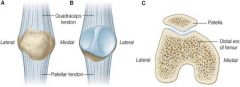
- Sesamoid Bone
- Articulates with femoral condyles - Usually has a smooth articular side which may become pathologically roughened (quandramalasia patella) and cause pain walking up and down stairs - Quadriceps muscles pass over the patella into the tibial tuberosity - Patella slides up and down in its groove and may dislocate laterally in juveniles if the medial quadratus is too weak to pull the patella correctly |
|
|
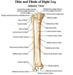
Find these on a tibia:
- Medial & lateral condyles - Intercondylar area - Intercondylar eminence - Tibial tuberosity - Soleal line - Medial, lateral & posterior surfaces - Anterior, medial & interosseous borders - Facet for fibula - Medial malleolus |
|
|
|
Describe the shaft of the tibia
|
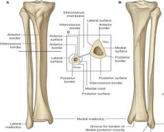
- 3 borders - anterior, medial and interosseous
- 3 surfaces = medial, lateral and posterior |
|
|
Briefly describe the pathology of Osgood-Schlatter's disease
|
- Pain over the tibia tuberosity with overuse of the quadriceps muscles during the growing phase
- Usually in young males - Can get necrosis and avulsion of tendon |
|
|
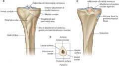
Describe the proximal tibia
|
|
|
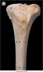
Label this diagram
|
1 Anterior border
4 Anterolateral tibial (Gerdy) tubercle important for attachment of iliotibial tract 5 Interosseous border 6 Lateral condyle 7 Lateral surface 8 Medial border 9 Medial condyle 10 Medial surface 13 Intercondylar eminence tubercles 14 Tibial tuberosity |
|
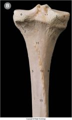
Label this diagram
|
2 Articular facet for fibula
3 Groove for semimembranosus 5 Inter-osseous border 6 Lateral condyle 8 Medial border 9 Medial condyle 11 Posterior surface 12 Soleal line 13 Inter-condylar eminence tubercles 15 Vertical line |
|
|
Describe the features of the proximal fibula
|
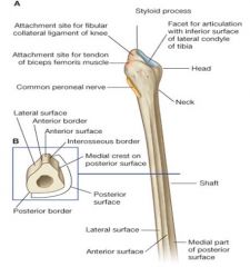
- No weight-bearing function so the shaft can be used for bone grafts
- Lateral collateral ligament inserts into its head - Common peroneal nerve wraps around the neck of the fibula to beomce superficial in the anterior compartment of the leg → commonly injured e.g. when hit by car |
|
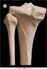
Label this diagram of the posterior proximal fibula
|
1 apex of head –styloid process
4 head of fibula 8 lateral condyle of tibia 12 superior tibiofibular joint |
|
|
Label this diagram of the posterior distal tibal and fibula
|
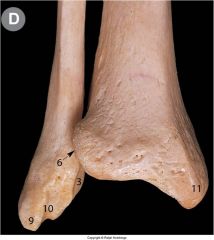
3 articular facet
6 inferior tibiofibular joint 9 lateral malleolus 10 malleolus fossa 11 medial malleolus |
|
|
Describe the bones of the foot
|
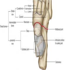
- 7 tarsal bones = calcaneus, talus, navicular, cuboid, medial, intermediate and lateral cuneiforms
- Talus has a head, body and neck, articulates with the tibia at the trochlea (or body) of the talus, and articular processes either side articulate with the malleoli of the tibia and fibula - Only the head of the talus can be seen from the inferior surface of the foot |
|
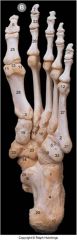
Label this diagram
|
4 calcaneus
5 cuboid 13 head of 1st metatarsal 14 head of talus 15 intermediate cuneiform 16 lateral cuneiform 19 medial cuneiform 23 navicular 24 neck of talus 28 shaft of 1st metatarsal 31 tuberosity of 5th metatarsal 33 tuberosity of navicular |
|
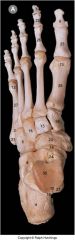
Label this diagram
|
1 anterior tubercle of calcaneus
4 calcaneus 5 cuboid 8 groove flexor hallucis longus 9 groove for peroneus longus 14 head of talus 15 intermediate cuneiform 16 lateral cuneiform 19 medial cuneiform 23 navicular 29 talar shelf 31 tuberosity base 5th metatarsal 33 tuberosity of navicular |
|
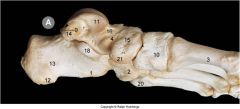
Label this diagram
|
1 calcaneal tubercle
2 cuboid 3 1st metatarsal 4 head of talus 10 medial cuneiform 11 talus 13 calcaneus 15 navicular 16 neck of talus 18 sustentaculum tali 20 tuberosity of 5th metatarsal |