![]()
![]()
![]()
Use LEFT and RIGHT arrow keys to navigate between flashcards;
Use UP and DOWN arrow keys to flip the card;
H to show hint;
A reads text to speech;
224 Cards in this Set
- Front
- Back
|
What are the 5 cellular elements of bone? |
osteoblasts osteocytes lining cells (inactive osteoblasts) osteoclasts |
|
|
What is the matrix made up of that is known as Osteoid? |
It's organic extracellular matrix made of glycoproteins and collagen fibres |
|
|
Osteoid rapidly undergoes mineralization by depositing __________________crystals to form bone |
calcium hydroxyapatite |
|
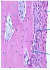
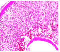
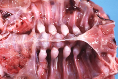
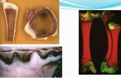
What is this? What is each layer? |
This is periosteum. The outer fibrous layer has blood vessels and nerves. The inner osteogenic layer contains many spindle-shaped osteoprogenitor cells. The inner-most layer is a continuous layer of osteoblasts that line the bone surface. |
|
|
There are two types of bone based on the degree of maturity. What are they? |
1. Woven bone - this is immature bone present during fetal development and in the early stages of active remodelling (bone repair) 2. Lamellar bone - this is mature bone. |
|
|
How are collagen fibres arranged in woven bone and lamellar bone? |
Woven bone - collagen fibres are short and randomly arranged is a criss-cross/woven pattern. Seen in some disease states and with fracture calluses Lamellar bone - collagen fibres are arranged in a parallel pattern. |
|
|
What type of bone are cortical/cancellous bones? |
Can be either woven or lamellar depending on the state of maturity |
|
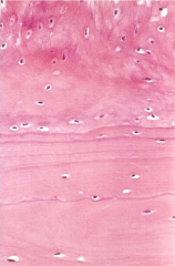
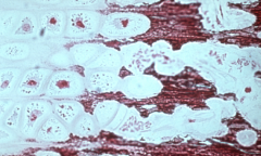
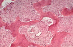
Where's the lamellar bone? Where's the woven bone? |
The woven bone (top) is deposited on the surface of the pre-existing lamellar bone (bottom) |
|
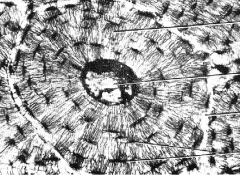
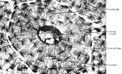
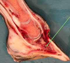
name each part |
|
|
|
During fetal life, bone formation occurs in 2 ways, both of which involve replacement of connective tissue by bone. What are these 2 ways? |
1. Intramembranous ossification 2. Endochondral ossification |
|
|
Where does intramembranous ossification occur? Name a bone made in this way. |
Occurs within "membranes" of condensed primitive mesenchymal tissues. Flat bones of the skull |
|
|
How does endochondral ossification occur? Name a bone made in this way. |
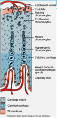
Bone develops from a cartilagenous model (hyaline cartilage) that is subsequently replaced by osseous tissue present in the ossification centers. The cartilage becomes mineralized as the cells move upwards. The hairpin arterioles under the growth plates supply oxygen for the osteoblasts. Occurs in the majority of bones in the skeleton (limbs, vertebral column, pelvis, base of skull) |
|
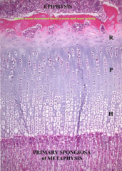
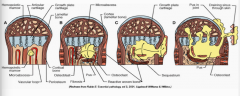
Name each layer of the Physis |
Reserve or resting zone - chondroblasts not really doing anything Proliferating zone - cells multiply, accumulate glycogen, produce matrix, and form columns. Hypertrophic zone - maturation, mineralisation, and apoptosis of chondrocytes Primary spongiosa - actual bone now. Blood vessels from periosteum bring in osteogenic cells. |
|
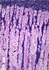
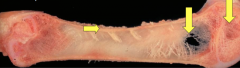
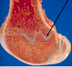
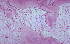
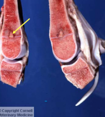
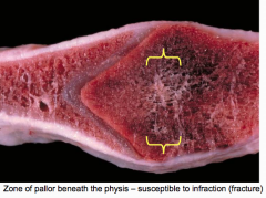
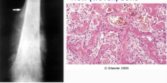
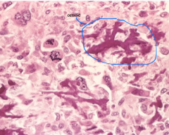
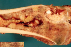
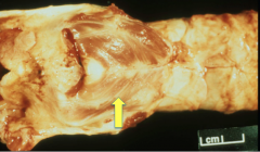
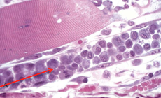
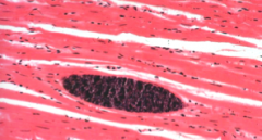
What is this? What cell type should no be here? What are the arrows pointing to? |
Primary spongiosa. There should be no surviving chondrocytes in the metaphysis. The arrows are showing bone deposits on basophilic spicules of cartilage that form a lattice. This lattice is mineralised matrix. |
|
|
Any interference with mineralization results in... |
osteodystrophy |
|
|
What is an essential step in endochondral ossification? |
Mineralization of cartilage matrix in hypertrophic zone of physis |
|
|
What is the most active form of Vitamin D (as part of bone composition)? |
1,25 dihydroxycholecalciferol is the most active form. |
|
|
Explain the conversion of vitamin D to 1,25- dihydrocholecalciferol in the body. What facilitates this conversion? |
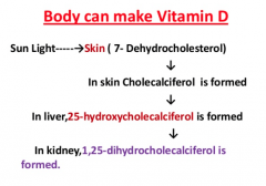
parathyroid hormone (PTH)
|
|
|
Where else does PTH act besides the vitamin D chain?
|
In the intestine to promote Ca and P absorption, and in the bone and cartilage to promote mineralization |
|
|
What is the overall goal of PTH in the body? |
To directly or indirectly raise plasma Ca concentrations. This occurs via the kidneys, intestines, or bone. |
|
|
How does PTH affect the kidneys? |
Increases tubular resorption of Ca and excretion of P. Increases formation of active form of Vitamin D |
|
|
How does PTH affect bone? |
Increases osteoclast activity and numbers |
|
|
What is calcitonin? |
The antagonist of PTH. Has the opposite effect. It decreases kidney resorption of Ca and decreases osteoclast activity in bone. It's produced by the parafollicular cells (C cells) of the thyroid gland. |
|
|
What occurs with chondrodysplasia? |
Membranous appositional growth is normal, but growth of cartilage is abnormal. This results in premature closure of growth plates and decreased length of long bones. Breed associated and hereditary (autosomal recessive) |
|
|
What are some examples of beef and sheep breeds with chondrodysplasia? |
Angus, Hereford, Dexters Suffolks, Hampshires (spider lambs) |
|
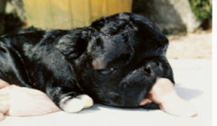
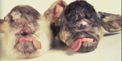
This is a "Dexter Bulldog". What is the actual name of this? |
Congenital lethal chondrodysplasia. Calves are often aborted and have a large head, short muzzle, short vertebral column, and marked micromelia (short legs) |
|
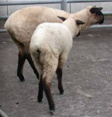
What does this lamb have? |
Spider lamb chondrodysplasia |
|
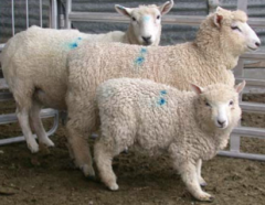
What is this lamb? |
A Texel lamb with Chondrodysplasia. (Varus deformity + dwarfism) |
|
|
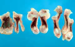
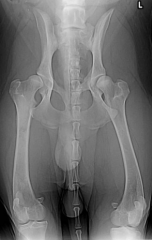
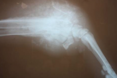
Chondrodysplasia in the long bones of dogs |
|
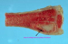
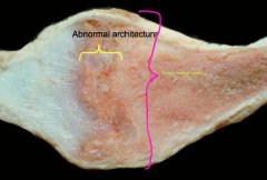
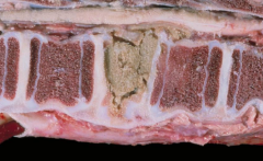
How do we know this is localised chondrodysplasia? |
there is a plug of cartilage that isn't getting mineralized |
|
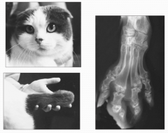
What kind of cat is this and what is the disease? |
Scottish fold cats Osteochondrodysplasia |
|
|
What is the actual name for "Wobbler Syndrome"? |
Cervico-vertebral stenotic myelopathy. It's a localized skeletal dysplasia seen in horses and large breed dogs. |
|
|
Wobbler syndrome results in ________ or __________ of the cervical spinal cord |
dynamic or static compression dynamic = cord lesion static = no cord lesion |
|
|
cervical vetrebral stenotic myelopathy in a Doberman |
|
|
When axons get swollen with wobbler's, what do they change into? |
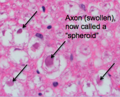
Axons swell up and become spheroids. |
|
|
What else will you see on histopath with Wobblers? |
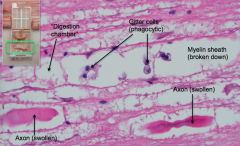
The myelin sheath will break down, you will see Gitter cells (phagocytic), and there will be digestion chambers |
|
|
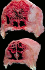
What is osteopetrosis? |
A congenital developmental abnormality where the animal has defective osteoclast function, so the bone has poor remodelling. This makes the bones very fragile so it's mostly a lethal trait. Occurs in angus, simmental cattle. Appaloosa and peruvian paso horses. White tailed deer |
|
|
Why do animals with osteopetrosis often present with aplastic anemia? |
There is no space for hematopoietic tissues of the bone marrow to grow. |
|
|
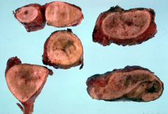
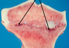
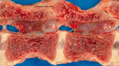
osteopetrosis (metaphyses filled with dense primary spongiosa) |
|
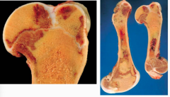
|
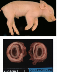
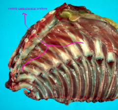
Congenital cortical hyperostosis of pigs. - Limbs appear swollen due to excessive trabecular bone deposition. Most are born dead. Inherited autosomal recessive abnormality (like Caffey's disease in people and monkeys) |
|
|
hyperostosis, pig |
|
|
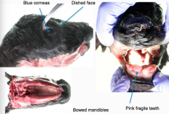
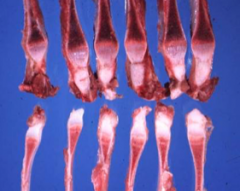
What is osteogenesis imperfecta? |
A hereditary disease of decreased bone mass (osteopenia). Defects in one of both genes that code for Type I collagen occur, with effects seen in bone, teeth, and cornea. |
|
|
Are the growth plates affected with osteogenesis imperfecta? Which animals get the disease? |
Growth plates are NOT affected (collagen in hyaline cartilage of growth plates is Type ll, not type l). Affects calves, kittens, lambs, puppies |
|
|
How do teeth appear with osteogenesis imperfecta? |
Pink and brittle with a thin crown that easily fractures. The dentinal tubules are short and the dentin is disorganized. |
|
|
What are the 4 main lesions seen with osteogenesis imperfecta? |
1. Multiple fractures that often occur in utero (callus formation) 2. Brittle bone 3. Dental fractures and pink teeth 4. Blue corneas |
|
|
osteogenesis imperfecta case at UCVM. Also saw a very domed cranial vault with several skull fractures, and osteal calluses on the ribs from fractured ribs. |
|
|
What does Dysraphia mean? |
Neural tube closure defect |
|
|
Why does dysraphia occur? Which animals are affected? |
Due to a defective interaction between neuroepithelium, the notochord, and mesenchyme in the fetus at specific closure sites. Occurs in cattle, horses, dogs (English Bulldogs), sheep, and cats. Genetic defect |
|
|
If neural tube defects occur in the skull, what is it called? What about in the spine? |
skull - Cranium Bifidium spine - Spina Bifida (usually lumbar area) |
|
|
What is the least severe type of neural tube closure defect? What is it called if the meninges are herniated? What about the meninges + cord? What is is called if the dorsal column fails to form? |
Least severe: Spina bifida occulta (vertebral arches are open, but everything else is in place). Meninges herniated: Meningocele Meninges + cord herniated: Myelomeningocele Entire dorsal column fails to form: Myeloschisis |
|
|
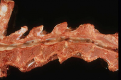
Kitten with myeloschisis |
|
|
What do the following terms mean? Amelia Hemimelia Polymelia Phocomelia Micromelia Syndactylia Polydactylia |
Amelia - absence of limb(s) Hemimelia - absence of the distal half of the limb Polymelia - supernumerary limbs Phocomelia - absence of the proximal portion(s) of limb(s) Micromelia - abnormally small or short limb(s) Syndactylia - fusion of digits Polydactylia - supernumerary digits |
|
|
Syndactyly |
|
|
Polydactyly |
|
|
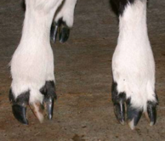
Brachygnathia inferior |
|
|
Define the following terms: Lordosis Kyphosis Scoliosis Kyphoscholiosis |
Lordosis - ventral deviation of the vertebral column Kyphosis - dorsal deviation of the vertebral column Scoliosis - lateral deviation of the vertebral column Kyphoscholiosis - dorso-lateral deviation of the vertebral column |
|
|
lordosis |
|
|
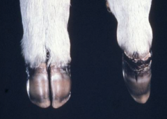
If an angular limb deformity has lateral deviation, what is it called? What about medial deviation? |
lateral = valgus medial = varus |
|
|
valgus |
|
|
Metabolic bone diseases are also known as... |
osteodystrophies |
|
|
What causes osteodystrophies? |
Nutritional or hormonal imbalances cause disturbed bone growth, modelling, or remodelling. MBD is characterized by failure of production of bone matrix, it's mineralization, or it's maintenance |
|
|
What nutritional imbalances can cause MBD? |
Vitamins C or D Ca P protein |
|
|
What hormonal imbalances can cause MBD? |
PTH, calcitonin, estrogens, or corticosteroids |
|
|
What toxins can cause MBD? |
lead poisoning fluoride poisoning aluminum hypervitaminosis A |
|
|
Can different forms of MBD coexist in the same individual? |
yes |
|
|
MBD's are traditionally classified as what 4 diseases? |
Osteoporosis Rickets Osteomalacia Fibrous osteodystrophy |
|
|
What is osteoporosis? |
reduced mass of normal quality bone tissue of clinical severity. Causes porous, thin, brittle bones |
|
|
What is osteopenia? |
reduced mass of normal quality bone tissue that can be clinically silent |
|
|
Name 4 of the 7 causes of osteoporosis |
1. Starvation 2. Specific deficiency (Ca, P, Cu, Vit C) 3. GI parasitism (trichostrongyles or ostertagia) 4. Malabsorption syndrome in dogs 5. Disuse 6. Endocrine associated 7. Senile osteoporosis |
|
|
What is the consequence of osteoporosis? What's an additional consequence only seen in sheep? |
Pathological fractures and epiphyseal separation Can also cause hypocalcemic crises in weaned lambs and lactating ewes |
|
|
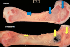
osteoporosis in a pig |
|
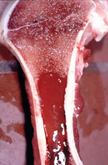
What is this? What features give it away? |
Serous atrophy of bone marrow (osteopenia) - will see metaphyseal growth arrest lines, widely spaced trabeculae, reduced cortical and trabecular bone in diaphysis, and deficient bone formation. |
|
|
Serous atrophy of marrow means there is a specific deficiency of what 3 things? |
Ca P Cu |
|
|
What's the difference between Ricketts and Osteomalacia? |
Same thing, only difference is the age they occur at. Ricketts = young animals Osteomalacia = adults |
|
|
What 3 deficiencies cause ricketts/osteomalacia? |
Vitamin D deficiency Phosphorous deficiency Calcium deficiency or Ca/P imbalance (birds only) |
|
|
What 2 features will you see to identify rickets/osteomalacia? |
1. decreased bone mineralisation (SOFT BONES) 2. endochondral ossification failure (thickened growth plates) |
|
|
What animals most often encounter Vitamin D deficiency (which can then lead to rickets) |
Camelids are most susceptible, then sheep, then cattle. Most common in pastured animals in regions where animals are not housed for winter, but the winter sun is low on the horizon (NZ, south Aus, Scotland) |
|
|
Which animals are most susceptible to phosphorous deficiency? Is this common? |
Cattle are most susceptible, then sheep. Horses, dogs, and cats are resistant. Uncommon, but is recognized in animals that graze phosphorus-deficient pastures |
|
|
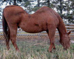
rickets (phosphorus deficiency in steer) |
|
|
thickened growth plate due to rickets |
|
|
Trabecular disruption with replacement by vascular fibrous connective tissue due to rickets |
|
these knobbly costro-chondral junctions are called what? Which disease? |
Rachitic rosary. Rickets |
|
|
osteodystrophic lines - foal with rickets |
|
|
Rachitic rosary in a chicken with rickets |
|
|
Lesions patterns with rickets may give you a clue as to which deficiency was present... |
P deficiency or Ca excess: thickened hypertrophic zone in growth plates, chondrocytes in regular columns on histo, reduced mineralization but normal vasculature, reduced osteoid, decreased osteoclasts and increased osteoblasts. Ca or Vit D deficiency: thickened and disorganized zone of proliferation, thin hypertrophic zone, poor vascularization, irregular basal area with lots of osteoid and thin trabeculae that may be collapsed, increased osteoclasts. |
|
|
What is the pathogenesis of osteomalacia? |
Failure of mineralization of osteoid due to Vitamin D or P deficiency. Unmineralized osteoid is resistant to osteoclastic resorption so it accumulates in the bone. Pathologic fractures and deformities (lordosis, kyphosis, and scoliosis) occur. A disease of grown animals! Unlike ricketts. |
|
|
osteomalacia |
|
|
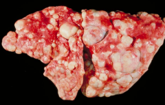
What is fibrous osteodystrophy? |
Relatively common MBD characterized by extensive bone resorption and replacement by fibrous connective tissue and poorly mineralized immature bone (woven bone). Results from persistently high PTH levels (due to hyperparathyroidism) |
|
|
Which animals get fibrous osteodystrophy? |
Horses, dogs, cats, pigs, reptiles, monkeys |
|
|
What's the difference between traumatic bone fractures and pathologic bone fractures? |
traumatic - normal bone broken by excessive force pathologic - abnormal bone broken by minimal trauma or normal weight bearing |
|
|
What is an avulsed fracture? |
Caused by the pull of a ligament/muscle tendon at its insertion into bone (fragment of bone tears away from the main mass) |
|
What initial event can cause this? |
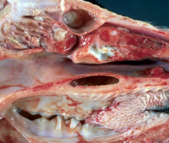
vertebral abscess resulting in pathologic fracture of the vertebral body and focal compression of the spinal cord. Common sequel of tail biting in pigs. |
|
|
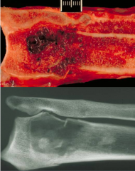
pathologic fracture, osteosarcoma, dog |
|
|
Describe the 4 steps of fracture repair |
1. Blood clot forms in fracture site 2. Collagen and fibroblasts infiltrate 3. Blood vessels and cartilage are laid down. Woven bone fills in the area. Osteoclasts break down broken bone pieces |
|
|
4 complications associated with bone fractures |
1. Bone necrosis and formation of sequestrum 2. Nonunion. Can lead to pseudoarthrosis/false joint formation 3. Osteomyelitis 4. Cachexia |
|
|
What is a common source of osteomyelitis in neonates? |
Omphalophlebitis |
|
|
What animals most commonly get bacteremia or septicemia? |
young farm animals |
|
|
Where does hematogenous infection usually localize in piglets? |
vertebral bodies mainly |
|
|
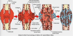
Why is the metaphysis the ideal spot for organisms to set up and create embolisms? |
The vascular features including fenestrated capillaries, hair pin loops of vessel, and sinusoidal sludging |
|
|
Describe how these little microabscesses grow and proliferate |
|
|
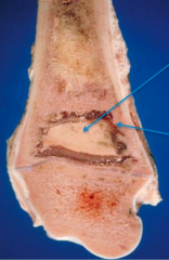
What's the difference between an involucrum and a sequestrum? |
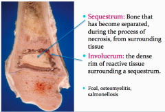
involucrum = dense collar of reactive bone encapsulating the cavity that contains necrotic material sequestrum = mummified necrotic bone surround by exudate |
|
|
embolic osteomyelitis |
|
|
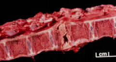
vertebral osteomyelitis and necrosis (calf) |
|
|
pedal osteomyelitis |
|
|
purulent osteomyelitis |
|
|
osteomyelitis of the growth plate and epiphysis |
|
What bacteria causes this? What is the disease called? |
Actinomyces bovis Bovine actinomycosis - aka lumpy jaw |
|
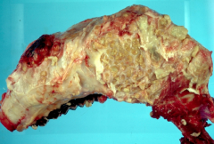
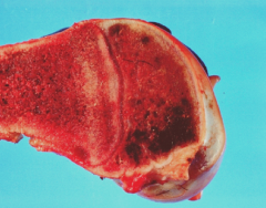
This is lumpy jaw. Describe the bone changes |
chronic pyogranulomatous osteomyelitis |
|
|
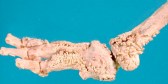
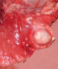
dog limb with hypertrophic pulmonary osteopathy |
|
|
Describe the disease Hypertrophic pulmonary osteoarthropathy or osteopathy |
Also called "Marie's disease", it's sporadically reported in humans and dogs. It usually occurs to individuals that have an intra-thoracic space-occupying mass (tumour or abscess). Painful swelling of the limbs occurs, caused by periosteal bone proliferation (hyperostosis) in long bones (usually the digits are spared). May be due to altered circulation or stimulation of the Vagus nerve. |
|
|
Are the bone changes permanent with hypertrophic pulmonary osteopathy? |
no. Bone changes can regress if the space-occupying lesion in the thoracic cavity is removed. |
|
|
Besides an intra-thoracic mass causing hypertrophic pulmonary osteopathy, what other problems can cause it? |
young dogs with rhabdomyosarcomas of the urinary bladder, or in mares with ovarian tumors |
|
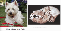
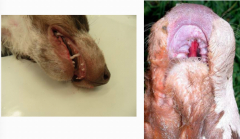
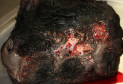
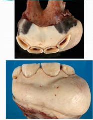
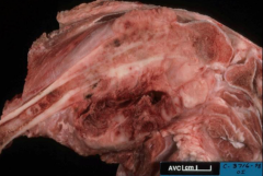
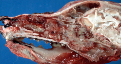
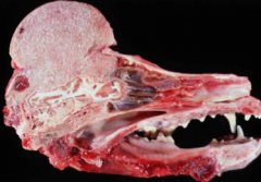
What disease does this dog have? |
Canine craniomandibular osteopathy (Lion Jaw). It's a proliferative disorder confined to the bones of the skull, especially the mandibles, occipital, and temporal bones. Most common in Westies where a genetic link is suspected. Usually seen at 4-7 months old where dogs will be unable to open their mouth or will show jaw discomfort. |
|
|
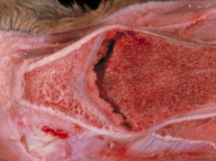
What is a disease of young, fast-growing dogs or large and giant breeds where the initial lesion is neutrophilic osteomyelitis of metaphyses? Describe the bone features. |
Metaphyseal osteopathy (aka canine hypertrophic osteodystrophy) - hemorrhagic, fibrinous, necrotic bone with collapse of long, thin primary trabeculae. |
|
|
metaphyseal osteopathy |
|
|
metaphyseal osteopathy |
|
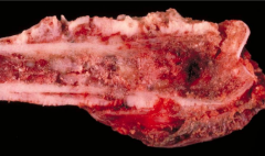
What disease is this? Describe your reasoning. |
Metaphyseal osteopathy. - chronic lesion with periosteal and extra-periosteal deposition of woven bone forming a metaphyseal bone collar |
|
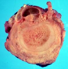
This dog has patchy to diffuse densities within the diaphyseal medullary canal. What disease is this? |
Canine panosteitis. - Occurs in young rapidly growing large breed dogs (German Shepherds), + mini schnauzers and scotties. - waxing and waning for several month with mild to severe lameness - diagnose with rads |
|
|
Are primary or secondary tumors more common with bone neoplasia? |
primary tumors |
|
|
Which animals get malignant tumours most commonly? Which animals get benign tumours most commonly? |
Malignant - dogs Benign - horses, cattle, and other domestics Cats are 50/50 |
|
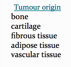
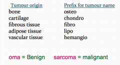
For each of the following tumour origins, give the prefix for the tumour name
|
|
|
|
Why is accurate diagnosis of bone tumours important? How do you diagnose bone tumours? |
An accurate diagnosis is very important for proper prognosis. Do not rely on microscopic features alone! Interpretation of slides is fraught with difficulty. Also use signalment, history, and radiographs. Always include a sample from an area of lysis. |
|
|
New woven bone. You can see Codman's triangle and sunburst pattern of periosteal new bone |
|
|
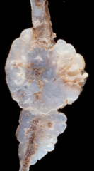
osteoma - benign, uncommon, smoothly contoured, well differentiated, compresses adjacent structures, and seen in the skull. |
|
|
osteoma |
|
|
ossifying fibroma - seen in the rostral mandible of horses under a year old. Made up of fibroblasts, osteoblasts, and woven bone. Will mature into osteoma |
|
|
What is the most common primary neoplasm of dogs and cats? |
Osteogenic sarcoma (osteosarcoma)! |
|
|
Describe osteogenic sarcoma/osteosarcoma |
- Rapidly progressive - Early hematogenous metastasis, usually to the lungs. - 14-22 weeks survival time |
|
|
Osteogenic sarcoma is classified into 6 histological categories. Name 4 of these 6 |
1. Poorly differentiated (aggressive, lysis, fractures). The worst type 2. Osteoblastic 3. Chondroblastic 4. Fibroblastic (least aggressive) 5. Telangiectatic (most aggresive) 6. Giant cell |
|
|
Describe where osteosarcoma is most commonly seen on the body |
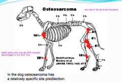
Distal radius and ulna get 28% of cases. The hock also gets a lot. The rule is "close to the knee and away from the elbow". |
|
|
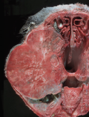
osteosarcoma |
|
|
osteosarcoma that has metastasized to the lungs |
|
|
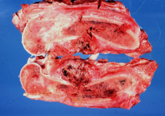
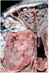
osteosarcoma in a femur |
|
|
osteosarcoma + pathological fracture |
|
|
osteosarcoma + lytic lesion |
|
which histiologic classification of osteosarcoma is this? |
Osteoblastic osteosarcoma. Malignant osteoblasts and bone production occurring. |
|
|
fibroblastic osteosarcoma |
|
|
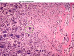
giant cell osteosarcoma |
|
|
Telangiectatic osteosarcoma |
|
|
2 types of cartilage-forming tumours |
Chondroma and osteochondroma |
|
|
What's the difference between a chondroma and osteochondroma? |
Chondroma - benign, firm smooth hard nodules made of hyaline cartilage, appears on flat bones and ribs. Osteochondroma - multiple, benign, cartilage capped masses found in the metaphyseal region (just below the growth plate) and are continuous with the medullary cavity. |
|
|
What do you call osteochondroma tumours when there are many of them? |
multiple cartilagenous exostoses OR osteochondromatosis |
|
|
Chondrosarcoma account for approximately ____% of _______ bone tumours in dogs. It involves which bone type most commonly? |
Accounts for approximately 10% of primary bone tumours. Involves flat bones (ribs, turbinates, pelvis) |
|
|
chondrosarcoma of the nasal turbinates |
|
|
chondrosarcoma |
|
|
chondrosarcoma of a cat rib |
|
|
Chondrosarcoma that has metastasized to the lungs. There's no way to tell this apart from an osteosarcoma with only this photo. Either would be correct on test. |
|
What signalment of animal does this usually affect? What is the cellular appearance? How can you treat it? |
Maxillary fibrosarcoma - seen in middle aged, large breed dogs - will have a benign cellular appearance - responds to radiation-hyperthermia therapy - these are why a proper diagnosis is so important. You can treat these ones! Don't euthanize! |
|
|
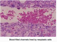
Where does hemangiosarcoma most commonly establish? |
On the ends of long bones |
|
|
plasma cell myeloma - axial skeleton usually affected |
|
|
malignant lymphoma (lymphosarcoma) |
|
|
enzootic bovine leukosis |
|
|
How common are secondary tumours (metastatic to bone)? |
Rare and under-diagnosed |
|
|
How do secondary tumours arise? Are carcinomas or sarcomas more common? Where are the primary tumours usually at in the body (many options...)? Where do they usually set up (3)? |
- Arise via hematogenous spread or direct extension. - Carcinomas > sarcomas - Come from mammary gland, thyroid, SCC, prostate, ovary, lung - Establish in the ribs, vertebrae, and proximal long bones |
|
|
Cats are unique (of course). Where do secondary bone tumours usually establish themselves? |
distal limb bones |
|
|
2 genetic diseases that indirectly affect the skeleton |
1. lysosomal storage diseases (mucopolysaccharidoses and gangliosidoses) 2. congenital erythropoietic porphyria |
|
|
Which animal mainly gets mucopolysaccharidoses and gangliosidosis? |
cats |
|
|
Which animal mainly gets congenital erythropoietic porphyria? How does this present? What other 3 animals can get it? |
cattle - will see brown teeth and photodynamic dermatitis due to uroporphyrin and coproporphyrin accumulation in bone and tissues (inc. skin). - pigs, cats, and rats can also get it |
|
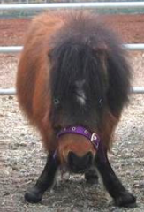
WTF caused these lambs to look like Alan |
Veratrum californicum toxicity (an alkaloid plant) |
|
|
2 types of muscle atrophy |
Denervation atrophy Physiologic atrophy |
|
|
3 types of physiologic atrophy |
1. Disuse atrophy 2. Atrophy of cachexia/malnutrition/old age 3. Atrophy of endocrine disease |
|
|
What occurs with denervation atrophy? |
Characterized by rapid atrophy. Long standing denervation may result in fibrosis and fat infiltration (steatosis). Type l and ll affected. |
|
|
What muscle fibres are affected with the 3 types of physiologic atrophy? |
All affect mainly type ll fibres |
|
|
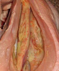
Horse with laryngeal hemiplegia. The atrophied muscle has pale, fibrotic, collapsed muscle. Damage to left recurrent laryngeal nerve, causing atrophy of the left cricoarytenoideus muscle. Creates Roarer Horses |
|
|
What occurs with muscle atrophy? |
Increase in size, but not number of muscle fibre cells. Normal response to increased work load |
|
|
What occurs with muscle hyperplasia? |
Increase in the number of muscle fibre cells. Seen in beef cattle (Angus, Santa's, Charolais). Inherited defect in the myostatin gene, which normally limits muscle growth. Leads to double muscling. Dystocia risk. |
|
|
What is a common sequel to myofiber injury, regardless of the cause? |
Degeneration/necrosis |
|
|
If muscle degeneration/necrosis is reversible, there is preservation of what? What is this injury called? |
Reversible if there is preservation of the basal lamina and the satellite cells. Non-disruptive injury. |
|
|
If muscle degeneration/necrosis is irreversible, what happens? What is this injury called? |
Necrosis will follow. Disruptive injury. |
|
|
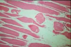
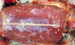
Muscle from a pig with Stress Syndrome. - often occurs on the way to slaughter and leads to condemned carcasses (like dark cutters in cattle) - myofiber degeneration. Characterized by swelling, hyalinization, loss of striations, and fragmentation |
|
|
Segmental necrosis due to monensin toxicity - both skeletal and cardiac muscle affected |
|
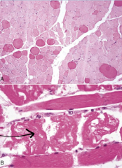
What is a common sequel to muscle degeneration and necrosis? As seen here. |
Calcification - starts as mitochondrial calcium overload, which can be detected grossly as white, chalky, gritty foci scattered throughout the affected muscle |
|
|
In a non-disruptive injury, _________ cells proliferate and migrate into the damaged myofibre. These become ______ which fuse to form multinucleated cellular bands. |
satellite myoblasts |
|
|
Myofiber regeneration is completed in how many weeks? |
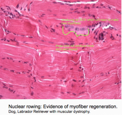
2-3 weeks. Although central nuclei and nuclear rowing may persist beyond this time. |
|
|
***What is a monophasic muscle injury? |
A one-off injury and necrosis, followed by invasion of macrophages. Division of satellite cells only. |
|
|
***What is a polyphasic muscle injury? |
On going injury. Will see necrosis with regeneration |
|
|
Monophasic injury |
|
|
White muscle disease/Nutritional myopathy occurs with what deficiency? |
Vitamin E/Selenium deficiency |
|
|
Explain the signalment of animals that get white muscle disease. What's the pathogenesis? |
Young animals that are rapidly growing are doing a lot of muscle building. Oxidative damage occurs in these muscles due to lack of enzymes that require Vit E and selenium for proper function (ex. glutathione peroxidase/reductase). Calcification of muscle occurs. Affects both skeletal and cardiac muscle |
|
|
Pigs with white muscle can co-exist with other Vit E/Selenium syndromes. Name 2. |
Hepatosis dietetica Mulberry heart disease |
|
|
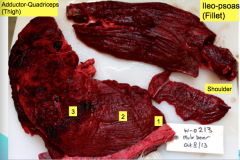
Which muscles are most often affected in white muscle disease?
Which muscle get the most severe lesions? |
Intercostal muscles and diaphragm (type l fibres) often affected, even if others aren't Most severe lesions in thigh and shoulder muscles |
|
|
Lamb with nutritional myopathy |
|
|
foal with nutritional myopathy |
|
|
4 types of exertional myopathies |
1. azoturia 2. tying up 3. capture myopathy 4. compartment syndrome |
|
|
How frequently does azoturia occur? It is the acute form of what equine problem? |
Relatively infrequent Severe and acute form of equine exertional rhabdomyolysis |
|
|
Horses that develop azoturia seem to have what type of underlying problem? |
underlying metabolic myopathy (equine polysaccharide storage myopathy). Likely genetically predisposed. |
|
|
What type of diet increases the liklihood of a horse getting azoturia? |
A high fat, high fibre, low starch, low sugar diet |
|
|
What muscles are most affected by azoturia? |
gluteal, femoral, lumbar muscles |
|
|
What is most common cause of death with azoturia? |
Myoglobinuric nephrosis |
|
|
Describe the lesions of capture myopathy |
Muscle degeneration-necrosis-hemorrhage. Occasional ruptured tendon. Myoglobinuric necrosis may occur |
|
|
capture myopathy in a mule deer buck |
|
|
capture myopathy - note the intense hemorrhaging in the muscles |
|
|
What is compartment syndrome? |
Ichemic damage that occurs in muscles surrounded by heavy aponeurosis (connective tissue sheaths) or by bone sheaths (non-expandable compartments) - seen in well-conditioned athletes |
|
|
In vet med, compartment syndrome is typically observed in the _______ muscles of poultry following vigorous wing flapping. This myopathy is called ________ |
supracoracoid muscles deep pectoral myopathy |
|
|
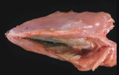
deep pectoral myopathy in chicken |
|
|
injection site necrosis, chicken |
|
|
3 presentations of traumatic myopathies in vet med |
Downer syndrome Equine post-anesthetic myopathy Crush syndrome |
|
|
What is downer syndrome? Explain the pathogenesis |
Ischemic necrosis of ventral and limb muscles following prolonged recumbancy. Weight of body --> increased pressure on vasculature --> edema --> more pressure --> ischemic necrosis - most common in cows |
|
|
Equine post-anesthetic myopathy occurs in _____% of cases where general anesthesia is used |
3-6% |
|
|
What's the difference between crush syndrome and downer syndrome? |
Same pathogenesis, but crush syndrome is caused by acute trauma. Downer syndrome is due to prolonged events. |
|
|
Inflammation of muscle is called... |
myositis |
|
|
Malignant edema is a type of myositis. What causes malignant edema (gas gangrene)? |
Acute fatal infection of wounds by gram-positive, anaerobic bacilli-shaped bacteria. They all exist in the environment as resistant spores and once they proliferate the tissues, are highly toxigenic. Includes Cl. septicum, Cl. perfringens, Cl. novyi, Cl. sordelli, and Cl. chauvoei species. |
|
|
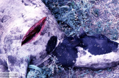
Black leg is a fatal necrotizing myositis of ruminants caused by _______ |
Clostridium chauvoei |
|
|
What is the pathogenesis of blackleg? |
Spores in soil --> ingestion --> spores in muscle (latent) --> muscle injury/hemorrhage --> local hypoxia --> germination of spores in muscle --> exotoxin release --> edema/myonecrosis --> emphysema --> generalized toxemia --> death in 24 hours |
|
|
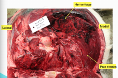
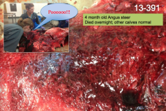
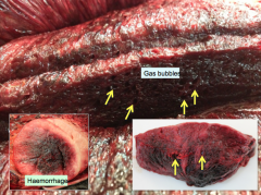
blackleg - muscle appears diffusely hemorrhagic and edematous with pockets of gas formation. Gas smells like rancid butter (ew) |
|
|
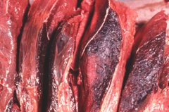
blackleg |
|
|
blackleg |
|
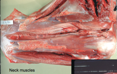
What bacteria caused this? |
Clostridium chauvoei (blackleg) |
|
|
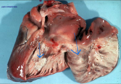
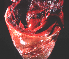
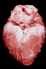
heart lesions seen with blackleg (Clostridium chauvoei) |
|
|
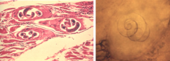
What species of parasite causes parasitic myositis? |
- Trichinella (remember that one that made you not want to eat bacon anymore?) - Cysticercus (larvae of Taenia tapeworm) - Neospora caninum (causes sarcocystosis) |
|
|
Where is the habitat for the Trichinella larvae? |
skeletal muscle. Becomes encysted in muscle. Muscles of the tongue, masseter, diaphragm, intercostals, and ocular muscles are commonly affected. zoonotic! |
|
|
Trichenosis |
|
|
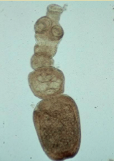
Echinococcus multilocularis - causes cysticercosis |
|
|
The cysticercus is the _______ of a Taenia tapeworm |
larval stage |
|
|
Cysticercosis is zoonotic! How do people get it? |
By ingesting Taenia eggs, not from ingesting cysticerci |
|
|
Cysticercosis |
|
|
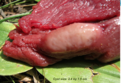
Cysticercosis - Moose measles (found in a moose) |
|
|
What is sarcocystosis? |
A protozoal disease affecting the striated muscle of cattle, sheep, and poultry. Carnivores (dogs, cats, humans) are the definitive host |
|
|
In sarcocystosis, what are the thin walled cysts filled with? |
bradyzoites |
|
|
What species can infect muscle and heart in the bovine fetus and cause abortion?
|
Neospora caninum |
|
|
Sarcocystitis |
|
|
Sarcocystitis in a Mallard |
|
|
2 primary neoplasias of muscle |
Rhabdomyoma Rhabdomyosarcoma |
|
|
Compare rhabdomyoma and rhabdomyosarcoma |
Rhabdomyoma is benign and congenital. It usually originates in the heart (65%) and commonly affects cattle, sheep, and pigs Rhabdomyosarcoma is aggressive and frequently metastasizes. Sometimes it arises from sites with no striated muscle (kidney, bladder, meninges). Embryonal rhabdomyosarcomas usually involving the head or neck occur in young animals. IHC diagnoses it. |