![]()
![]()
![]()
Use LEFT and RIGHT arrow keys to navigate between flashcards;
Use UP and DOWN arrow keys to flip the card;
H to show hint;
A reads text to speech;
85 Cards in this Set
- Front
- Back
|
Components of the Integumentary System |
1) Epidermis and dermis 2) Hypodermis 3) Thick and thin skin 4) Skin color 5) Skin Marking 6) Hair and nails 7) Cutaneous glands 8) Skin disorders |
|
|
Skin is |
Largest organ (15% of body weight) Thickness of various layers of skin varies - Normally 1-2mm - Dermis may thicken up to 6mm - Stratum Corneum layer (dead cells) increased - calluses on hands and feet |
|
|
Epidermis |
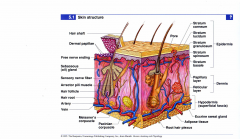
Keratinized stratified squamous epithelium |
|
|
Hypodermis |
Subcutaneous fat and blood vessels |
|
|
Functions of the skin |
1) Resistance to trauma and infection -- Acid mantle (ph 4-6) 2) Barrier to Ultraviolet light 3) Vitamin D synthesis 4) Sensory receptors 5) Thermoregulation 6) Excretion 7) Nonverbal communication 8) Appearance |
|
|
Cells of the epidermis |
1) Stem cells - Undifferentiated cell in basal layer 2) Keratinocytes - most of the skin cells 3) Melanocytes - synthesize pigment that shields from UV 4) Sensory (cutaneous) receptors 5) Dendritic (langerhans) cells - macrophages guard agains pathogens |
|
|
Keratin |
Fibrous, structural protein - Beaks, baleen, claws, hooves, feathers, scales, silk, fingernails, hair - White/Transparent |
|
|
Cutaneous Receptors - Mechanoreceptors |
1) Respond to deformation 2) Touch, proprioception, discrimination 3) Meissner's and Pacinian corpuscles |
|
|
Cutaneous Receptors - Thermoreceptors |
Hot or cold receptors |
|
|
Cutaneous Receptors - Nociceptors |
Pain receptors |
|
|
Cutaneous Receptors - Chemoreceptors |
Damage |
|
|
Stratum Basale |

Single layer of stem cells on basement membrane - Basal Lamina |
|
|
Cell types in Stratum Basale |
1) Karatinocytes - constant mitosis and subsequent apoptosis 2) Melanocytes - distribute melanin through cell processes - melanin picked up by keratinocytes 3) Merkel cells are touch receptors for somatosensory afferents |
|
|
Stratum Spinosum |
Several layers of living keratinocytes - Inflated with keratin |
|
|
Stratum Spinosum contains |
Dendritic (langerhans) cells - macrophages from bone marrow that migrate to the epidermis - 800 cells/ millimeter - helps protect body against pathogens by "presenting" them to the immune system |
|
|
Stratum Granulosum |

3 to 5 layers flat keratinocytes - Cells undergo apoptosis and lose nuclei and organelles |
|
|
Stratum Granulosum produces |
Lipid filled vesicles - Lamellar Bodies - that release a glycolipid by exocytosis to waterproof the skin - forms a barrier between surface cells and deeper layers of the epidermis - cuts off surface strata from nutrient supply |
|
|
Stratum Lucidum |

Thin translucent zone seen only in thick skin (palms and soles) - Keratinocytes are packed with eleidin - a precursor to keratin - It does not stain well - Cells have no nucleus or organelles |
|
|
Stratum Corneum |
15 to 20 layers of dead keratinized cells Caluses: Increase in thickness of stratum corner due to chronic friction Desquamation: Shedding of dead cells from surface |
|
|
Superficial and deep skin layers |
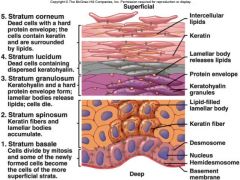
Superficial layers 1) Stratum Corneum 2) Stratum Lucidum 3) Stratum Granulosum Deep layers 1) Stratum Spinosum 2) Straum Basale |
|
|
Life history of keratinocytes |
1) Produced by stem cells in stratum basal - Daughter cells through mitosis 2) New cells push others toward surface - cells grow flat and fill with vesicles and pigment 3) Cells filled with keratin and melanin - forms water barrier 4) Cells die and exfoliate 5) 28 to 30 days timeline |
|
|
Skin thickness |
Varies by location - Eyelids thinest - Palmar and plantar surfaces thickest Primarily dependent on thickness of stratum corner |
|
|
Genetic Factors |
1) Melanin production: 16 genes for skin - default setting from conception 2) Thickness - By location - Changes after grafting |
|
|
Environmental factors |
1) Melanin production: UV light for skin - Immediate increase in production - Quick return to default - Tan lost in 2 to 4 weeks 2) Thickness: > fraction - > Stratified layer - Decreased speed of transit through layers |
|
|
Friction Blisters |
1) Separation of epidermis layers 2) Usually at stratum spinosum 3) Fill with tissue fluid and cell plasma 4) Blood blisters indicate damage to dermis |
|
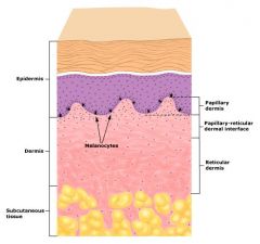
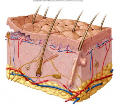
Dermis - Connective tissue layer |
1) Thickness = 0.6mm to 3mm 2) Composition - Collagen, elastic and reticular fibroblasts - Conservative: Low turnover rate 3) Dermal papillae - extensions of the dermis into the epidermis - forms the ridges of the fingerprints 4) Layers - Papillary layer - Reticular layer is deeper part of dermis |
|
|
Dermis is impregnated with |
1) Sensory neuron receptors 2) Fibroblasts 3) Blood vessels 4) Hair follicles 5) Sweat glands 6) Arrector pili muscles 7) Immune cells and mast cells ***Tattoos: Ink injected into upper layers |
|
|
Aging Skin |
1) Thinner, less collagen, elastin and GAGs 2) Stretch marks: Torn dermis 3) Wrinkles: Collagen and elastin damage 4) Age spots: sun damage, excess melanin production |
|
|
Hypodermis |
1) Subcutaneous tissue/ superficial fascia 2) Mostly adipose Hypodermis injection (SubQ) - Highly vascular |
|
|
Hypodermic functions |
1) Energy reservoir 2) Thermal insulation 3) Appearance 4) Protection from shearing |
|
|
Wound Healing |
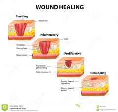
1) Hemostasis: Clotting, vasoconstriction 2) Inflammation: Cytokines, immune cells, macrophage cleanup 3) Proliferation (granulation): Angiogenesis, Fibroplasia, Granulation tissue, Epithelial regenration 4) Remodeling: Wolff's law - Bony tissue adapts to the forces exerted on them |
|
|
Abnormal skin colors |
1) Cyanosis
2) Erythema 3) Jaundice 4) Bronzing 5) Pallor 6) Albinism 7) Hematoma |
|
|
Cyanosis |
Blueness from deficiency of oxygen in the circulating blood (cold weather) e.g. Blue lips |
|
|
Erythema |
Redness due to dilated cutaneous vessels (anger, sunburn, embarrassment) |
|
|
Jaundice |
Yellowing of skin and sclera due to excess bilirubin in blood (liver disease) |
|
|
Bronzing |
Golden-brown color of Addison Ds - (deficiency of glucocorticoid hormone - Adrenal glands) |
|
|
Pallor |
Pale color from lack of blood flow (shock, rage) |
|
|
Albinism |
A genetic lack of melanin |
|
|
Hematoma |
A bruise (visible clotted blood) |
|
|
Skin Markings - Hemangiomas (Birthmarks) |
Discolored skin caused by benign tumors of dermal blood capillaries - strawberry birthmarks disappear in childhood - port wine birthmarks lasts for life |
|
|
Skin markings - Freckles |
Piebald suntan from irregular distribution of melanocyte (genetic) |
|
|
Skin markings - Nevi (moles) |
1) Clusters of Melanocytes 2) Benign Neoplasms 3) Monitor for change |
|
|
Keratoses |
1) Actinic: Precancer - Sun damage - Crusty 2) Seborrheic: Benign - Age related - " Liver spots" |
|
|
Psoriasis |
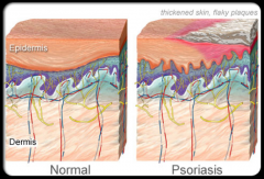
1) Chronic inflammation of skin 2) Autoimmune 3) Genetic component 4) Overproduction of keratinocytes |
|
|
Scarring |
1) Collagen plug formed by Fibroblasts 2) Replaces normal dermis and epidermis 3) Subsequent remodeling 4) No hair, sweat glands, basal layer 5) Keloid: Hypertrophic collagen in scarring - Raised thickened tissue |
|
|
Characteristics of human hair |
1) Hair - composed of hard keratin - disulfide bridges between molecules 2) Hair found almost everywhere - differences between sexes, or individuals is difference in texture, color and shape of hair |
|
|
Body hair types |
1) Lanugo - fine, unpigmented fetal hair 2) Vellus - fine, unpigmented hair of children and women 3) Terminal hair - coarse, long, pigmented hair or scalp and skin |
|
|
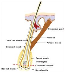
Structure of hair |

Hair shaft is filament of keratinized cells - shaft = above skin - root = within follicle - in cross section: medulla, cortex and cuticle
|
|
|
Hair follicle |

Follicle is oblique tube within the skin - bulb is where hair originates - vascular tissue (papilla) in bulb provides nutrients - continuous with epidermal layer |
|
|
Structure of Hair follicle |
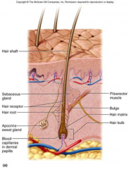
1) Epithelial root sheath 2) Connective tissue root sheath 3) Hair receptors entwine each follicle 4) Piloerector muscle - goose bumps |
|
|
Hair texture |
Cross-sectional shape determines body 1) Round = straight 2) Oval = wavy 3) Flat = kinky Denaturable: set and perm |
|
|
Hair Color |
1) Eumelanin: Black and Brown 2) Pheomelanin: Orange and Yellow 3) Gray/White: Decreased Melanin - Age - Stress - Illness |
|
|
Hair Cycle |
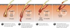
3 repeating cycles 1) Anagen is growth stage (90% scalp follicles) - Lasts 6 to 8 years in young adults 2) Catagen is shrinking follicle - Lasts 2 to 3 weeks 3) Telogen is resting stage - Lasts 1 to 3 months New Anagen phase: Old hair pushed out |
|
|
Thinning or Baldness |
Alopecia |
|
|
Pattern Baldness |
Genetic 1) Sex influenced trait. Dominant in males and recessive in females Hormonal 2) Testosterone sensitivity |
|
|
Hirsutism |
Excessive hair growth - hormone imbalance (ovary or adrenal cortex problem) |
|
|
Functions of hair |
1) Body hair - too thin to provide warmth - alerts us to parasites crawling on skin 2) Scalp hair - heat retention and sunburn cover 3) Beard, pubic and axillary hair - indicates sexual maturity and help distribute sexual scents 4) Guard hairs and eyelashes - prevent FB from getting into nostrils, ear canals or eyes 5) Expression of emotions with eyebrows |
|
|
Nails |
Derivative of stratum corneum - Densely packed cells filled with hard keratin |
|
|
Nail growth |
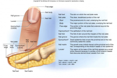
1) New cells added by mitosis in the germinal matrix 2) Nail plate is visible part of nail Damage to Germinal matrix: Loss of nail and regrowth |
|
|
Nail growth rate |
1) Fingernails = 1 mm per week 2) Toenails = 1 mm per month |
|
|
Nail Pathologies |
1) Vitamin A, D, B or Mineral Fe, Ca deficiency: Dry, brittle, misshapen, discolored nails 2) Fungus: Thick, crumbly, irregular, discolored - Tx: topical (ineffective) or systemic (liver toxicity) |
|
|
Sweat Glands |
Eccrine glands Density varies: 1) Palms - 370/sq cm 2) Forehead - 170/sq cm 3) Back - 60/sq cm |
|
|
Sweat gland functions |
1) Thermoregulation 2) Excretion 3) Protection |
|
|
Sweat glands containts |
1) Primarily water and salt 2) Urea 3) Lactic acid pH = 4-6 - Hostile environment for bacterias, viruses |
|
|
Sebaceous glands (exocrine gland) |
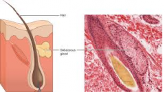
1) Highest concentration in face and scalp 2) Absent in palms and soles |
|
|
Sebaceous glands function |

1) Produces oily secretion 2) Lubricant for skin and hair 3) Bacteria Fodder 4) Bloakage associated with acne |
|
|
Apocrine glands |
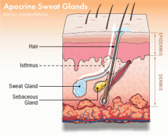
1) Type of sweat gland associated with certain hair follicles 2) Located in axillary, pubic, nasal, nipples, ears |
|
|
Apocrine glands functions |
1) Bacterial digestion of exudate produces scent 2) Produces secretion in response to emotions, stress, sex 3) Contributes to body odor 4) Pheromones |
|
|
Ceruminous Glands |
1) Found only in external ear canal 2) Their secretion combines with sebum to produce ear wax 3) Waterproofing - keeps the eardrum flexible 4) Bitterness repels mites and other pests |
|
|
Mammary glands |
1) Subcutaneous structures in both males and females 2) Secondary sex characteristics 3) Estrogen induces glandular (Apocrine) development 4) Testosterone inhibits development 5) Oxytocin induces expression of milk |
|
|
Oxytocin |
1) Neurohypophyseal (Pituitary) Hormone 2) Released into bloodstream |
|
|
Oxytocin roles |
1) Sex 2) Lactation 3) Childbirth 4) Wound healing 5) Social interactions |
|
|
Skin Cancer |
Induced by UV rays of the sun |
|
|
Skin cancer types |
1) Basal cell carcinoma (least dangerous) 2) Squamous cell carcinoma 3) Malignant melanoma (most deadly) |
|
|
Basal cell carcinoma |
1) Least dangerous 2) Arises from stratum basal and invades dermis |
|
|
Squamous cell carcinoma |
1) Arises from keratinocytes in stratum spinosum 2) Metastasis to the lymph nodes can be lethal |
|
|
Malignant melanoma |
1) Most deadly 2) Arises from melanocytes of a preexisting mole 3) ABCD - Asymmetry, irregular Border, Color mixed and Diameter over 6mm |
|
|
First Degree burn |

1) Epidermis only, Basal lamina intact 2) Sunburn |
|
|
Second Degree burn |

1) Extends into Dermis 2) Blistering, dermal damage, repair |
|
|
Third Degree burn |
1) Entire Dermis 2) Scarring, contraction, infection, grading & Dermal destruction |
|
|
Fourth Degree burn |
1) Subcutaneous, muscle, bone 2) Surrounding tissue and vascular loss, amputation |
|
|
Third-degree burns require |
Skin grafts
|
|
|
Graft options |
1) Autograft - Tissue from patient 2) Isograft - Tissue from identical twin 3) Culdtured keratinocyte patches |
|
|
Temporary grafts |
1) Immune response 2) Homograft (allograft) - from unrelated person 3) Heterograft (xenograft) - from another species 4) Amnion from after birth 5) Artificial skin from silicone and collagen |
|
|
Pressure ulcers (Decubitus) |
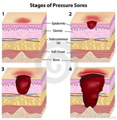
1) Stage 1 - redness after pressure removed 2) Stage 2 - damage to, but not thru dermis 3) Stage 3 - damage thru dermis into SubQ fat 4) Stage 4 - damage beyond fascia into underlying muscle, bone, ligament, tendon |