![]()
![]()
![]()
Use LEFT and RIGHT arrow keys to navigate between flashcards;
Use UP and DOWN arrow keys to flip the card;
H to show hint;
A reads text to speech;
53 Cards in this Set
- Front
- Back
|
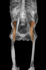
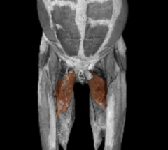
Psoas Major Origin: Transverse process and vertebral bodies of lumbar vertebrae Insertion: Lesser trochanter of the femur Action: Flexes hip joint; lateral rotation of the thigh; flexion of trunk when suppine |
|
|
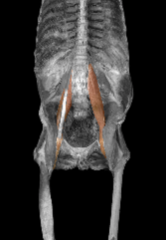
Iliacus Origin: Iliac fossa & sacrum Insertion:Lesser trochanter of femur Action: Flex hip & lateral rotation of thigh |
|
|
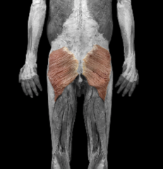
Gluteus Maximus Origin: Iliac crest, sacrum, coccyx, & aponeurosis of sacrospinalis Insertion: Iliotibial tract, lateral part of linea aspera, & gluteal tuberosity Action: Extends and laterally rotates thigh at hip joint; locks knee during extension RMA: Extends torso |
|
|
Gluteus Minimus Origin: Ilium Insertion: Greater trochanter Action: Abduction & medial rotation of the thigh at the hip joint |
|
|
Tensor Fasciae Latae Origin: Iliac crest Insertion: Tibia (Iliotibial tract) Action: Flexes and abducts thigh at hip joint |
|
|
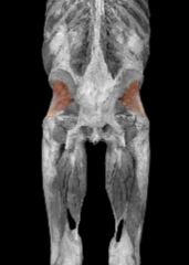
Piriformis Origin: Anterior sacrum Insertion: Superior border of greater trochanter Action: Lateral rotation and abduction of the thigh at hip joint |
|
|
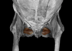
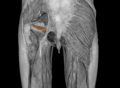
Obturator Internus Origin: Inner surface of oburator foramen, pubis, & ischium Insertion: Medial surface of greater trochanter Action: Lateral rotation and abduction of the thigh at hip joint |
|
|
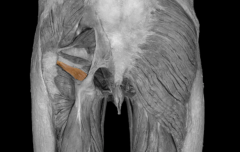
Obturator Externus Origin: Outer surface of obturator membrane Insertion: Deep depression inferior to greater trochantor (trochanteric fossa) of femur Action: Lateral rotation and abduction of the thigh at hip joint |
|
|
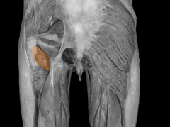
Superior Gemellus Origin: Ischial spine Insertion: Medial surface of greater trochanter Action: Lateral rotation and abduction of the thigh at hip joint |
|
|
Inferior Gemellus Origin: Ischial tuberosity Insertion: Medial surface of greater trochanter Action: Lateral rotation and abduction of the thigh at hip joint |
|
|
Quadratus Femoris Origin: Ischial tuberosity Insertion: Superior to intertrochanteric crest (posterior femur) Action: Lateral rotation of the thigh at hip joint; stabilization of hip joint |
|
|
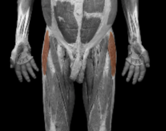
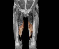
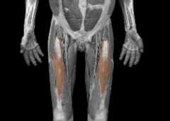
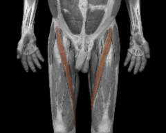
Adductor Longus Origin: Pubic crest and pubic symphysis Insertion: Linea aspera of femur Action: Adduction, flexion, and rotation of the thigh at hip joint RMA: Extends thigh at hip joint |
|
|
Adductor Brevis Origin: Inferior ramus of pubis Insertion: Superior half of the linea aspera of the femur Action: Adduction, flexion, and rotation of the thigh at hip joint RMA: Extends thigh at hip joint |
|
|
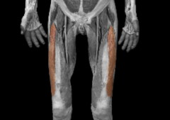
Adductor Magnus Origin: Inferior ramus of pubis and ischium to ischial tuberosity Insertion: linea aspera Action: Adduction and rotation of the thigh at hip joint. Anterior part flexes thigh; Posterior part extends thigh |
|
|
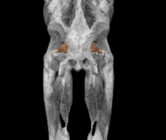
Pectineus Origin: Superior ramus of pubis Insertion: Between lesser trochanter and linea aspera Action: Flexion and adduction of the thigh at the hip joint |
|
|
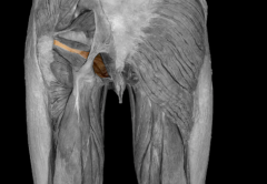
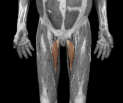
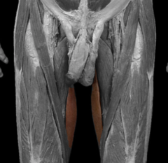
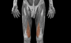
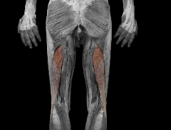
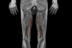
Gracilis Origin: Body and inferior ramus of pubis Insertion: Medial surface of body of tibia Action: Adducts thigh at hip joint, medially rotates thigh, and flexes leg at knee joint |
|
|
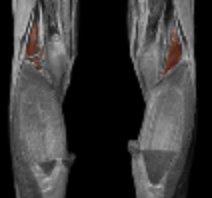
Rectus Femoris Origin: Anterior inferior iliac spine Insertion: Patella (via. quadriceps tendon) and tibial tuberosity (via patellar ligament) Action: Extend leg at knee. Flexes thigh at hip |
|
|
Vastus Lateralis Origin: Greater trochanter and linea aspera of femur Insertion: Patella (via. quadriceps tendon) and tibial tuberosity (via patellar ligament) Action: Extend leg at the knee joint |
|
|
Vastus Medialis Origin: Linear aspera of femur Insertion: Patella (via. quadriceps tendon) and tibial tuberosity (via patellar ligament) Action: Extend leg at the knee joint |
|
|
Vastus Intermedius Origin: Anterior and lateral surfaces of the body of the femur Insertion: Patella (via. quadriceps tendon) and tibial tuberosity (via patellar ligament) Action: Extend leg at the knee joint |
|
|
Sattorius (crossing leg over knee) Origin: Anterior superior iliac spine Insertion: Medial surface of body of tibia Action: flexion of leg at knee, and weak flexion, abduction and lateral rotation of the thigh at the hip. |
|
|
Biceps femoris Origin: Long head - ischial tuberosity; short head - linea aspera of femur Insertion: Fibula head and lateral condyle of fibia |
|

|
Semitendinosus Origin: Ischial tuberosity Insertion: proximal part of medial surface of shaft of tibia Action: flexes leg at the knee and extends thigh at the hip joint |
|
|
Semimembranosus Origin: Ischial tuberosity Insertion: Medial condyle of tibia Action: Flexes leg at knee; extends thigh at hip joint |
|
|
What is the role of the patella? |
Increases leverage of the quadriceps femoris muscle; maintains the position of the tendon when the knee is bent; protects the knee joint |
|
|
Name the joints at the knee |
Patellofemoral joint; tibiofemoral joint |
|
|
What structure connects the tibia and the fibula |
An interosseus membrane |
|
|
Name the joints formed by the tibia and the fibula. |
Proximal tibiofibular joint; distal tibiofibular joint |
|
|
Name the structure that separates the muscle compartments in the leg. |
|
|
|
What is the common tendon of the 4 anterior compartment muscles? |
|
|
|
Why are the hamstrings extensors of the thigh and flexors of the leg? |
|
|
|
Describe the borders of the popliteal fossa. |
|
|
|
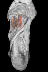
Tibialis anterior (anterior compartment) Origin: Lateral condyle and body of the tibia; interosseous membrane Insertion: Metatarsal I and first cuneiform (medial) Action: Dorsiflexion of the foot at the ankle. Supination of the foot at the intertarsal joints |
|
|
Extensor Halluces Longus (anterior compartment) Origin: Anterior surface of middle of the fibula; interosseous membrane Insertion: Distal phalanx of big toe Action: Dorsiflexion of the foot at the ankle, extension of the proximal phalanx of the great toe |
|
|
Extensor digitorum longus (anterior compartment) Origin: Lateral condyle of tibia, anterior surface of fibula, interosseous membrane Insertion: Middle and distal phalanges of toes II-V Action: Dorsiflexion of the foot at the ankle; extension of the proximal, middle and distal phalanges |
|

|
Fibularis Tertius (anterior compartment) Origin: Distal third of fibula, interosseous membrane Insertion: Base of metatarsal V Action: Dorsiflexes foot at ankle joint and pronation of the foot at the intertarsal joints. |
|
|
Fibularis Longus (lateral compartment) Origin: Head and body of fibula Insertion: Metatarsal I and first cuneiform Action: Plantar flexion of the foot at the ankle joint, pronation at the intertarsal joints |
|
|
Fibularis Brevis (lateral compartment) Origin: Distal half of body of fibula Insertion: Base of metatarsal V Action: Plantar flexion of foot at the ankle joint, pronation at the intertarsal joints |
|
|
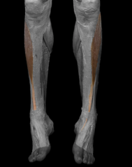
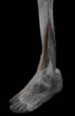
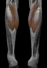
Gastrocnemius (superficial compartment) Origin: Lateral and medial condyles of femur and capsule of knee Insertion: Calcaneus (by the achilles/calcaneal tendon) Action: Plantar flexes foot at ankle and flexion of leg at knee joint |
|

|
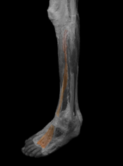
Solues (superficial compartment) Origin: Head of fibula and medial border of tibia Insertion: Calcaneus (by the achilles/calcaneal tendon) Action: Plantar flexes foot at ankle |
|
|
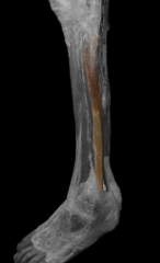
Plantaris (superficial compartment) Origin: Lateral epicondyle of femur Insertion: (by the achilles/calcaneal tendon) Action: Plantar flexes foot at ankle and flexion of the leg at the knee. |
|
|
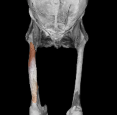
Popliteus (deep compartment) Origin: Lateral condyle of the femur Insertion: Proximal tibia Action: Flexes leg at knee joint and medially rotates tibia to unlock extended knee |
|
|
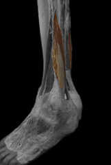
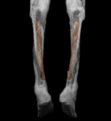
Tibialis Posterior (deep compartment) Origin: Proximal tibia, fibula, interosseous membrane Insertion: Metatarsals II-IV, navicular, the 3 cuneiforms Action: Plantar flexion of the foot at the ankle and supination of the foot at the intertarsal joints |
|
|
Flexor Digitorum Longus (deep compartment) Origin: Middle third of posterior surface of tibia Insertion: Distal phalanges of II-V Action: Plantar flexion of the foot at the ankle, flexion of proximal, middle and distal phalanges |
|
|
Flexor Hallucis Longus (deep compartment) Origin: Inferior 2/3rds of the posterior of the fibula Insertion: Distal phalanx of the great toe Action: Plantar flexion of the foot at the ankle joint flexion of the proximal and distal phalanges of the big toe |
|
|
Extensor Retinaculum: Superior and inferior - hold the tendons of the ankle firmly to the ankle. |
|
|
Extensor Hallucis Brevis Origin: Calcaneus and inferior extensor retinaculum Insertion: Proximal phalanx of big toe Action: extends big toe at metatarsophalangeal joint. |
|
|
Extensor Digitorum Brevis Origin: calcaneus and inferior extensor retinaculum Insertion: Middle phalanges of toes II-IV Action: Extension of toes II-V at interphalangeal joints |
|
|
Abductor Digiti Minimi Origin: Calcaneus, plantar aponeurosis, and flexor retinaculum Insertion: Lateral side of proximal phalanx of little toe Action: Abduction and flexion of the little toe at the metatarsophalangeal joint. |
|
|
Abductor Hallucis Origin: Calcaneus, plantar aponeurosis, flexor retinaculum Insertion: Medial side of proximal phalanx of big toe Action: Abducts and flexes big toe |
|
|
Dorsal Interossei Origin: Adjacent side of all metatarsals Insertion: Proximal phalanges of both sides of toe II and the lateral sides of toes III-IV Action: Abduction and flexion of toes II-IV and extension of toes at interphalangeal joints |
|

|
Adductor hallucis Origin: Metatarsals of II-IV, ligaments of metatarsals III-V, tendon of fibularis longus Insertion: Lateral side of proximal phalanx of big toe Action: Adduction and flexion of big toe at the metatarsophalangeal joint. |
|
|
Plantar Interossei Origin: Metatarsals III-V Insertion: Medial side of proximal phalanges of toes III-V Action: Adduction and flexion of the proximal metatarsophalangeal joints, and extension of the toes at the interphalangeal joints. |