![]()
![]()
![]()
Use LEFT and RIGHT arrow keys to navigate between flashcards;
Use UP and DOWN arrow keys to flip the card;
H to show hint;
A reads text to speech;
142 Cards in this Set
- Front
- Back
|
What is it called when a disease is caused by fungus? |
Mycoses |
|
|
What part of the body is affected by the following mycoses? -superficial -cutaneous -subcutaneous -systemic |
•Superficial– hair, skin, nails •Cutaneous– deep layers of skin, hair, nails •Subcutaneous– muscle, connective tissue •Systemic/Opportunistic– specific organs, all tissues |
|
|
Do fungi have chlorophyll? |
No |
|
|
What sugar molecule is located in the rigid cell walls of fungi? |
Chitin |
|
|
What pH do fungi prefer? |
Neutral, but can tolerate wide range |
|
|
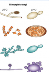
Optimal temperature for growth? For dimorphic yeast? |
30C generally 37C for dimorphic yeast |
|
|
Where do fungi obtain their nutrients? |
They absorb it from the environment... they do not contain chlorophyll |
|
|
Hyphae refers to.. Mycelium refers to.. |
Hyphae are filaments that are the microscopic units of fungi. They are either septate or aseptate. Mycelium are the intertwined hyphae. |
|
|
Where do the two types of mycelium grow |
Vegetative (THALLUS) mycelium grow on or in a substrate and absorbs the nutrients Reproductive (AERIAL) mycelium grow out of the substrate or agar and produce the fruiting bodies which make the reproductive structures: SEXUAL: spores ASEXUAL: conidia |
|
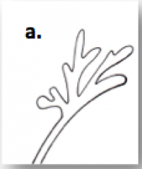
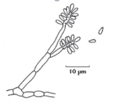
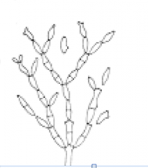
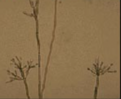
What general fungal structure is this? What is the name of its shape? |
Favic chandeliers: resemble antlers of a deer, bluntends, and branched. Seen with Trichophyton schoenleinii. |
|
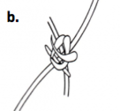
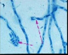
What general fungal structure is this? What is the name of its shape? |
Nodular organs: knotsof twisted hyphae. Seen with Microsporumferrugineum. |
|
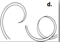
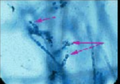
What general fungal structure is this? What is the name of its shape? |
Racquet hyphae –resemble tennis racquets with smaller end attached to large end of an adjacentclub-shaped hyphae. Seen with Trichophytonajelloi. |
|
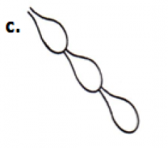
What general fungal structure is this? What is the name of its shape? |
Spiral hyphae – coiled or corkscrew-like turns inhyphae. Seen with Chrysosporiumsp. |
|
What general fungal structure is this? What is the name of its shape? |
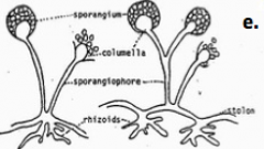
Rhizoids –root-like structures that may be located at the base of a sporangiophore or internodallyalong the hyphae. Seen some members ofthe Zygomycetes. |
|
|
What two terms are used to describe the pigmentation in fungal structures? |
Hyalinehyphae-nonpigmented orlightly pigmented Dematiaceous-darklypigmented |
|
|
Mature zygospore. Sexual reproduction that involves the fission to 2 identical cellsarising from the same hypha |
|
|
Ascus with ascospores. Sexual reproduction that involves sexual spores in a round saclike ascus that usuallycontains 2-8 spores |
|
|
What are perfect fungi? |
Fungithatexhibit a sexual phase |
|
|
What are imperfecti fungi? |
Fungithatdo not exhibit a sexual phase. Reproduction involves only mitosis. |
|
|
Are conidia sexual or asexual spores? |
Asexual spores that are produced singly or multiply in chains or clusters |
|
|
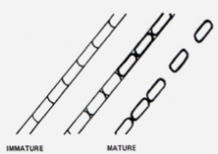
What are the three types of conidia produced directly from vegetative mycelium? |
1. Blastoconidia: daughter cells bud off 2. Chlamydoconidia: thick walled, made in unfavorable conditions 3. Arthroconidia: formed by fragmenation of mycelia |
|
|
What are the three basic types of chlamydoconidia? |
1. Terminal: on the hyphae tip 2. Intercalary: within the hyphae tip 3. Sessile: on the side |
|
|
What type of conidia are formed during unfavorable conditions? |
Chlamydoconidia |
|
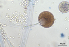
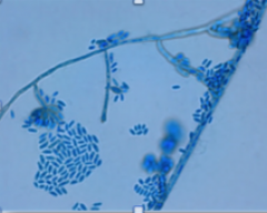
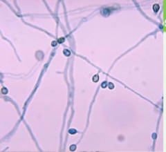
Name this conidia |
Blastoconidia |
|
Name this conidia type |
Arthroconidia |
|
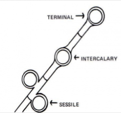
Name this conidia type |
Chlamydoconidia |
|
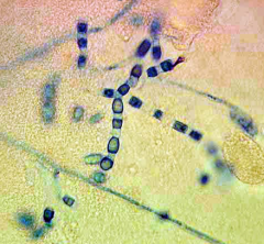
What kind of conidia does this fungus have? Are those sexual or asexual structures? |
Arthroconidia. Conidia = Asexual Example: Coccidoides immitis |
|
|
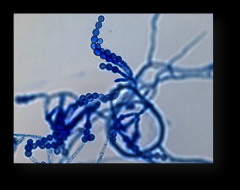
What are the different types of aerial mycelium? |
Sporangiospores Phialoconidia Annelloconidia Macroconidia Microconidia |
|
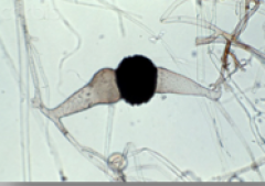
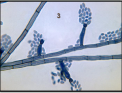
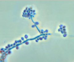
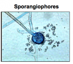
What kind of aerial mycelia is that? What fungus is this? |
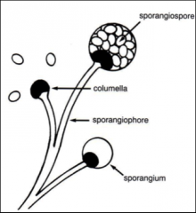
Sporangiospore Mucor sp. |
|
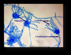
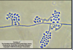
What kind of aerial mycelia is seen here? What fungus is this? |
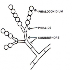
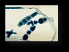
Phialoconidia Ex. Penicillium sp |
|
What kind of aerial mycelia is seen here? What fungus is this? |
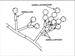
Annelloconidia Ex. Scopulariopsissp. |
|
What kind of aerial mycelia is seen here? What fungus is this? |

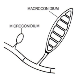
Macroconidia Epidermophytonsp. |
|
What kind of aerial mycelia is seen here? What fungus is this? |
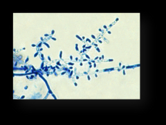
Microconidia Trichophyton tonsurans |
|
|
What is the proper way to collect a skin or nail scraping? |
Clean with 70% isopropanol first. KOH prep |
|
|
What media promotes hyphal and blasto conidia formation |
Cornmeal Tween 8- Agar (CMT) |
|
|
What would you observe when looking at Candidaalbicans on CMT agar |
pseudohyphae and chlamydoconidia in Candidaalbicans |
|
|
What are Nigerseed or Birdseed agar used for? |
Isolationof Cryptococcusneoformans |
|
|
What kind of growth does PotatoDextrose Agar (PDA) stimulate? |
Stimulatesspore formation and pigmentation |
|
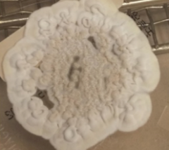
What is the colony morphology topography |
Verrucose: colonies have a wrinkled, convoluted surface |
|
|
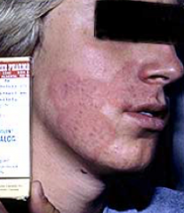
Pityriasis versicolor is also called _______. What organism causes it? |
Ringworm: on chest and back skin Malassezia furfur |
|
|
Tinea nigra is also called _________. what organism causes it? |
Ringworm: on palms of hands Hortaea werneckii |
|
|
What is the spaghetti and meatballs fungus? |
Malassezia furfur |
|
|
Which fungi cause otomycoses? |
Aspergillus niger Penicillium Mucor Rhizopus spp. |
|
|
What fungus causes Black piedra (Tropical climates)? |
Piedraia hortae |
|
|
What fungus causes white piedra? |
Trichosporon beigeilii (a.k.a.cutaneum) |
|
|
What does anthropophilic, zoophilic and geophilic mean? |
Anthropophilic –found primarily in humans• Zoophilic –found primarily in animals such as cats and dogs (man easily infected)• Geophilic–found primarily in soil |
|
|
What do dermatophytes use for their nitrogen source? |
Dermatophytesbreak down and utilize keratin as a source of nitrogen (but unabletopenetrate the subcutaneous tissue) |
|
|
Which fungi will have a positive wood's lamp on a hair sample> |
Microsporum spp. (canis and audouinii) Ectothrix hair |
|

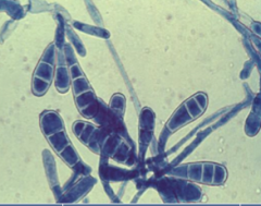
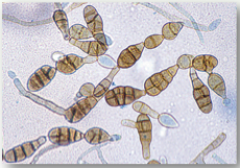
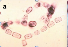
ID species and conidia types and charaacteristics |
Microsporum macroconidia: •Numerous •Rough walled •Elliptical/spindle •Either thin or thick walled •Usually3-7 cells inside |
|
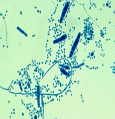
ID species and conidia types and characteristics |
Epidermophyton Macroconidia: •Numerous •Smooth and thin walled•Club shaped •Usually3-4 cells inside Microconidia: absent |
|
|
Trichophyton Macroconidia: •Usuallyrare•Smooth•Pencil•Thin•Usually 3-8 Microconidia: •Numerousor few•Round,oval, or club•Singly/grapelikeclusters |
|
|
What area do these fungi infect? Tinea capitis Tinea corporis Tinea barbae Tinea cruris Tinea pedis Tinea unguium |
Tinea capitis: Head/scalp Tinea corporis: body Tinea barbae: face Tinea cruris: groin Tinea pedis: feet Tinea unguium: nails |
|
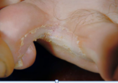
What causes this Dermatophytosis: Tinea pedis |
Epidermophyton floccosum |
|
What causes this Dermatophytosis: Tinea corporis |
Microsporum canis |
|
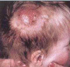
What causes this Dermatophytosis: Tinea capitis |
Trichophyton rubrum |
|
|
What three yeasts cause most of the human yeast infection? |
Candida albicans Cryptococcusneoformans Geotrichumcandidum |
|
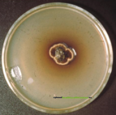
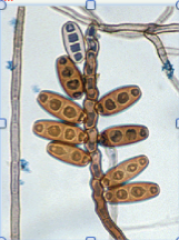
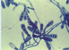
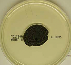
ID species |
This is a plate culture growingthe fungus Piedraiahortae,thecausativeagent for Black Piedra, a superficial fungal infection of the hair shaft.Infections are usually localized to the scalp but may also be seen on hairs ofthe beard, moustache and pubic hair. |
|
|
What are fungus that cause subcutaneous mycoses? |
•Causative fungiare soil saprophytes |
|
|
What are the four diseases (clinical classifications) of subcutaneous mycoses that we talked about? |
•Chromoblastomycosis •Mycetoma (eumycotic) •Phaeohyphomycosis •Sporotrichosis |
|
|
What color are dematiaceous molds |
•BROWN – BLACKPIGMENTATION |
|
|
What are two fungus-like bacteria that cause subcutaneous mycoses? |
•Actinomycetes •Norcardia species |
|
|
Which three dematiaceous molds cause chromoblastomycosis? |
1.Cladophialophora carrionii 2.Fonsecaea pedrosoi 3.Phialophora verrucosa |
|
|
Chromobastomycosis Microscopic Morphology: Cladosporium- Acrotheca- Phialophora- |
•Cladosporium – elliptical conidia inchains •Acrotheca – “bottle brush” •"flowers in a vase” - phialophora |
|
structure? common to what group of fungi? |
phialophora. common to dematacious molds that cause Chromoblastomycoses. |
|
structure? common to what group of fungi? |
acrotheca "bottle brush" common to dematacious molds that cause Chromoblastomycoses. |
|
structure? common to what group of fungi? |
cladosporium- •elliptical conidia in chains. common to dematacious molds that cause Chromoblastomycoses. |
|
what general group will exhibit this microscopic morphology? hint: called flowers in vase or bottle brush... |
Dematiaceous molds that cause chromoblastomycosis |
|
|
NOTE: subcutaneous mycoses are... |
These are chronic, localized infections of the skin and subcutaneous tissue following the traumatic implantation of the aetiologic agent. The causative fungi are all soil saprophytes. |
|
|
Direct microscopic lab diagnosis of chromoblastomycosis is commonly done usually which two stains? |
•Skin scrapings should be examined using 10%KOH and or calcofluor white mounts; •Tissue sectionsshould be stained using H&E, PAS or GMS |
|
|
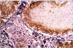
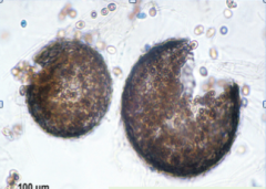
Direct microscopic lab diagnosis of chromoblastomycosis is commonly done using skin scrapings or direct tissue exam. What key feature will you see in infected tissue? |
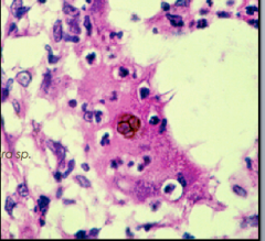
•Sclerotic bodies – black dots - in biopsied tissue |
|
ID this fungi. What disease does it cause? |
Phialaphora sp. Chromoblastomycosis (which is a subcutaneous mycoses) |
|
|
Which disease is characterized by a triad of symptoms: 1)Tumor-like swelling of tissue 2)Sinus tract (tunnel) 3)Granulomatous drainage – containaggregates of fungal hyphae = granulesor grains that are white,yellow, red, or black |
Mycetoma (eumycotic) Note: Microscopy shows yellowish browngranules containing spores and hyphae. |
|
|
What are the causative agents of mycetoma (eumycotic)? |
1. Pseudallescheriaboydii (Sexual)/Scedosporium apiospermum (asexual) 2. Acremonium 3. Fusarium 4. Madurella 5. Exophiala (NOT... its in her notes, but these cause phaeohyphomycoses) |
|
what's this? |
Yellow-brown to black, spherical cleistothecia, the sexual form P. body. Agent of EUMYCETOMA |
|
|
what is the hallmark of a mycetoma? |
Granules in exudate |
|
ID genus |
Acremonium spp. Caused Mycetoma Septate. Unbranched, taperedconidiophores, closely packed balls of sickle or elliptical conidia. |
|
|
How is Phaeohyphomycosis different from Chromoblastomycosis and Mycetoma? |
This type of subcutaneous mycoses does not involve sclerotic bodies or granules in tissue. |
|
Phaeohyphomycosis characterized by yeast-like cells or hyphae seenin the tissue. What is this? |
Exophialajeanselmei hyphae in walls of 'cyst'. Phaeohyphomycosis |
|
|
What are 5 causative agents of phaeohyphomycosis? |
Alternaria spp. Bipolaris spp. Curvularia spp. Exophialajeanselmei Exophialadermatitidis |
|

ID genus. What is the clinical classification of the disease it causes? |
Alternariaspp. showing branched,‘zig zag’ or alternating brick wall arrangement of conidia with short conical beaks. Casuses phaeohyphomycosis |
|
|
Which fungi has a brick wall arrangement of its conidia? |
Alternia spp. |
|
ID genus. What is the clinical classification of the disease it causes? |
Bipolaris spp. Showing bending conidia that are oval and thick-walled with 4-5 septations Casuses phaeohyphomycosis |
|
Genus? What is the clinical classification of the disease it causes? |
Curvularia spp. Casuses phaeohyphomycosis |
|
Genus? species? What is the clinical classification of the disease it causes? |
Exophialajeanselmei Casuses phaeohyphomycosis |
|
ID genus. What is the clinical classification of the disease it causes? |
Exophialadermatitidis Casuses phaeohyphomycosis |
|
|
What is the term for "rose gardener’s" disease? |
Sporotrichosis Etiological agent: Sporothrix schenckii Subcutaneous infection |
|
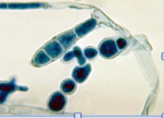
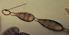
ID |
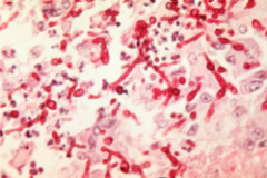
Sporothrix schenckii yeast forms |
|
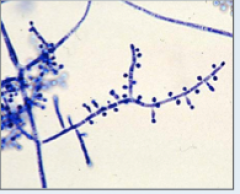
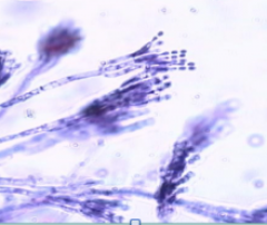
ID |
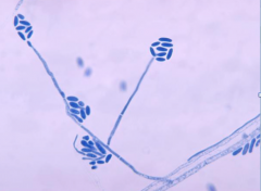
A photomicrograph showing theconidiophores and ‘flower-like- conidial arrangement of the fungusSporothrix schenckii. |
|
|
Nameat least three genus of fungi known to be fungal opportunists. |
a. Blastomyces b. Coccidioides c. Histoplasma d. Paracoccidioides e. Penicilliun |
|
A. B. C. D. |
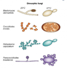
Blastomycesdermatitidis Coccidioidesimmitis Histoplasmacapsulatum Paracoccidioidesbrasiliensis |
|
|
Are all systemic mycoses caused by fast or slow growers? Where do they usually infect? |
SLOW growing dimorphs primarily infecting the lungs |
|
|
What laboratory conditions does one need to deal with systemic infections caused by fungi? |
Need a BSL-3 b/c thefungi are inherently virulent, extremely infective |
|
|
Most common transmission sources for systemic infections caused by fungi? |
Transmissionsources include: •Soil(Dust) •Decayingvegetation •Birdand bat droppings |
|
|
What fungi causes Gilchrist’s Disease? What is the scientific name for the disease? |
Blastomyces dermatitidis Blastomycosis |
|
|
What fungi causes "Valley Fever" or "Desert Fever"? What is the scientific name for the disease? |
Coccidioides immitis & C. posadasii Coccidioidomycosis |
|
|
What fungi causes "Darling’s", "Cave Disease", "Spelunker’s Disease"? What is the scientific name for the disease? |
Histoplasma capsulatum Histoplasmosis |
|
|
What fungi causes "South American Blastomycosis"? What is the scientific name for the disease? |
Paracoccidioides brasiliensis Paracoccidiodomycosis |
|
|
Where is Gilchrist's Disease found? |
Blastomycosis: Endemicin central and southeastern parts of US: Mississippi & Ohio River valleys,and Great Lakes |
|
ID. Note: "lollipop" forms |

Blastomycesdermatitidismoldform lives with decaying organic matter: leaves & wood Causes Gilchrist |
|
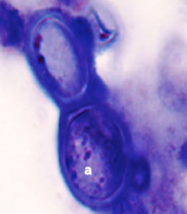
ID. Note: budding form |
Blastomycesdermatitidis. Warmerbody temperature signals spore transformation to broad-based budding yeast Causes Gilchrist |
|
|
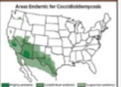
Where is Valley Fever found? What is the scientific name for it? |

Arizona,south central California (San Joaquin Valley), Nevada, New Mexico, parts ofUtah, western half of Texas. "Coccidioidomycosis" |
|
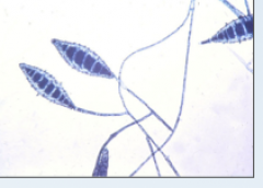
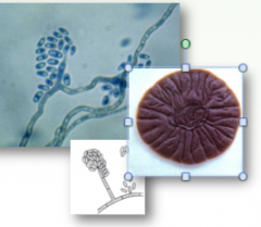
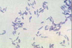
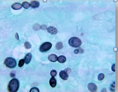
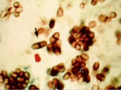
What are these? What do they develop into? What is this fungi and what disease does it cause? |
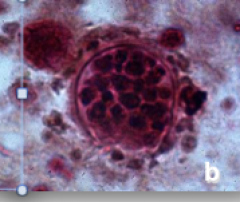
Arthroconidiadevelop into spherulescontaining endospores. Coccidioides = Valley Fever. |
|
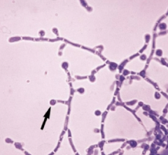
What are these structures and what is this fungus? |
Microscopy shows typicalsingle-celled, hyaline, rectangular to barrel-shaped, alternate arthroconidia. Coccidioides = Valley Fever. |
|
ID genus. |
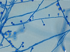
Histoplasma Hyaline,sepatatehyphae with unicellular macroconidia |
|
|
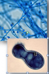
Which two yeast forms of the fungi that cause systemic infection look identical? |
Histoplasma and Blastomyces Budding yeast form |
|
|
Where in the world is Histoplasma found? |
Worldwide,though most common in North and Central AmericaqInUS: Central and Eastern states |
|
|
What fungi causes "Darling’s Disease"? What is the scientific name for the disease? |
Histoplasma Histoplasmosis |
|
|
Where in nature is the fungus that causes Darlings disease found? What are other names for the disease |
Bat guano Spelunkers, cave, etc |
|
|
Which mycoses affects males at a much higher rate? A 3:1 ratio? |
South American Blastomycosis caused by Paracoccidiodesbrasiliensis (Systemic) . |
|
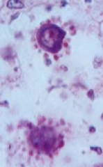
ID |
Paracoccidiodes brasiliensis Hyaline,septatehyphae w/intercalary & terminal chlamydoconidia |
|
ID |
P. brasiliensis intissue, showing the ‘ship’swheel’appearance |
|
|
Cutaneousskin test for a delayed type hypersensitivity reaction using the antigen __________ for the diagnosis of Paracoccidiodesbrailiensis. |
paracoccidioidin |
|
|
Four examples of aseptate opportunists: What microscopic structure do they have in common |
Absidiaspp. Mucorspp. Rhizopusspp. Cunninghamellaspp. |
|
What is the general name for this structure? |
Poroconidia SeptateOpportunists have these |
|
|
Fungal opportunist characteristics: |
Rapid Growers: 4-5 days Saprobic: live on decaying organic matter Airborne Must be repeatedly isolated from multiple patient specimens |
|
What is the general name for this structure? |
Phialoconidia |
|
|
What are the Dermatiaceous Septate opportunists? |
DERMATIACEOUS Alternariaspp. Aureobasidiumspp. Bipolarisspp. Cladosporiumspp. Epicoccumspp. |
|
|
What are the Hyaline Septate opportunists? |
HYALINE Chrysosporium spp. Acremoniumspp. Penicillium spp. Fusarium spp. Aspergillus spp. C.A.P.F.A. |
|
|
What is the onlydimorph of the genus Penicillium? What does it cause? |
Penicilliummarneffei Systemic Penicilliosis |
|
ID |
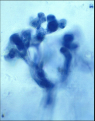
P. marneffei: typicalyeast-like cells with a central septa, ellipsoidal |
|
ID |
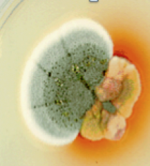
Cultureshowing a common green saprophytic Penicillium sp.w/typical yellow-pink colony w/distinctive red diffusablepigment of Penicilliummarneffei; |
|
ID |
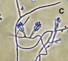
Phialides andconidia of P. marneffei. |
|
|
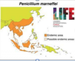
What geographic area is systemic penicilliosis endemic to? |
Endemicto southern China and SE Asia |
|
|
What is the 3rd most common opportunisticinfection in AIDS patients in endemic areas (China and SE Asia) |
systemic penicilliosis caused by Penicilliummarneffei |
|
|
What is the prognosis of patients with systemic penicilliosis? |
Poor prognosis: Cutaneouslesions are frequently present Usuallydisseminated with multi-organ involvement Usuallyfatal |
|
Whats this?! ID: |
Favic chandelier hyphae as seen in Trichophyton schloenleinii |
|
|
Growth of what structures does Cornmeal Tween Agar promote? What genus are being targeted? |
Promotes hyphal and blastoconidia formation Candida |
|
|
Which wet prep stain is used to observe for capsules around yeast? Which yeast is this especially good for? |
India Ink Cryptococcus neoformans |
|
|
Which wet prep dissolveskeratin, enhances visualization of fungal elements |
10%KOH |
|
|
Which histological stain is often used for fungi? |
Gomori MethenamineSilver (GMS) stain |
|
|
Which yeast infects hair "within"... endothrix |
Candida albicans |
|
|
Which fungi did we say had positive wood's lamp results? |
Microsporum (canis, audonii) Trichophyton (rubrum) |
|
|
What are the three genera of dermatophytes? |
1. Trichophyton 2. Microsporum 3. Epidermophyton |
|
|
Out of the three genera of dermatophytes, which one does not have microconidia |
Epidermophyton |
|
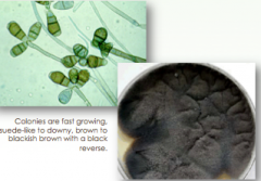
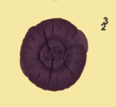
ID Causative agent of what general category of mycoses? |
Cladophialophora carrionii •Black, velvety texture•Compact Chromoblastomycosis (Subcutaneous) |
|
ID Causative agent of what general category of mycoses? |
Fonsecaea pedrosoi •Black-olive green,suede-downy-like•Flat to heaped and folded Chromoblastomycosis (Subcutaneous) |
|
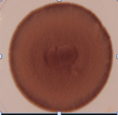
ID Causative agent of what general category of mycoses? |
Phialophora verrucosa •Black-brown-olive green , suede texture Chromoblastomycosis (Subcutaneous) |
|
|
Scedosporium apiosperum (asexual) Causes mycetoma |
|
|
Exophiala jeanselmei |
|
|
Fonsecaea pedrosoi |
|
|
Phialophora verrucosum |
|
|
Cladosporium carrionii |