![]()
![]()
![]()
Use LEFT and RIGHT arrow keys to navigate between flashcards;
Use UP and DOWN arrow keys to flip the card;
H to show hint;
A reads text to speech;
79 Cards in this Set
- Front
- Back
|
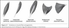
Types and arrangements of skeletal muscle fibers |
Parallel: Fibers are parallel in arrangement. |
|
|
skeletal muscle fibers diagram |
|
|
|
Tendon |
Thick dense fibrous tissues, band like structure, originate from muscles and attached them to bones. |
|
|
Ligament |
Thick dense fibrous tissues, band like structure which binds the two bones and helps in the formation of joint. |
|
|
Aponeurosis |
Thick sheet of connective tissue originate from the muscles. |
|
|
|
The attachment of the muscle which remains more stationary during during movement is termed the origin. In the limbs the proximal attachment is the origin. |
|
|
Insertion of muscle |
The movable part of the muscle during contraction is termed the insertion. In the limbs the distal attachment is the insertion. |
|
|
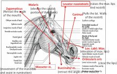
Muscles of the head |
1. Caninus |
|
|
Muscles of the head diagram |
|
|
|
Caninus |
|
|
|
Levator nasolabialis |
It is thin in the ox and in the horse divided into two thick part which originate from the rostral part of the frontal bone and insert in the maxillary lip and nostril. It raise the maxillary lip and lateral portion of the nostril. |
|
|
Levator labii maxillaris |
Thin muscle in the ruminant and dorsal to the caninus muscle causes elevation of maxillary lip and muzzle. |
|
|
Depressor labii maxillaris |
Thin muscle in the ruminant and ventral to the caninus muscle causes retraction of maxillary lip and muzzle. |
|
|
Depressor labii mandibularis |
It is a thin muscle layer originating beneath the masseter muscle at the caudal portion of the cheek. It causes depression of the mandibular lips and skin of the chin. |
|
|
Orbicularis oris |
It is located between the skin and mucous membrane and act as a sphincter muscle of the lip. Action: To close the lips. |
|
|
Buccinator |
It is more well developed in ruminants than in horses. It is broad, flat muscles which forms the major portion of cheek. Action: Retract (take back) the angle of the of the mouth. |
|
|
Zygomaticus |
is a strong muscle in ruminants. It extends from ventral surface of the eye to the maxillary lips. Action: Retract (take back) the angle of the of the mouth. |
|
|
Malaris |
located ventral and rostral to the eye. The muscle is well developed in ox than the horse. Action: Elevate the caudal portion of the cheek.
|
|
|
Masseter muscle |
Strong, broad, flat muscle located on the lateral surface of the ramus of the mandible. Action: To close the jaw, movement of mandible and assisting in rumination. |
|
|
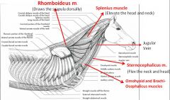
Muscles of the neck region |
1. Brachiocephalicus |
|
|
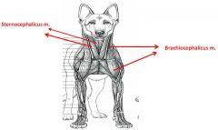
Brachiocephalicus |
is a thin muscle which extends along the side of the neck from the head to the arm. Action:It depresses the head and full the forelimb forward. |
|
|
Sternocephalicus |
It is the ventral muscle of the neck and extend from sternum to the angle of the mandible and mastoid process of the head. Action: Flex the neck and head. |
|
|
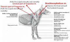
Cervical Part of the Trapezius |
is a wide, undivided, flat and triangular muscles of the neck and thorax. Action: The cervical part pulls the scapula dorsad and craniad.
The thoracic part pulls the scapula dorsad and caudad. |
|
|
Splenius |
It is a thin, large, flat, triangular muscle which lies on the lateral surface of the neck dorsal to the level of the cervical vertebra. Action: Elevate the head and neck. |
|
|
Rhomboideus |
The rhomboideus arises on the nuchal ligament from the second cervical to the fifth thoracic vertebrae. It is attached to the deep surface of the scapular cartilage. Action: Draws the scapula dorsally. |
|
|
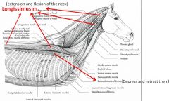
Iliocostalis muscle |
is a strong, segmented muscle which extends across the series of ribs in contact with the lateral edges of the longissimus muscles. Action: Depress and retract the ribs and helps in expiration. |
|
|
Longissimus muscle |
This muscle lies medial to the intercostales muscle and extend from the atlas bone to the lumber region. It is the largest and longest muscles in the body. It is the most powerful extensor and flexor muscles of the vertebral column. |
|
|
Muscles of the neck region diagram |
|
|
|
Muscles of the neck region diagram |
|
|
|
Muscles of the neck region diagram |
|
|
|
Muscles of the neck region diagram |
|
|
|
Muscles of the Thoracic Region |
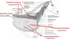
1. Levatores costarum |
|
|
Levatores costarum |
They constitute a series of small muscles which overlie the vertebral ends of the intercostal space. |
|
|
Intercostales externi |
This muscle occupied the external part of the intercostal space, extending from the levatores costarum to the sternal extremity of the rib. |
|
|
Intercostales interni |
They located underneath of the intercostales externi.
|
|
|
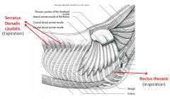
Rectus thoracis |
This and straight muscle lies on the cranioventral aspect of the thorax. |
|
|
Transversus thoracis |
Flat muscles and situated on the thoracic surface of the sternum. |
|
|
Retractor costae |
It is a thin muscle located in the angle formed by the last rib and the ends of the lumbar transverse processes. |
|
|
Serratus dorsalis caudalis |
In ox it is very poorly developed and present 3-4 digitations. |
|
|
Muscles of the Thoracic Region diagram |
|
|
|
Muscles of the Thoracic Region diagram |
|
|
|
Diaphragm |
It is a broad, unpaired muscles which forms a partition between the thoracic and abdominal cavities. In outline it has some resemblance palm-leaf fan. The thoracic surface it is strongly convex while the abdominal surface is concave. Its outer part is muscular while central part is tendinous. |
|
|
Diaphragm Attachments |
Costal Part: Cartilages of the 8th-10th ribs in horse. |
|
|
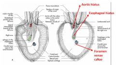
Openings and content of the diaphragm |
The aortic hiatus: is an interval between the two crura and ventral to the last thoracic vertebra. It contains descending aorta, right vena azygos, and cisterna chyli. |
|
|
Diaphragm diagram |
|
|
|
Abdominal muscles General functions |
In general abdominal press which causes compression of abdominal viscera leading to:
micturition (urination) parturition (giving birth) and expiration (exhale)
|
|
|
Abdominal muscles |
1. Obliquus externus abdominis |
|
|
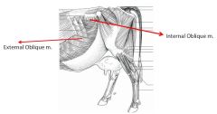
Obliquus externus abdominis |
It is the most extensive of the abdominal muscles. It is a broad sheath, irregularly triangual in shape, whose fibers directed caudad and ventrad, but which in the area of the paralumbar fossa are seen to pass in a horizontal direction. |
|
|
Abdominal muscles diagram |
|
|
|
Obliquus internus abdominis |
is situated beneath the obliquus externus abdominis muscle. Its fiber directed ventrad, craniad and mediad. It occupies the entire flank region from the coxal tuber to the last rib. |
|
|
Transversus abdominis |
As a muscular sheet it is located deep to the Obliquus internus abdominis and rectus abdominis. |
|
|
Rectus Abdominis |
It is confined to the ventral abdominal wall. It extend from the sternum to the pubis. It is a straight muscle. |
|
|
Cremaster muscle |
of male may be regarded as detached portion of the obliquus abdominis externus muscle which separates as slip of fleshy tissue to enter the inguinal canal. |
|
|
Abdominal tunic |
This is a sheet of elastic tissue which assists the muscles in supporting the great weight of the abdominal viscera. It is practically coextensive with the obliquus externus abdominis which it covers. Ventrally it is thick and is intimately adherent to the aponeurosis (flat tendon) of the muscles. Laterally it become thinner and is more easily separated
|
|
|
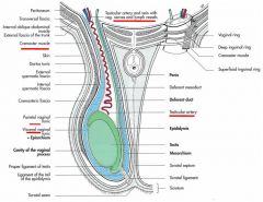
Inguinal canal |
Inguinal canal or space is a term applied to an oblique passage through the caudal part of the abdominal wall. It begins at the deep inguinal ring and ends at the superficial inguinal ring. Canal in between the deep and superficial inguinal ring formed the inguinal canal. This canal is formed by the obliquus internus abdominis muscles. |
|
|
Inguinal canal contents |
In the male: Contains (i)Spermatic cord, (ii)the vaginal tunic, (iii) the cremaster muscles, (iv) the external pudendal artery, (v) the inguinal lymph vessels, and (vi) branches of ilioinguinal and genitofemoral nerve. |
|
|
Inguinal canal diagram |
|
|
|
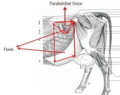
Flank |
The soft lateral abdominal wall formed by the obliquus abdominis externus and internus, and transversus abdominis muscle is known as flank. |
|
|
Flank diagram |
|
|
|
Paralumbar fossa |
The triangular depression of the upper lateral abdominal wall is called paralumbar fossa. The cranial border is marked by the last rib, caudal border extend up to the tuber coxae, dorsal border is at the label of the transverse processes of the lumbar vertebrae. The ventral border is marked by the upper part of the abdominal muscles.
|
|
|
Clinical importance of Flank and Paralumbar fossa |
Flank: For the gastrointestinal, reproductive and urinary tract operation.
Paralumbar fossa: Operation related to the stomach and intestines. |
|
|
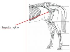
Prepubic tendon |
Prepubic tendon is the tendon of insertion of two recti abdominis muscles but also furnishes the attachment of obliquus abdominis muscle, gracilis and pectinous muscles. It is attached to the cranial part of the pubic bones
|
|
|
Prepubic diagram |
|
|
|
Linea alba |
It is a median fibrous raphe (line) extends from the xiphoid cartilage to the prepubic tendon. It is formed chiefly by the junction of the aponeuroses of the obliquus abdominis externus, internus and transversus abdominis. Slightly caudal to its middle is the umbilicus. |
|
|
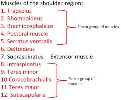
Muscles of the forelimb |
|
|
|
Muscles of the arm |
1. Biceps brachii- Flexor |
|
|
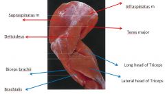
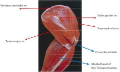
Shoulder muscles lateral view |
|
|
|
Shoulder muscles medial view |
|
|
|
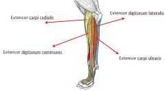
Muscles of the forearm (extensor) |
Extensor group:
1. Extensor carpi radialis |
|
|
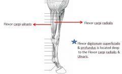
Muscles of the forearm (flexor) |
Flexor muscles:
|
|
|
Muscles of the forearm lateral view |
|
|
|
Muscles of the forearm medial view |
|
|
|
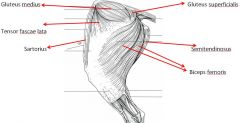
Muscles of the hip and thigh |
1. Gluteus superficialis |
|
|
Muscles of the pelvic limb |
|
|
|
Muscles of the pelvic limb |
|
|
|
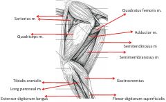
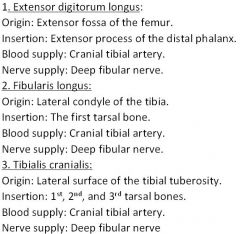
Muscles of the leg (extensor) |
Extensor muscles:
|
|
|
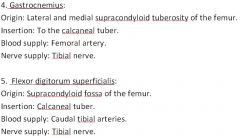
Muscles of the leg (flexor) |
Flexor muscles:
|
|
|
5 Muscles of Hindlimb |
|
|
|
5 Muscles of Hindlimb |
|