![]()
![]()
![]()
Use LEFT and RIGHT arrow keys to navigate between flashcards;
Use UP and DOWN arrow keys to flip the card;
H to show hint;
A reads text to speech;
310 Cards in this Set
- Front
- Back
|
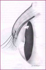
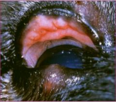
Increased Scleral Show |
|
|
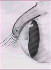
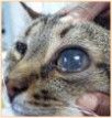
Micropthalmos |
|
|
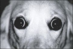
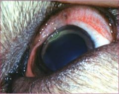
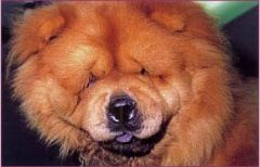
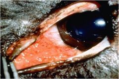
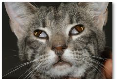
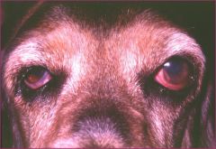
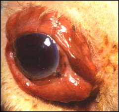
Bilateral Exopthalmos Increased Scleral Show - Stressed Look |
|
|
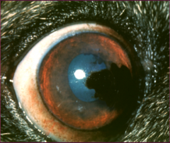
proptosis |
|
|
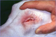
Ankyloblepharon |
|

|
Ankyloblepharon |
|
|
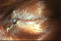
Eyelid Agenesis |
|
|
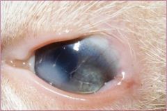
Dermoid |
|
|
Trichiasis |
|
|
Distichiasis |
|
|
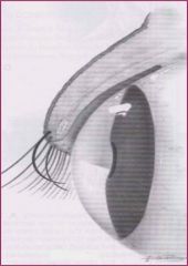
Ectopic Cilia |
|
|
Ectopic Cilia with Concurrent Corneal Ulcer |
|
|
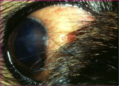
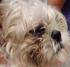
Pigmentary Keratitis |
|
|
Nasal Trichiasis |
|
|
Entropion |
|
|
Ectropion |
|
|
Combined Entropion-Ectropion |
|
|
Entropion |
|
|
Blepharitis |
|
|
Chalazion |
|
|
Multifocal Granulomas - Blepharitis |
|
|
Conjunctivitis with Mucopurulent Discharge |
|
|
Chemosis |
|
|
Epiphora |
|
|
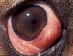
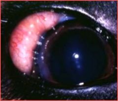
Conjunctival Hyperemia |
|
|
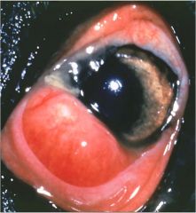
Scleral Hyperemia |
|
|
Conjunctival Emphysema |
|
|
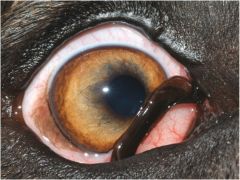
Conunctival Hemorrhage |
|
|
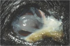
Conunctival Tissue Proliferation - Swelling/Mass |
|
|
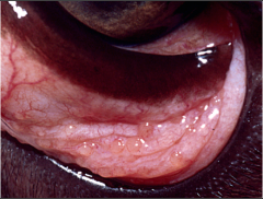
Conjunctival Tissue Proliferation - Follicle Formation |
|
|
Symblepharon |
|
|
Symblepharon |
|
|
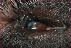
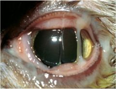
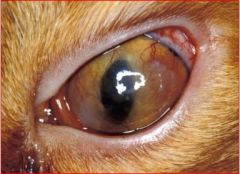
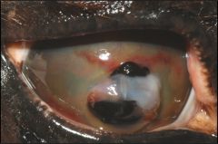
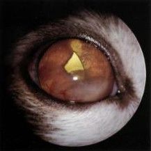
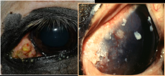
Corneal Sequestrum |
|
|
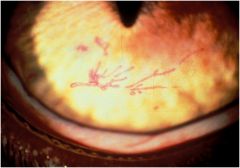
Dendritic Corneal Lesions - Branching Ulcers or Erosions |
|
|
Follicles in Abnormal Location - Anterior Surface of Third Eyelid |
|
|
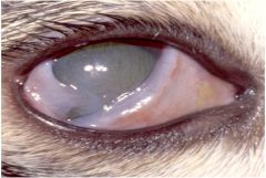
KCS |
|
|
KCS |
|
|
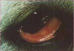
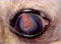
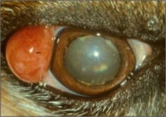
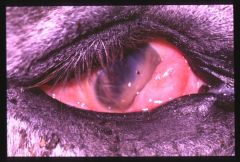
Nictitans Gland Prolapse |
|
|
Nictitans Gland Prolapse |
|

|
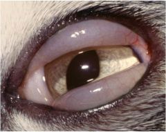
Nictitans Elevation |
|
|
Scrolled Cartilage |
|
|
KCS |
|
|
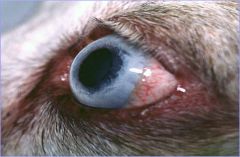
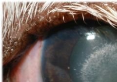
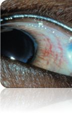
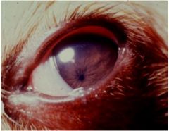
Superficial Vessels (Trees) on Cornea |
|
|
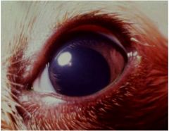
Deep Vessels (Hedge) on Cornea |
|
|
Superficial Vessels (Trees) on Cornea |
|
|
Deep Vessels (Hedges) on Cornea |
|
|
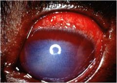
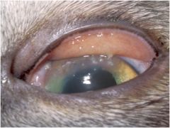
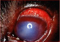
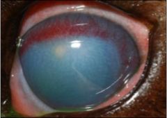
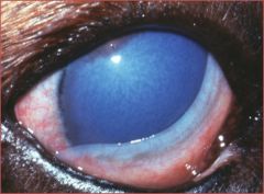
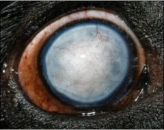
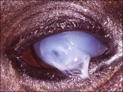
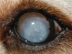
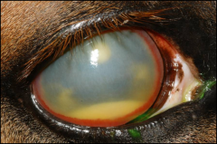
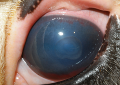
Corneal Edema |
|
|
Corneal Edema |
|
|
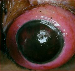
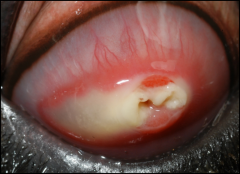
Corneal Malacia |
|
|
Corneal Scar Tissue |
|
|
Corneal Scar Tissue |
|
|
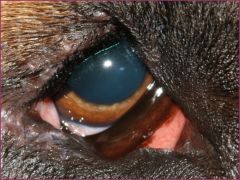
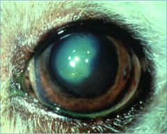
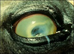
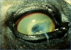
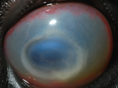
Corneal Lipid/Mineral Deposition |
|
|
Corneal Lipid/Mineral Deposition |
|
|
Corneal Lipid Dystrophy |
|
|
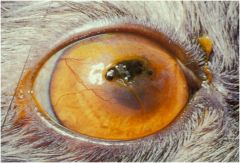
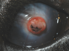
Corneal Pigment (Pigmentary Keratitis?) |
|
|
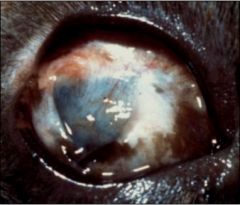
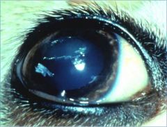
Corneal Rupture with Pigment |
|
|
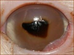
Corneal Pigment (Cat Sequestrum?) |
|
|
Keratic Precipitates |
|
|
Corneal Cellular Infiltrates |
|
|
Chronic Superficial Keratitis - Pannus |
|
|
Chronic Superficial Keratitis - Pannus |
|
|
Dermoid = Choristoma |
|
|
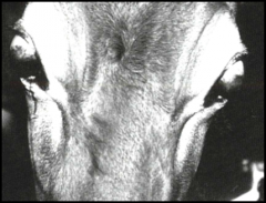
Dysautonomia |
|
|
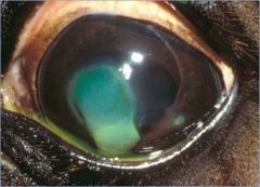
Indolent Ulcer |
|
|
Indolent Ulcer |
|

|
Stromal Ulcer |
|
|
Stromal Ulcer |
|
|
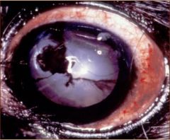
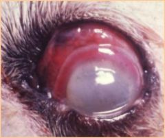
Melting (Deep) Corneal Ulcer |
|
|
Melting (Deep) Corneal Ulcer |
|
|
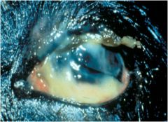
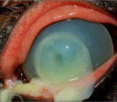
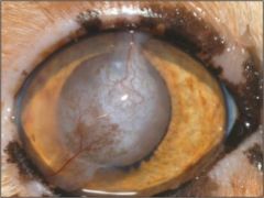
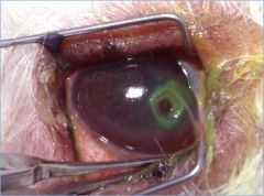
Desmetocele |
|
|
Desmetocele |
|
|
Positive Seidel Test |
|
|
Conjunctival Graft |
|
|
Scleral Coloboma |
|
|
Nodular Granulomatous (NGE) |
|
|
Nodular Granulomatous (NGE) |
|
|
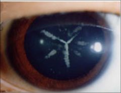
Corneal Y Sutures |
|
|
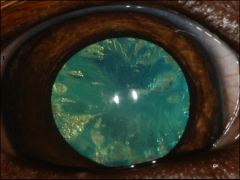
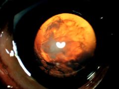
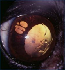
Cataract with Vacuoles (Diabetic) |
|
|
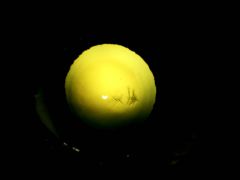
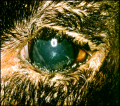
Nuclear Sclerosis |
|
|
Nuclear Sclerosis |
|
|
Microphakia |
|
|
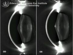
Lenticonus |
|
|
PHPV or PHTVL |
|
|
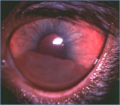
Incipient Cataract |
|
|
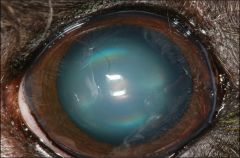
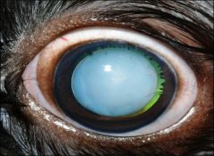
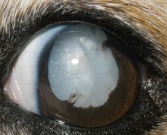
Immature Cataract |
|
|
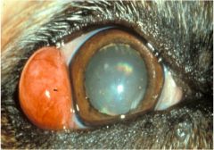
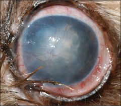
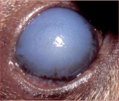
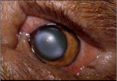
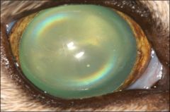
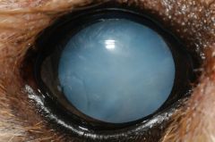
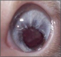
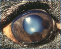
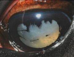
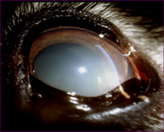
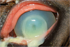
Mature Cataract |
|
|
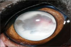
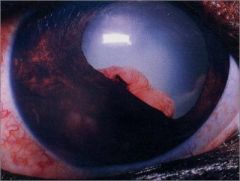
Hypermature Cataract |
|
|
Morganian Cataract |
|
|
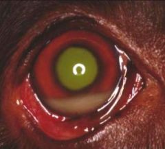
Miosis |
|
|
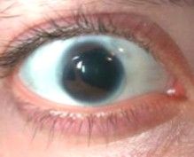
Mydriasis |
|
|
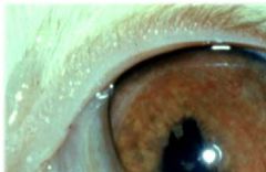
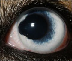
Heterochromia Iridis |
|
|
Heterochromia Iridum |
|
|
Heterochromia Iridis |
|
|
Iris Coloboma |
|
|
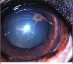
Persistent Pupillary Membrane (PPM) |
|
|
Dyscoria (Improper formation of the iris) |
|
|
Corectopia |
|
|
Aniridia - absence of the iris |
|
|
Polycoria - more than one pupillary opening in the iris |
|
|
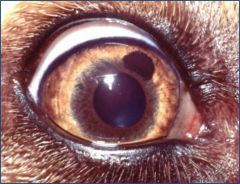
Iris Atrophy |
|
|
Iris Atrophy |
|
|
Melanocytoma (Uveal Cyst) |
|
|
Iris Melanoma |
|
|
Ciliary Body Adenoma/Adenocarcinoma |
|
|
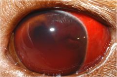
Anterior Tract Uveitis due to Lymphosarc |
|
|
Uveitis |
|
|
Uveitis |
|
|
Aqueous Flare |
|
|
Anterior Uveitis |
|
|
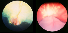
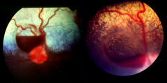
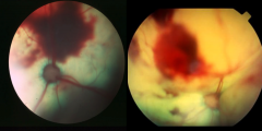
Retinal Detachment (Ballooned around optic N) |
|
|
Enopthalmitis |
|
|
Panopthalmitis |
|
|
Liquid Aqueous (d/t hyperlipidemia) |
|
|
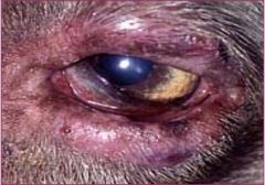
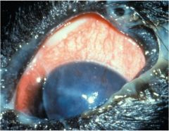
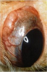
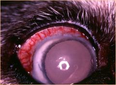
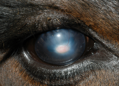
Glaucoma |
|
|
Episcleritis |
|
|
Episceral Hyperemia |
|
|
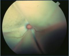
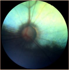
Cupping of Optic N. (Glaucoma) |
|
|
Open Iridiocorneal Angle |
|
|
Closed Iridocorneal Angle |
|
|
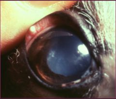
Glaucoma |
|
|
Enucleation |
|
|
Pthisis Bulbi |
|
|
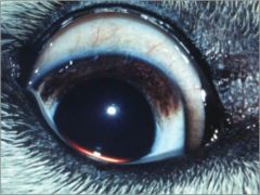
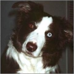
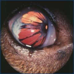
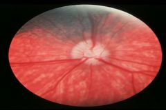
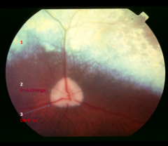
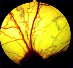
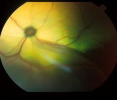
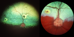
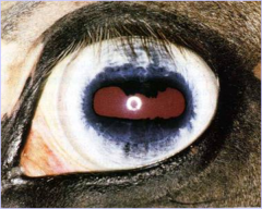
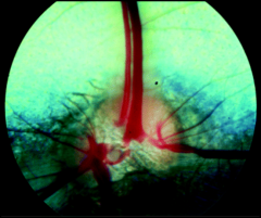
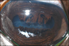
Blue Eyed Dog: Pigmented Choroid, Lack of Tapetum, Retinal Vessels, Choroidal Vessels, and Scleral Show through Choroid |
|
|
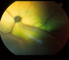
1. Tapetum Lucidum 2. Choroidal Vessels 3. Retinal Vessels |
|
|
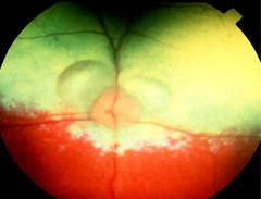
Normal Variations of Pigmentation in Choroid |
|
|
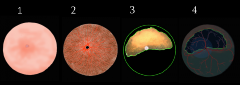
1. Sclera 2. Choroid 3. Tapetum 4. Retina |
|
|
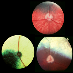
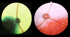
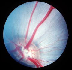
Normal Dog: Vessels Anastamose |
|
|
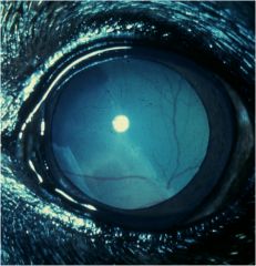
8-12wk Puppy with Blue Tapetum |
|
|
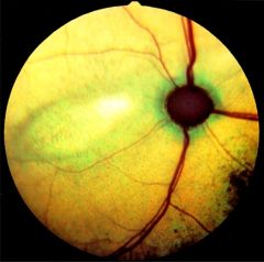
Normal Cat: Vessels to edge of Optic N. and stop |
|
|
Horse: Oval optic N, Tiny retinal vessels across optic N, No vessels across tapetum |
|
|
Horse Fundus Pattern |
Paurangiotic vascular pattern |
|
|
Cat/Dog fundus pattern |
Holangiotic Pattern |
|
|
Cow |
|
|
Pig |
|

|
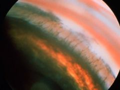
Tapetal Hyporeflectivity *Addition of something |
|
|
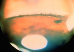
Tapetal Hypereflectivity *Subtraction of something |
|
|
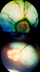
Retinal Detachment (Hypereflective) |
|
|
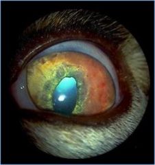
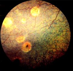
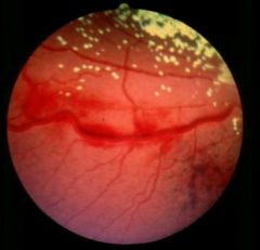
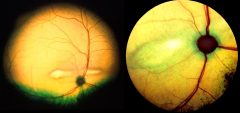
Focal Tapetal Hypereflectivity: Healed chorioretinitis lesions |
|
|
Retinal Folds (Dysplasia): Hyporeflective |
|
|
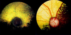
Retinal Detachment (Hyporeflective) |
|
|
Retinal Detachment (small): Hyporeflective |
|
|
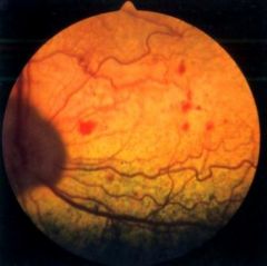
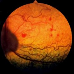
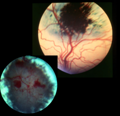
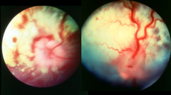
Retinal Hemorrhages (Hyporeflective) |
|
|
Pre-Retinal Hemorrhage |
|
|
Superficial Intraretinal Hemorrhage |
|
|
Deep Intraretinal Hemorrhage |
|
|
Subretinal Hemorrhage |
|
|
Retinal Detachment |
|
|
Retinal Detachment |
|
|
Retinal Dysplasia |
|
|
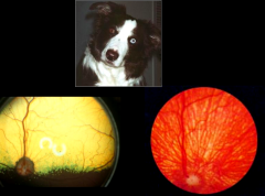
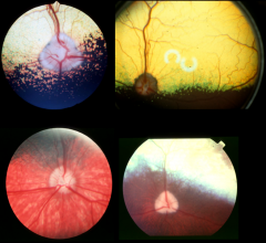
Collie Eye Anomaly |
|
|
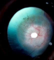
Retinal Degeneration/Atrophy |
|
|
Focal Retinal Degeneration (Taurine Deficiency) |
|
|
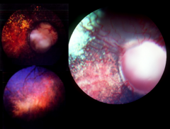
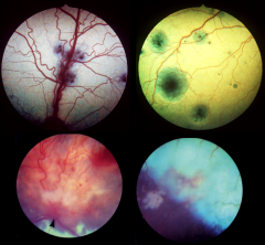
Active Chorioretinitis |
|
|
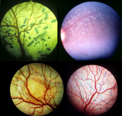
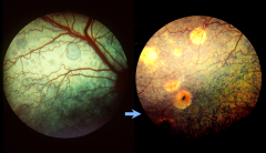
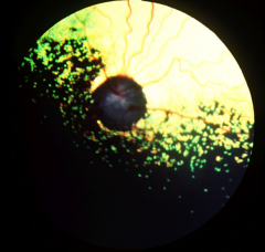
Distemper: Gold Medallion Lesions; Can go from a hyporeflectivemedallion to goldmedallion lesions (shows they had distemper at some time) |
|
|
Inactive Chorioretinitis |
|
|
Fundic Neoplasia |
|
|
Optic Nerve Coloboma (part of Collie Eye Anomaly) |
|
|
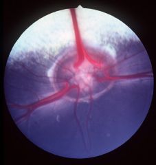
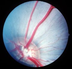
Appearance of Optic N with Glaucoma |
|
|
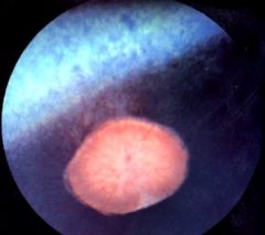
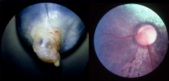
Optic Nerve Hypoplasia |
|
|
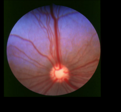
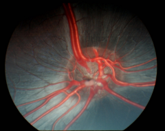
Papilledema (swelling of Optic N) |
|
|
Optic Neuritis |
|
|
Persistent Hyaloid (PHPV/PHTVL) |
|
|
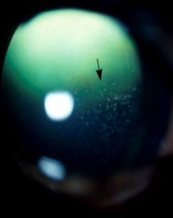
Asteroid Hyalosis (Vitreous Degeneration); Ca + P + Lipid |
|
|
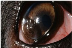
Normal Horse Eye |
|
|
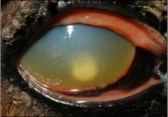
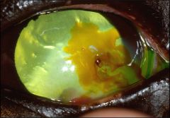
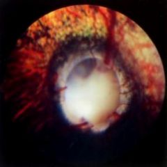
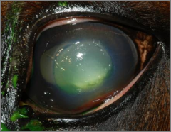
Keratomalacia |
|
|
Keratomalacia |
|
|
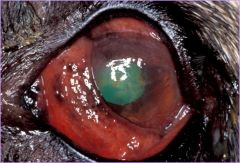
Corneal Ulcer with Farrow Ring |
|
|
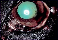
Equine Stromal Abscess |
|
|
Equine Ocular SCC |
|
|
Normal Pig Fundus |
|
|
Sheep |
|
|
Esotropia |
|
|
Exophtalmos caused by Lymphosarcoma |
|
|
Entropion |
|
|
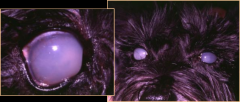
Pinkeye |
|
|
Pinkeye |
|
|
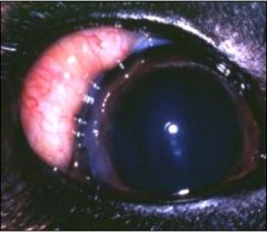
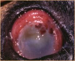
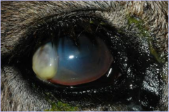
Corneal Ulcer with Cellular Infiltrate, Ciliary Flush, and maybe Malacia |
|
|
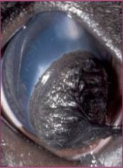
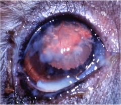
Perforation - Iris Prolapse |
|
|
Corneal Scar |
|
|
Camelid |
|
|
Llama |
|
|
What structures are contained within the intraconal space? |
connective tissue, extraocular muscles, nerves, blood vessels, fat, smooth muscle |
|
|
What is the endorbita/periorbita? |
fibrous connective tissue, next to bone of orbital wall that encircles the extraocular muscles - is the boundary between the intraconal/extraconal spaces |
|
|
Name the extraocular muscles? |
superior oblique, dorsal rectus, medial rectus, inferior oblique, ventral rectus, retractor bulbi, lateral rectus |
|
|
exophthalmos |
globe is too rostral |
|
|
enophthalmos |
globe is too caudal |
|
|
proptosis
|
equator of globe anterior to palpebral fissure |
|
|
T/F: A dog with an orbital abscess is painful on opening the mouth. |
True |
|
|
T/F: A dog with masticatory muscle myositis is painful on opening the mouth. |
True |
|
|
How can you differentiate between masticatory muscle myositis and extraocular muscle myositis? |
MMM - painful, elevated 3rd eyelid, facial muscle inflammation; EMM - non-painful, no elevation of 3rd eyelid, scleral show, common in Goldens |
|
|
T/F: Orbital neoplasia is usually benign in cats and dogs. |
False |
|
|
Positive prognostic indicators for proptosis? |
positive consenual PLR, voluntary movement of globe, +/- pupil size -- miosis means parasympathetic innervation intact |
|
|
Negative prognostic indicators for proptosis? |
transected optic nerve, chronic proptosis (>48 hrs), hyphema (indicates scleral rupture, severe uveal trauma), corneo-scleral laceration, rupture of >3 extraocular muscles, doliocephalic or cat, complete bony orbit (cows/horses) |
|
|
How long should you leave in a tarsorrhaphy for proptosis? |
2-3wks |
|
|
Proptosis replacement complications? |
lagophthalmos (unable to fully close lids), KCS, strabismus, blindness, phthisis bulbi (due to uveitis) |
|
|
enucleation |
removal of globe only |
|
|
exenteration |
removal of globe + orbital contents |
|
|
evisceration |
removal of intraocular contents only |
|
|
Functions of eyelids |
protection, tear film (production, distribution, drainage), oxygen exchange (conjunctiva) |
|
|
ankyloblepharon |
delayed/incomplete eyelid opening -- normal is 10-15 days of age |
|
|
Etiology of ankyloblepharon |
dogs - staph spp., cats - chlamydophila or FHV-1 |
|
|
Eyelid coloboma |
partial palpebral fissue absence |
|
|
Consequences of eyelid coloboma |
inability to blink normally, exposure keratitis, conjuctivitis, other congenital ocular diseases |
|
|
Breeds commonly affected by dermoids |
ST. bernard, GSD, Dalmation |
|
|
trichiasis |
normal hair (on face or eyelid) that contacts the cornea -- causes mild irritation or pigmentation; common in soft-coated breeds |
|
|
distichiasis |
hair that originates in the Meibomian gland -- grows through the duct of the gland and contacts the cornea (common in Cockers) |
|
|
Ectopic cilia |
follicle is within Meibomian gland & growth occurs through the palpebral conjunctiva (usually causes an ulcer or significant pain & ocular irritation) |
|
|
What are the components of brachycephalic ocular syndrome? |
macropalpebral fissures + medial trichiasis + medial lower lid entropion + pigmentary keratitis |
|
|
most common eyelid tumor in dog |
benign adenoma |
|
|
most common eyelid tumor in cat |
SCC |
|
|
most common eyelid tumor in horses |
SCC, Sarcoid |
|
|
most common eyelid tumor in cattle |
SCC |
|
|
Breeds commonly affected by cherry eye |
Beagles, Cocker Spaniels, Bostons, Poodles, other brachycephalic breeds |
|
|
Reasons for protrusion of third eyelid |
conformation (breed-related enophlathmos), non-pigmented leading edge (can be deceiving), decrease in orbital contents (dehydration, emaciation, phthisis bulbi), increase in orbital contents (neoplasia, abscess, hematoma), sympathetic denervation (horner's syndrome), ocular pain with retractor bulbi contraction, tetanus |
|
|
Breeds predisposed to scrolled 3rd eyelid cartilage |
large breed dogs |
|
|
Most common tumor of third eyelid in domestic animals? |
SCC |
|
|
3 Layers of tear film? |
Lipid, Aqueous, Mucous |
|
|
What produces the lipid component of the tear film? |
Meibomian glands |
|
|
What is the function of the lipid component of the tear film? |
stabilize and prevents evaporation of the aqueous layer |
|
|
What produces the aqueous component of the tear film? |
orbital (lacrimal) gland & gland of the third eyelid |
|
|
What is the function of the aqueous portion of the tear film? |
provides corneal nutrition, removes waste products |
|
|
What produces the mucous component of the tear film? |
conjunctival goblet cells |
|
|
What is the function of the mucous component of the tear film? |
interface of tear film with hydrophobic cornea, contains secretory IgA |
|
|
What are common causes of KCS? |
Immune-mediated (#1 cause), Congenital (Pug, Yorkies), Drug-induced (anesthesia, atropine), drug toxicity (sulfas), irradiation, surgical (removal of nictitans), systemic disease (distemper) |
|
|
T/F: A cat with a low Schirmer Tear test value OU has KCS. |
False: low values may be normal, interpret with clinical |
|
|
How long do you need to administer cyclosporine topically for KCS before you should expect to see a response? |
6 weeks |
|
|
What drug can be used to treat neurogenic KCS? |
Pilocarpine |
|
|
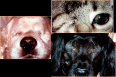
How can you differentiate neurogenic KCS from immune-mediated KCS? |
with neurogenic KCS, ipsilateral nose will also be dry (parasympathetic innervation is interrupted) |
|
|
Differentials for epiphora? |
excessive lacrimation - due to irritation (distichia, FB, tumor, entropion, etc.) or drainage abnormality - obstructions (NL system), imperforate punctum, dacryocystitis |
|
|
What is infection of the nasolacrimal ducts called? |
dacrycocystitis |
|
|
What is the limbus? |
junction of cornea and sclera |
|
|
Signs of conjunctival disease? |
chemosis*, hyperemia*, lymphoid hyperplasia, mucopurulent exudate, emphysema, hemorrhage, pruritis, epiphora |
|
|
Most common infectious cause of chemosis in the cat? |
Chlamydophila felis |
|
|
How do you distinguish between conjunctival hyperemia and scleral injection? |
conjunctival -superficial, tortuous vessels, branching, blanch with epi, indicative of superficial disease***, scleral - deeper, straighter, less branching, immobile, blanch slowly with epi, indicative of deep disease*** |
|
|
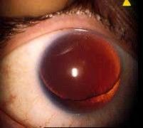
Causes of hyphema |
trauma - strangulation, CSF tap; septicemia, vasculitis, coagulopathies |
|
|
Non-infectious causes of conjunctivitis in the dog |
pannus, KCS, entropion, FB |
|
|
Non-infectious causes of conjunctivitis in the horse |
immune-mediated, keratitis, uveitis |
|
|
Infectious causes of conjunctivitis are most common in what two species? |
feline & ruminants |
|
|
What is a good topical treatment option for chalmydophila felis conjunctivitis? |
topical tetracyclines |
|
|
Clinical signs of FHV-1 conjunctivitis |
hyperemia, sneezing, nasal discharge, keratitis |
|
|
Ophthalmic abnormalities with FHV-1 |
dendritic corneal lesions - branching ulcers or erosions |
|
|
Best antiviral agent to use in cats |
Famciclovir*** |
|
|
Treatment for allergic conjunctivitis |
flush out allergens, topical antihistamines (ketotifen) |
|
|
Viral causes of conjunctivitis in dogs |
***Distemper (#1), Adenovirus, Herpesvirus |
|
|
Layers of the cornea body |
epithelium, stroma, Descemet's membrane, endothelium |
|
|
What maintains the clarity of the cornea? |
absence of blood vessels, deturgescence (relative dehydration of cornea), regular arrangement of collagen lamellae in the stroma, absence of pigment, relatively acellular |
|
|
Causes of deep corneal vessels (ciliary flush) |
deep keratitis, scleritis, uveitis, glaucoma |
|
|
Primary causes of corneal edema |
endothelial degeneration, endothelial dystrophy - primary endothelial problem, non-painful, uninflamed, normal IOP |
|
|
Secondary causes of corneal edema |
Glaucoma, uveitis, lens luxation; secondary endothelial problem, painful*, abnormal IOP, flare present |
|
|
What are keratic precipitates? |
clumps of fibrin and WBC's in the anterior chamber - indicative of prior or current uveitis |
|
|
Chronic superficial keratitis is also known as what? |
Pannus |
|
|
Clinical signs of pannus |
reddened plaque, ventrolateral cornea, bilateral*** (not always symmetrical) |
|
|
What breeds are predisposed to pannus? |
German Shepherds & Greyhounds |
|
|
How do you treat pannus? |
topical steroids + cyclosporine |
|
|
How long should it take for an uncomplicated ulcer to heal? |
7-10 days with appropriate therapy (TAB ointment QID) |
|
|
Three reasons for a persistent ulcer |
Underlying cause is still present, ulcer is indolent, ulcer is infected |
|
|
T/F: Primary corneal infections are common in cats |
True: FHV-1 |
|
|
Spontaneous Chronic Corneal Epithelial Defect AKA what? |
Indolent ulcer, Boxer ulcer, chronic, superficial ulcer, redundant epithelium, stromal problem |
|
|
Three treatments for SCCED (indolent ulcers) |
Q-tip debridement, Grid keratotomy, Burr debridement |
|
|
Why shouldn't you perform a grid keratotomy in a horse or cat? |
can impregnate primary corneal pathogens into the corneal stroma -- results in a corneal sequestrum |
|
|
Causes of melting or deep stromal ulcers |
Endogenous proteinases (leukocytes, corneal epithelial cells, stromal fibroblasts), Infection - collagenase production from Pseudomonas or beta-hemolytic strep spp., topical steroids - cause local immune suppression and potentiation of collagenases |
|
|
Treatment for melting ulcers |
topical antibiotics - Fluoroquinolones, Serum (contains proteinase inhibitors) |
|
|
T/F: Medications are usually not needed for corneal facets. |
True |
|
|
Topical antifungal medication used to treat fungal keratitis |
Cidofovir |
|
|
What is the lamina cribrosa? |
area of the sclera where the optic nerve exits |
|
|
What is the cause of nodular granulomatous episcleritis? What breeds is it common in? |
immune-mediated, Collies |
|
|
Potential post-op complications of cataract surgery |
uveitis, glaucoma, retinal detachment; less common - persistent corneal edema, synechiae, intraocular hemorrhage, secondary cataract formation, suture failure, corneal ulceration |
|
|
Two types of lens-induced uveitis |
Phacoclastic - lens capsule rupture, Phacolytic - leakage of abnormal lens proteins |
|
|
Breeds commonly affected with primary lens luxation*** |
Jack Russell, other terriers, Chinese crested, Shar-pei |
|
|
Incomplete resorption of iridal embryonal vasculature and mesenchymal tissues |
Persistent pupillary membranes |
|
|
Where do all PPMs originate from? |
iris collarette |
|
|
dyscoria |
abnormally shaped pupil |
|
|
corectopia |
pupil off center |
|
|
aniridia |
no iris |
|
|
polycoria |
more than one pupil |
|
|
Anterior segment dysgenesis components (AKA Merle ocular dysgenesis) |
iris colobomas, eccentric pupil (corectopia), PPMs, other abnormalities (microphthalmia, cataracts, microphakia, etc.) |
|
|
How can you differentiate between a uveal cyst and an iris melanocytoma/melanoma? |
uveal cyst will transilluminate - iris melanocytoma/melanoma will not! |
|
|
Most common primary uveal neoplasia |
Melanocytic iridal neoplasia (melanoma/melanocytoma) |
|
|
Second most common uveal neoplasia |
ciliary body adenoma/adenocarcinoma - 50/50 benign/malignant (can be pigmented or non-pigmented) |
|
|
Most common metastatic uveal neoplasia |
lymphosarcoma |
|
|
Clinical signs of anterior uveitis (7) |
ciliary flush, corneal edema, decreased IOP (due to decreased production of aqueous humor by CB), decreased vision, hyphema, hypopyon/fibrin, iris color change (rubeosis iridis), etc. |
|
|
What is rubeosis iridis? |
color change of iris - due to growth of vessels on the iris |
|
|
Clinical signs of posterior uveitis |
tapetal hyporeflectivity, granulomas, retinal changes (edema, detachment, hemorrhage), vitreous opacity |
|
|
Sequelae to uveitis |
cataract, synechia, iris atrophy, lens luxation, phthisis bulbi, iris bombe, secondary glaucoma |
|
|
Common infectious causes of uveitis in the dog |
viral (distemper, CAV-1), Tick-borne, fungal, bacterial (brucella, lyme), parasitic (dirofilaria), algal (prototheca), protozoal (toxoplasma, neospora) |
|
|
Common infectious causes of uveitis in the cat |
FeLV, FIV, FIP, Fungal, Toxoplasma, Bartonella |
|
|
Non-infectious causes of uveitis |
Hyperlipidemia, diabetic (phacolytic uveitis), immune-mediated (uveodermatologic syndrome, episcleritis), trauma |
|
|
Treatment of uveitis in the cat |
Anti-inflammatories, clindamycin |
|
|
increased IOP above which the optic nerve and retina can't function and ocular pathology results |
glaucoma |
|
|
what anesthetic drug increases IOP? |
ketamine |
|
|
Dog breeds affected with primary glaucoma*** |
Beagles, Bassett hounds, Cocker Spaniels, Bouvier des Flanders, Shih tzu, Akitas, Huskies |
|
|
Conventional route of aqueous outflow |
iridocorneal angle |
|
|
Unconventional route of aqueous outflow |
uveoscleral outflow |
|
|
#1 reason to measure intraocular pressure |
red eye |
|
|
Normal IOP in dogs |
12-25mmHg |
|
|
Normal IOP in cats |
12-27 mmHg |
|
|
Normal IOP in horses |
17-28 mmHg |
|
|
MOA of Carbonic anhydrase inhibitors |
decrease aqueous production |
|
|
MOA of beta blockers |
decrease aqueous production |
|
|
MOA of adrenergic agonists- alpha-2 agonists |
decrease aqueous production |
|
|
MOA of adrenergic agonists- epinephrine & and derivatives |
increase aqueous outflow |
|
|
MOA of cholinergic agonists |
increase aqueous outflow |
|
|
MOA of osmotics |
increase aqueous outflow |
|
|
MOA of prostaglandins |
both decrease aqueous production and increase aqueous outflow |
|
|
Pilocarpine MOA - indirect or direct stimulation of muscarinic receptors? |
direct - agonize AChR |
|
|
Carbachol MOA - indirect or direct stimulation of muscarinic receptors? |
direct - agonize AChR |
|
|
Demecarium MOA - indirect or direct stimulation of muscarinic receptors? |
indirect - inhibits ACh-esterase |
|
|
T/F: Beta-adrenergic blockers are not strong enough therapy for dogs with clinical glaucoma. |
True - used in conjunction with other medications, or used in fellow eye (prophylaxis) |
|
|
Contraindications for glycerol administration |
DM, cardiac disease, renal disease, dehydration |
|
|
T/F: Pupillary light reflex may be present with significant retinal dysfunction. |
True |