![]()
![]()
![]()
Use LEFT and RIGHT arrow keys to navigate between flashcards;
Use UP and DOWN arrow keys to flip the card;
H to show hint;
A reads text to speech;
142 Cards in this Set
- Front
- Back
- 3rd side (hint)
|
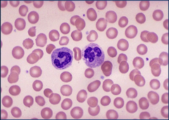
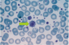
Banded (left) and segmented (right) neutrophil |
|
|
|
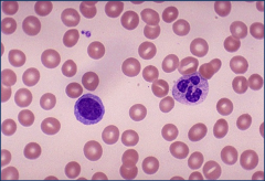
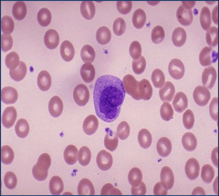
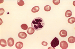
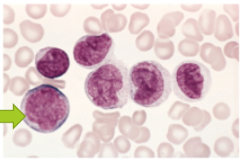
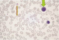
Lymphocyte (left) |
|
|
|
Eosinophil (right) |
|
|
|
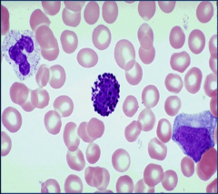
Basophil (center) |
|
|
|
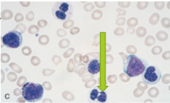
Monocyte |
|
|
|
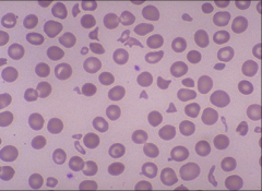
Reticulocytes |
|
|
|
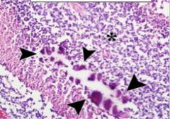
Marrow smear from a patient withhemolytic anemia. Increased numbers of maturing erythroidprogenitors (normoblasts) |
|
|
|
Spherocyte |
|
|
|
Bite cell (from G6PD deficiency) |
|
|
|
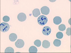
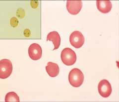
Sickle cells (showing anisocytosis and poikilocytosis) |
|
|
|
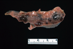
Autoinfarcted spleen remnant (from SCD) |
|
|
|
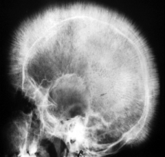
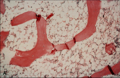
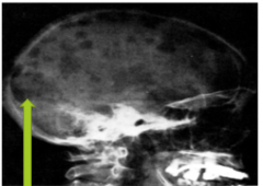
Crew cut appearance of skull (from beta thalassemia) |
|
|
|
Reticulocyte fragments (schistocytes) from microangiopathic anemia |
|
|
|
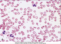
Hypersegmented neutrophil seen in megaloblastic anemia |
|
|
|
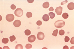
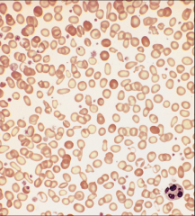
Iron deficiency anemia (hypochromic and small RBC's) |
|
|
|
Hypocellular bone marrow (from aplastic anemia) |
|
|
|
Thrombocytopenia (from aplastic anemia) |
|
|
|
Intravascular thrombi (from thrombotic microangiopathies) |
|
|
|
Fragmented RBC's (schistocytes) from hemolytic uremic syndrome |
|
|
|
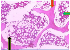
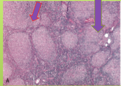
Bone marrow showing adipocytes (black arrow), bony trabeculae (red arrow), hematopoietic marrow elements (green arrow) |
|
|
|
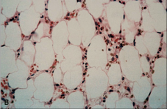
Hyperplastic bone marrow (fat cells disappear in this condition) |
|
|
|
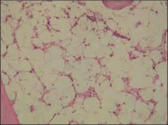
Hypocellular bone marrow (incr fat is seen) |
|
|
|
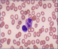
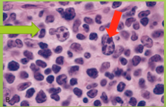
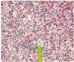
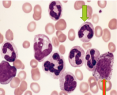
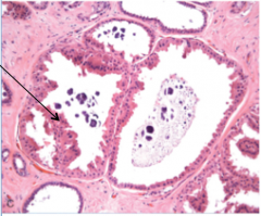
Reactive changes in neutrophils in agranulocytosis; you can see the Dohle bodies (arrow) which represent dilated ER |
|
|
|
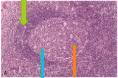
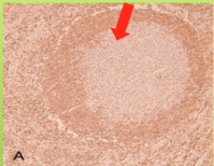
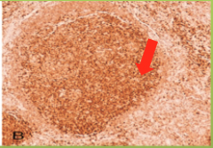
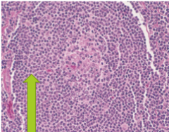
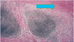
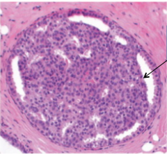
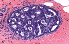
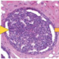
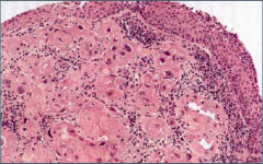
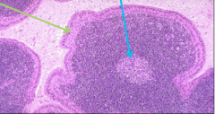
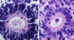
Reactive follicle (caused by follicular hyperplasia seen in chronic nonspecific lymphadenitis) showing dark staining mantle zone (green arrow), light zone (blue arrow), and dark zone (orange arrow) |
|
|
|
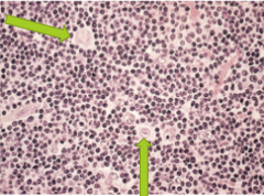
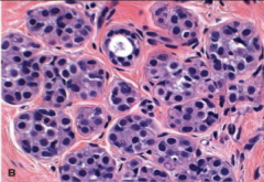
Reactive follicle (caused by follicular hyperplasia seen in chronic nonspecific lymphadenitis) showing mitotic figures & numerous macros (green arrow) containing phagocytosed apoptotic cells (tingible bodies). |
|
|
|
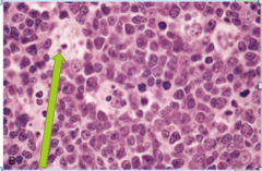
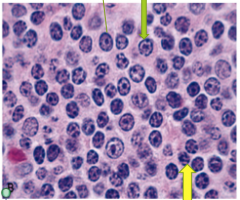
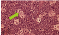
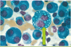
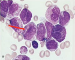
ALL (arrow points to lymphoblast which have little cytoplasm, lack peroxidase, small nucleoli, condensed nuclear c'tin) |
|
|
|
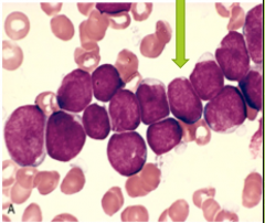
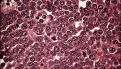
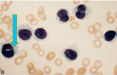
AML (arrow points to myeloblast which have more voluminous cytoplasm, prominent nucleoli, azurophilic cytoplasmic granules) |
|
|
|
SLL/CLL lymph node; arrow shows diffuseeffacement of nodal architecture |
|
|
|
SLL/CLL lymph node; green arrow shows pro lymphocytes (larger cells with centrally placed nucleoli) and yellow arrow shows small round lymphocytes |
|
|
|
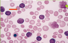
SLL/CLL; green arrow shows smudge cells and red arrow shows spherocytes |
|
|
|
Follicular lymphoma (arrow shows nodules) |
|
|
|
Arrow points to an absent BCL2, indicating it is not a follicular lymphoma |
|
|
|
Arrow points to BCL2, indicating it is a follicular lymphoma |
|
|
|
Follicular lymphoma; green arrow points to centrocytes, red arrow points to centroblasts |
|
|
|
DLBCL (arrow shows rapidly enlarging, symptomaticmass at a nodal or extra nodal site) |
|
|
|
DLBCL (arrow shows large and prominent nuclei) |
|
|
|
Burkitt lymphoma (arrow shows macros with clear space around them, representing 'starry-sky' appearance) |
|
|
|
Burkitt lymphoma (high mitotic index, multiple small nucleoli) |
|
|
|
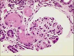
Skull in multiple myeloma; arrow shows punched out bone lesion |
|
|
|
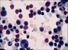
Bone marrow in multiple myeloma; plasma cells fill BM with arrow showing multiple nuclei |
|
|
|
Bone marrow in multiple myeloma; plasma cells fill BM with arrow showing cytoplasmic droplets containing Ig |
|
|
|
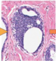
Mantle cell lymphoma; image shows neoplastic lymphoid cells surrounding a small, atrophic germinal center (a mantle zone pattern of growth) |
|
|
|
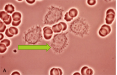
Hairy cell leukemia; arrow shows fine hair-like cytoplasmic projections and extensions |
|
|
|
Hairy cell leukemia; arrow shows stained smears, roundor folded nuclei & agranularcytoplasm |
|
|
|
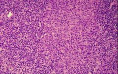
Hodgkin lymphoma; shows Reed Sternberg cells |
|
|
|
Hodgkin lymphoma; arrow shows mononuclear variant |
|
|
|
Nodular sclerosis (HL); arrow shows lacunar variant |
|
|
|
Hodgkin lymphoma; arrow shows lymphohistocytic variant |
|
|
|
Nodular sclerosis (HL); arrow shows collagen band dividing the lymphoid tissue into nodules |
|
|
|
Mixed cellularity (HL); arrow shows diagnostic RS cells |
|
|
|
Lymphocyte predominant (HL); arrows show lymphocytic and histocytic variants |
|
|
|
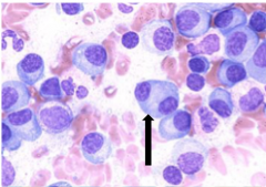
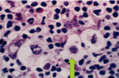
AML w/ t(15:17); arrow shows aurer rods |
|
|
|
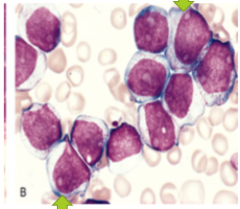
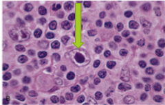
AML w/ monocytic differentiation; arrow shows monoblast |
|
|
|
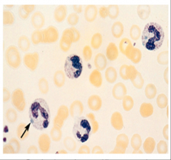
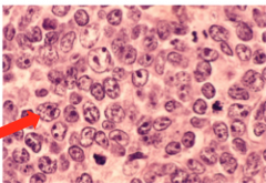
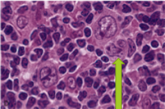
Myelodysplastic syndrome; arrow shows neutro with 2 nuclear lobes |
|
|
|
CML; green arrow shows mature neutrophil and red arrow shows immature neutrophil |
|
|
|
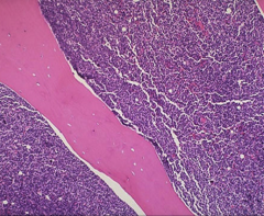
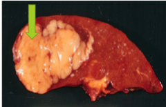
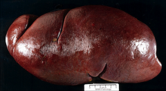
Polycythemia vera spent phase; massive splenomegaly due to EMH in the setting of advanced marrow myelofibrosis |
|
|
|
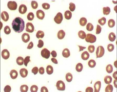
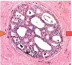
Essential thrombocytosis; peripheral blood smear shows markedthrombocytosis, including giant platelets |
|
|
|
Primary myelofibrosis; green arrow shows nucleated erythroid precursor and orange arrow shows tear drop shaped RBC's (dacryocytes) |
|
|
|
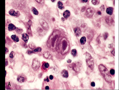
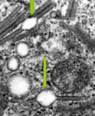
Langerhans histiocytosis; arrow shows birbeck granules |
Cd-1a positive and S-100 positive |
|
|
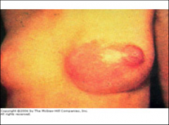
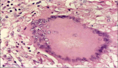
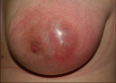
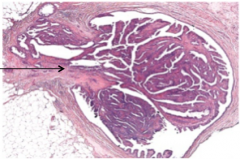
Acute mastitis |
|
|
|
Acute mastitis; black dots represent neutrophils |
|
|
|
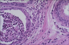
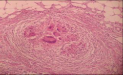
Granulomatous mastitis |
|
|
|
Granulomatous mastitis |
|
|
|
Periductal mastitis (Zuska disease) |
|
|
|
Mammary duct ectasia; arrow shows dilated ducts filled with lipid-laden macros |
|
|
|
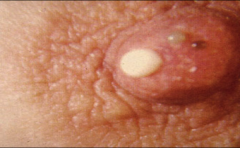
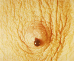
Mammary duct ectasia (white discharge from nipple) |
|
|
|
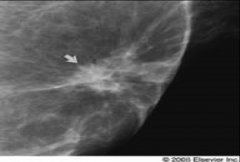
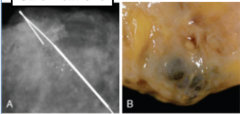
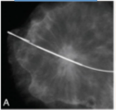
Fat necrosis; picture shows speculated lesion on mammogram |
|
|
|
Fat necrosis; picture shows liquefactive necrosis and foamy lipid filled macros |
|
|
|
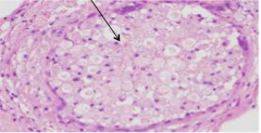
Non proliferative breast cyst (fibrocystic); arrow shows apocrine metaplasia |
|
|
|
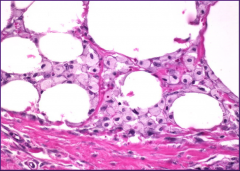
Non proliferative breast cyst (fibrocystic); picture shows calcifications |
|
|
|
Epithelial hyperplasia; arrow shows irregular slit-like lumen |
|
|
|
Sclerosing adenosis |
|
|
|
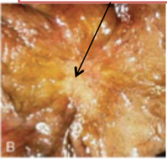
Complex sclerosing lesion; picture shows central scar |
|
|
|
Complex sclerosing lesion; arrow shows solid irregular mass |
|
|
|
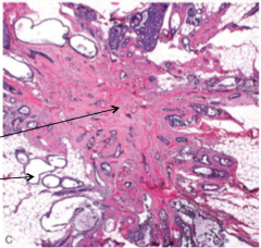
-Complex sclerosing lesion; -Top arrow shows central nidus of small tubules entrapped in dense fibrous stroma -Bottom arrow shows cysts/hyperplasia |
|
|
|
Papilloma; blood stained discharge |
|
|
|
Papilloma |
|
|
|
Atypical ductal hyperplasia |
|
|
|
Atypical lobular hyperplasia |
|
|
|
Normal/non proliferative breast |
|
|
|
Proliferative disease of breast |
|
|
|
Atypical hyperplasia of breast |
|
|
|
Carcinoma in situ of breast |
|
|
|
Invasive carcinoma of breast |
|
|
|
Fibroma; yellow arrow showing the fibrous tissue and green arrow showing squamous mucosa |
|
|
|
Fibroma; arrow shows ulcerated nodular fibroma |
|
|
|
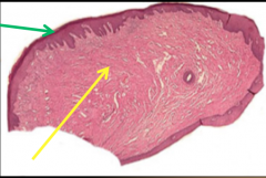
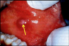
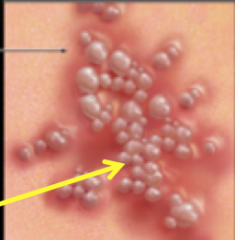
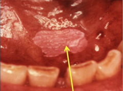
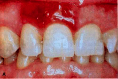
Aphthous ulcer (canker sores); top arrow shows hyperemic ulcer, bottom arrow shows narrow zone of erythema |
|
|
|
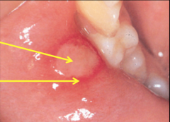
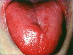
Glossitis |
|
|
|
Oral herpes; arrow shows abrupt onset of vesicles and ulcers |
|
|
|
Oral herpes; arrow shows abrupt onset of vesicles and ulcers |
|
|
|
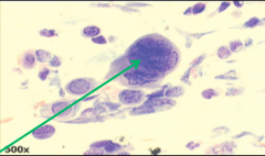
Oral herpes; arrow shows multinucleated cells from the Tzanck test |
|
|
|
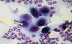
Oral herpes; arrow shows intranuclear inclusions cells from the Tzanck test |
|
|
|
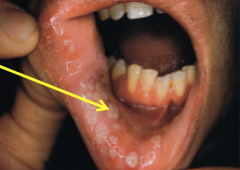
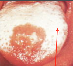
Oral candidiasis; arrow shows pseudomembrane |
|
|
|
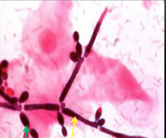
Oral candidiasis; green arrow shows oval yeast like budding cells and yellow arrow shows pseudohyphae |
|
|
|
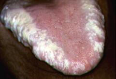
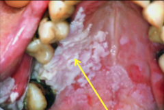
Hairy leukoplakia; white confluent patches on the lateral border of tongue |
|
|
|
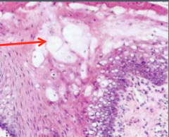
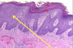
Hairy leukoplakia; arrow shows ballooning of squamous cells in upper epithelium |
|
|
|
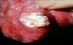
Leukoplakia; arrow shows homogenous area (uniformly white) |
|
|
|
Leukoplakia; arrow shows speckled leukoplakia (white and red) and also known as leukoerythroplakia |
|
|
|
Leukoplakia; arrow shows verrucousleukoplakia – corrugated/nodular |
|
|
|
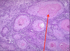
Leukoplakia; arrow shows hyperkeratosis and thickened, acanthoticepithelium |
|
|
|
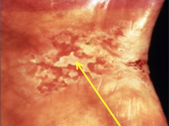
Erythroplakia; shows red velvet slightly depressed plaque |
|
|
|
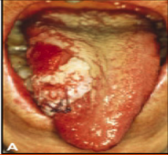
Oral cancer; picture shows proliferative mass |
|
|
|
Oral cancer; arrow shows keratin pearls |
|
|
|
Oral cancer; picture shows ulcerated mass |
|
|
|
Nasopharyngeal undifferentiated carcinoma; non neoplastic lymphoid cells with vesicular nuclei and prominent nucleoli in syncitial |
|
|
|
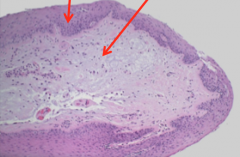
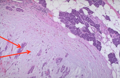
Larynx; left arrow shows keratotic, hyper plastic epithelium and right arrow shows loose myxoid CT core |
|
|
|
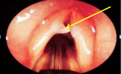
Larynx; arrow points to vocal cord nodules |
|
|
|
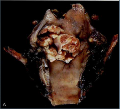
Carcinoma larynx |
|
|
|
Carcinoma larynx |
|
|
|
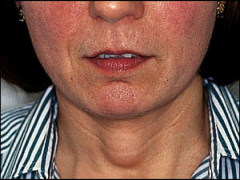
Branchial cyst |
|
|
|
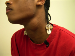
Thyroglossal cyst |
|
|
|
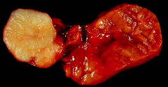
Paraganglioma (carotid body tumor); picture shows zellballen |
|
|
|
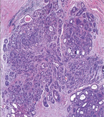
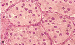
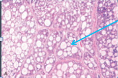
Pleomorphic adenoma; picture shows myxoidareas and chondroid areas |
|
|
|
Pleomorphic adenoma; top arrow shows epithelial/myoepithelial cells and bottom arrow shows mesenchyme like stroma |
|
|
|
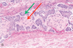
Warthin's tumor; green arrow shows double cell layer and blue arrow shows stroma (mature lymphoid follicles with germinal center) |
|
|
|
Mucoepidermoidcarcinoma; blue arrow shows mucuscells secreting mucus and red arrow shows squamous cells |
|
|
|
Mucoepidermoid carcinoma; picture shows mucicarminestains the mucin reddish pink |
|
|
|
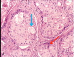
Adenoid cystic carcinoma; arrow shows small cells – tubular, solid &cribriform pattern |
|
|
|
Adenoid cystic carcinoma; red arrow shows the nerve which hasbeen invaded by malignant cells shown by green arrow |
|
|
|
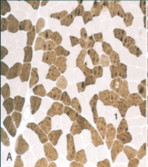
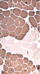
Normal muscle with checker board distribution of type 1 (light) and type 2(dark) fibers |
|
|
|
Type grouping of muscle fibers in re-innervation and regeneration |
|
|
|
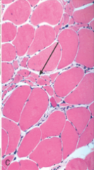
Clustered atrophic muscle fibers in denervation (arrow) |
|
|
|
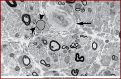
Peripheral neuropathy in adult onset of DM; arrow head shows loss of myelinated fiber, arrow shows thickeningof endoneurialvessel wall |
|
|
|
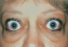
Propoptosis |
|
|
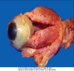
|
Propoptosis |
|
|
|
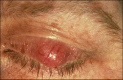
Chalazion; picture shows meibomian gland |
|
|
|
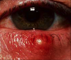
Stye; picture shows infected eyelid |
|
|
|
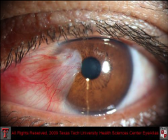
Pterygium (conjuctiva infection); fibrovasc CT invading cornea. Resembles a fan shape |
|
|
|
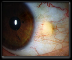
Pinguecla (conjuctiva infection); picture shows small yellow discoloration. No cornea invasion |
|
|
|
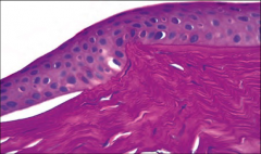
Keratinous; picture shows thinningofthe corneaw/ breaks in Bowman |
|
|
|
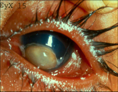
Keratomalacia; picture shows xerosis and bitot spot |
|
|
|
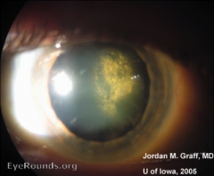
Lens cataract; picture shows glare |
|
|
|
Iris melanoma; picture shows pigmented mass |
|
|
|
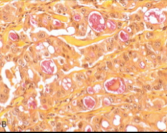
Epithelial melanoma cells; largecells, vesicular nucl; prominent nucleolus, melanin pigment+ |
|
|
|
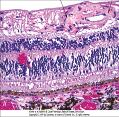
Hypertensive retinopathy; picture shows vein compressed where the sclerotic arteriole crosses over it |
|
|
|
Non-proliferativediabetic retinopathy; b/w the arrows shows tangleof abnormal vessels lying just beneath the internal limiting membrane of theretina |
|
|
|
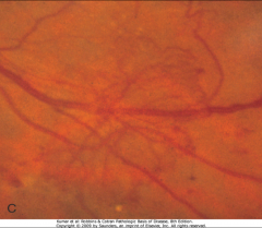
Proliferative diabetic retinopathy; picture is anex of intraretinalangiogenesis known asintraretinalmicroangiopathy(IRMA) |
|
|
|
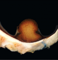
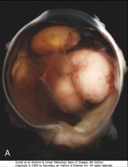
Retinoblastoma |
|
|
|
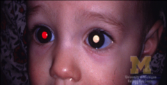
Retinoblastoma; red eye reflex seen in normal retina, white means there is something wrong |
|
|
|
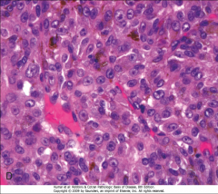
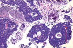
Retinoblastoma; red arrow shows Flexner-Wintersteinerrosettes, black arrow shows smallblue cells-round with hyperchromaticnuclei, scanty cytoplasm |
|
|
|
Retinoblastoma; shows necrosis and dystrophic calcification |
|
|
|
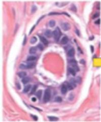
Flexner-Wintersteiner rosette; seen in retinoblastoma. Picture shows central lumen around which the cellsaggregate (Gives a flower appearance aka fleurretes) |
|