Reading...
![]()
Play button
![]()
Play button
![]()
Use LEFT and RIGHT arrow keys to navigate between flashcards;
Use UP and DOWN arrow keys to flip the card;
H to show hint;
A reads text to speech;
11 Cards in this Set
- Front
- Back
|
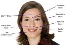
Nasolabial folds
|
|
|
|
Canthus of eye
|

|
|
|
outer canthus of eye
|
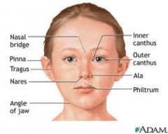
medial canthus (endocanthion)
lateral canthus (exocanthion) |
|
|
Periorbital edema
|
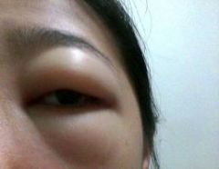
the appearance of swelling in the tissues around the eyes, called the orbits. It is almost exclusively caused by fluid buildup around the eyes, or periorbital edema. Minor lower periorbital edema is often called eye bags.
|
|
|
Cornea
|
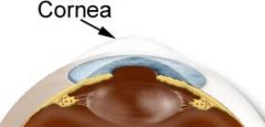
transparent front part of the eye that covers the iris, pupil, and anterior chamber. Together with the lens, the cornea refracts/ bends light rays entering the eye
|
|
|
Pupil
|
Where light enters the eye
Surrounded by the iris |
|
|
Limbus
|
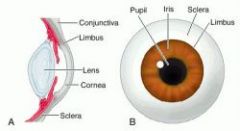
junction b/t the cornea and the sclera
|
|
|
iris
|

muscles of the iris control the aperature of the pupil and controls the amount of light that enters the retina
|
|
|
Lacrimal Puncta
|
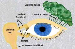
There are two lacrimal puncta in the medial (inside) portion of each eye.
Together, they function to collect tears produced by the lacrimal glands. The fluid is conveyed to the lacrimal sac, and thence via the nasolacrimal duct to the inner nose. |
|
|
Lacrimal Puncta
|
There are two lacrimal puncta in the medial (inside) portion of each eye.
Together, they function to collect tears produced by the lacrimal glands. The fluid is conveyed to the lacrimal sac, and thence via the nasolacrimal duct to the inner nose. |
|
|
Lacrimal Puncta
|

There are two lacrimal puncta in the medial (inside) portion of each eye.
Together, they function to collect tears produced by the lacrimal glands. The fluid is conveyed to the lacrimal sac, and thence via the nasolacrimal duct to the inner nose. |