![]()
![]()
![]()
Use LEFT and RIGHT arrow keys to navigate between flashcards;
Use UP and DOWN arrow keys to flip the card;
H to show hint;
A reads text to speech;
38 Cards in this Set
- Front
- Back
- 3rd side (hint)
|
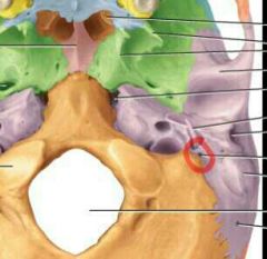
What bones make up the cranial bones? |
Frontal bone 2 parietal bones 2 temporal bones Occipital bone Sphenoid bone Ethmoid bone |
|
|
|
Which bones make up the facial bones? |
2 nasal bones 2 maxillae 2 zygomatic bones Mandible 2 lacrimal bones 2 palatine bones 2 inferior nasal conchae Vomer |
|
|
|
What three components make up the nasal septum? |
the Vomer Septal cartilage the perpendicular plate of the ethmoid bone |
|
|
|
Nasal septum |
The vertical partition that divides the left and right side of the nasal cavity |
|
|
|
How do the three components of enable septum work together? |
The anterior border of the vomer articulates with the septal cartilage, which is hyaline cartilage, to form the interior portion of the septum. The superior border of the vomer articulates with the perpendicular plate of the ethmoid bone to form the remainder of the nasal septum. |
|
|
|
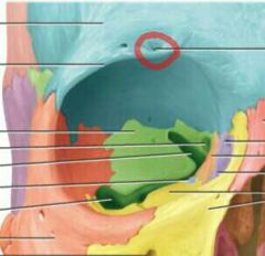
What are the seven bones that make up the orbital cavity? |
Frontal Sphenoid Ethmoid Palatine Zygomatic Lacrimal Maxilla |
Fella saw Mola entertain long pale zanthropods. |
|
|
What makes up the roof of the orbit? |
Parts of the frontal and sphenoid bones |
Roofs shouldn't fall. |
|
|
What makes up the lateral wall of the orbit? |
parts of the zygomatic and sphenoid bones |
Walls shouldn't zigzag |
|
|
What makes up the floor of the orbit |
Parts of the maxilla, zygomatic, and paletine bones |
|
|
|
What makes up the medial wall of the orbit? |
Parts of the maxilla, lacrimal, and sphenoid bones |
Floor placement might zigzag. |
|
|
Superior orbital fissure |
At the superior lateral angle of the apex |
|
|
|
Inferior orbital fissure |
At the junction of the lateral wall and floor |
|
|
|
Suture |
And in movable joint in most cases in an adult skull that holds most skull bone together. Sutures in the skulls of infants and children, however, often are movable and function as an important growth center in the developing skull |
|
|
|
Coronal suture |
Unites the frontal bone and both parietal bones |
|
|
|
Sagittal suture |
Unite the two parietal bones on the superior midline of the skull. The saggital suture is so named because in the infant, before the bones of the skull are firmly united, the suture and the fontanels associated with it resemble an arrow |
|
|
|
Lambdoid suture |
Unite the two parietal bones to the occipital bone. This suture is so named because of its resemblance to the capital Greek letter lambda, sutural bones may occur within the sagittal and lambdoid sutures. |
|
|
|
Squamous sutures |
Unites the parietal and temporal bones on the lateral aspect of the skull |
|
|
|
Paranasal sinuses |
Cavities within certain cranial and facial bones near the nasal cavity. they are most evident in a sagittal section of the school. They are lined with mucous membranes that are continuous with the lining of the nasal cavities. secretions produced by the mucous membrane of the paranasal sinuses drain into the lateral wall of the nasal cavity. |
|
|
|
Which bones make up the sinus cavity? |
Frontal Sphenoid Ethmoid Maxillae |
|
|
|
Anterior fontanelle |
The largest fontanelle, it is located at the midline among the two parietal bones and the frontal bone, and is roughly shaped like a daimond. And usually closes 18 to 24 months after birth. |
|
|
|
Posterior fontanelle |
Is located at the midline among the two parietal bones and the occipital bone. Because it is much smaller than the anterior fontanelle, it generally closes about two months after birth. |
|
|
|
Paired anterolateral fontanels |
Located laterally amongst the frontal, parietal, temporal, and sphenoid bones, are small and irregular in shape. Normally, they close about 3 months after birth |
|
|
|
Paired posterolateral fontanels |
Located laterally among the parietal, occispital, and temporal bones, are irregularly shaped. They begin to close one to two months after birth, but closure is generally not complete until 12 months. |
|
|
|
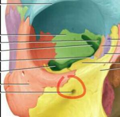
Carotid Foramen |
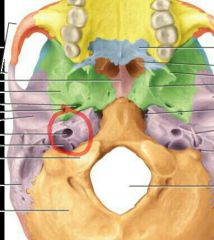
(relating to carotid artery in the neck) Petrous portion of temporal bone Structures passing through: internal carotid artery, sympathetic nerves for eyes |
|
|
|
Hypoglossal Foramen |
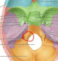
(Hypo - under, glossus - tongue) Superior to base of occispital condyles Structures passing through: cranial nerve XII , hypoglossal, branch of ascending pharyngeal artery |
|
|
|
Infraorbital foramen |
(Infra - below)
Inferior to orbit in maxilla Structures passing through: infraorbital nerve and blood vessels, branch of maxillary division of cranial nerve V (trigeminal) |
|
|
|
Jugular foramen |
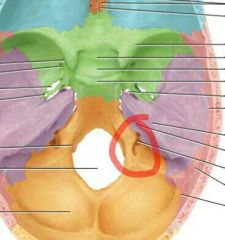
(Jugul - throat) Posterior to carotid canal between petrous portion of temporal bone and occipital bone Structures passing through: internal jugular vein; cranial nerves IX (glossopharyngeal), X (vagus), XI (accessory) |
|
|
|
Lacerum foramen |
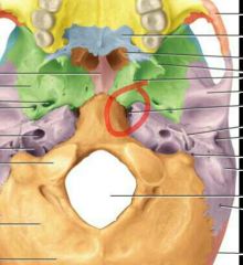
(Lacerum - lacerated) Bounded anteriorly by sphenoid bone, posteriorly by petrous portion of temporal bone, medially by sphenoid and occipital bones Structures passing through: branch of ascending pharyngeal artery |
|
|
|
Magnum |
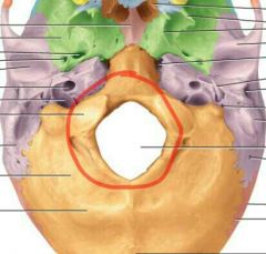
(Large) Occipital bone Structures that pass through: medulla oblongata and its membranes (meninges), cranial nerve XI (accessory), vertebral and spinal arteries |
|
|
|
Mandibular |
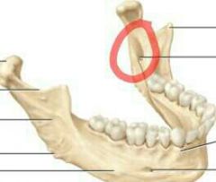
(Mand - to chew)
Medial surface of ramus of mandible Structures that pass through: inferior alveolar nerve and blood vessels Structures that pass through: inferior alveolar nerve and blood vessels |
|
|
|
Mastoid |
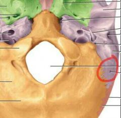
(Breast shaped) Posterior border of mastoid process of temporal bone Structures that pass through: emissary vein to transverse sinus, branch of occipital artery to dura mater |
|
|
|
Mental Foreman |
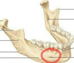
(Ment - chin)
Inferior to second premolar tooth in mandible Structures that pass through: mental nerve and vessels Structures that pass through: mental nerve and vessels |
|
|
|
Olfactory foramen |
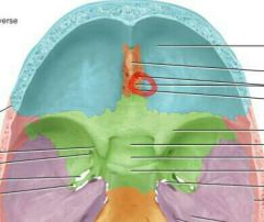
(Olfact - to smell)
Cribriform plate of ethmoid bone Structures that pass through: cranial nerve I (olfactory) Structures that pass through: cranial nerve I (olfactory) |
|
|
|
Optic foramen |
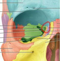
(Eye) Between superior and inferior portions of small wings of sephmoid bone Structures that pass through: cranial nerve II (optic), ophthalmic artery |
|
|
|
Ovale foramen |
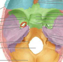
(Oval) Greater wing of sphenoid bone Structures that pass through: mandibular branch of cranial nerve V (trigeminal) |
|
|
|
Rotundrum foramen |

(Round) Junction of anterior and medial part of sphenoid bone Structures that pass through: maxillary branch of cranial nerve V (trigaminal) |
|
|
|
stylomastoid foramen |
(Stylo - stake or pole) Between styloid and mastoid processes of temporal bone Structures that pass through: cranial nerve VII (facial), stylomastoid artery |
|
|
|
Supraorbital foramen |
(Supra - above) Supraorbital margin of orbit in frontal bone Structures that pass through: supraorbital nerve and artery |
|