![]()
![]()
![]()
Use LEFT and RIGHT arrow keys to navigate between flashcards;
Use UP and DOWN arrow keys to flip the card;
H to show hint;
A reads text to speech;
142 Cards in this Set
- Front
- Back
- 3rd side (hint)
|
1 List vascular tumors? (6) |
1 Angiosarcoma |
|
|
|
1 List extraskeletal osseous and cartilaginous tumors? (3) |
1 Extraskeletal chondrosarcoma, extraskeletal osteosarcoma, myositis ossificans |
|
|
|
Extraskeletal chondrosarcoma |
1 well diff, myxoid, mesenchymal |
|
|
|
Extraskeletal osteosarcoma: (histologically identical to bone version) |
1 50s +, M=W |
|
|
|
Myositis ossificans |
1 young 2nd 3rd decades active males; upper arms and thighs, trauma |
|
|
1 List fibrohistiocytic tumors? (10) |
1 Atypical fibroxanthoma |
|
|
|
Atypical fibroxanthoma |
1 Superficial malignant fibrous histiocytoma; superficial form of pleomorphic MFH |
|
|
Dermatofibrosarcoma protuberans |
1 Bland monomorphic spindled cells in distinct storiform pattern involving dermis and subcutis (checkerboard pattern of fat infiltration) |
|
|
Fibrous histiocytoma |
1 Dermatofibroma |
|
|
Giant cell fibroblastoma: |
1 <5yrs male; dermal/subcutis of thigh, inguinal region or chest wall |
|
|
Jeuvenile xanthogranuloma |
1 infants (<6m), head and neck (50%) |
|
|
1 Considered soft tissue counterpart of PVNS? aka? |
1 GCT of tendon sheath, diffuse type aka malignant GCT of tendon sheath because may represent extra articular extension. |
|
|
Pleomorphic malignant fibrous histocytoma |
|
|
|
Tenosynovial GCT, diffuse type |
1 Proliferative synovitis, florid synovitis, extra-articular PVNS, PVN bursitis, malignant GCT of tendon sheath (as described earlier) |
|
|
Tenosynovial giant cell tumor, localized type |
1 Nodular tenosynovitis, giant cell tumor of tendon sheath |
|
|
Xanthoma |
1 skin and subcutis, hyperliproproteinemia (PBC or DM) |
|
|
1 Tendinous xanthoma associated with what AR disease? |
1 cerebrotendinous xanthomatosis |
|
|
|
List fibroblastic/myofibroblastic tumors ? (13) |
1 Calcifying aponeurotic fibroma |
|
|
|
Calficying aponeurotic fibroma |
1 kids and teens; hand/feet, <3cm |
|
|
Elastofibroma |
1 >55yrs W>>M (associated with repetitive manual labor) deep seated on back ribs 6, 7, 8 or scapula R>L |
|
|
/Fibroma of tendon sheath |
1 20-50 M>W, hand |
|
|
Fibromatosis |
1 adults; |
|
|
Facts bout fibromatosis? |
1 toward the side of the lesion |
|
|
|
Fibrosarcoma |
1 adult and juvenile/infantile |
|
|
Infantile fibrosarcoma |
1 hemagiopericytoma like pattern with dilated vessels and fibrin thrombi and myxoid changes |
|
|
Infantile digital fibromatosis |
1 Inclusion body fibromatosis |
|
|
Inflammatory myofibroblastic tumor |
1 (extrapulmonary) inflammatory pseudotumor, inflammatory fibrosarcoma and plasma cell granuloma |
|
|
Low grade fibromyxoid sarcoma |
1 young adultss (35) on trunk and proximal extremities |
|
|
Low grade myofibroblastic sarcoma |
1 adults M>F, thigh or H&N especially tongue |
|
|
Myofibroma 1 Patient population and location? |
What the f |
|
|
Myxoinflammatory fibroblastic sarcoma |
1 inflammatory myxohyaline tumor of distal extremities wiht virocytes or reed sternburg like cells and acral MFS |
|
|
Myxofibrosarcoma |
1 elderly (70-80), superficial > deep extremities lower> upper |
|
|
Nodular fasciitis |
1 young adults (20-50) upper extremities or trunk; rapidly growing but <3cm |
|
|
1 List lipomatous tumors? (4) |
1 Hibernoma |
|
|
|
Hibernoma |
1 young adults (25yrs) on shoulder mediastium, thigh, axilla and inguinal region |
|
|
Lipoblastoma |
1 lipoblastomatosis (because usually localized in subcutis) |
|
|
Lipoma |
1 50-60, M>W any location but usually upper half of body on trunk or neck |
|
|
Fibrolipoma 1.describe histolog including differrentiaton from spindle cell lipoma? |
1. prominent bundles of mature bland fibrous tissue traversing mature adipose lobules, does not have the vascular pattern and thick collen bundles of spindle cell lipoma. |
|
|
Chondroid lipoma |
1 women |
|
|
spindle cell lipoma |
1 shoulder and posterior neck of older men (>40) |
|
|
Pleomorphic lipoma |
1 spindle cell lipoma |
|
|
1 Multiple lipomas are associated with what 4 syndromes? describe each, multiple lipomas + ? |
1 |
|
|
|
Liposarcoma |
1 40-60 yrs M>W, lower extremities and retroperitoneum |
|
|
|
Well differentiated/atypical lipomatous tumor |
1 variably sized mature adipocytes separated by fibrous bands with atypical, multinucleated hyperchromatic stromal cells |
|
|
Myxoid |
1 younger adults than other liposarcs; myxoid on extremities almost never retroperitoneal |
|
|
Round cell |
1 extremities with frequent mets to retroperitoneum and lungs, very agressive |
|
|
Pleomorphic liposarcoma |
1 Radiation therapy or NF |
|
|
Dedifferentiated liposarcoma |
1 retroperitoneum and groin |
|
|
1 List lymphatic tumors? (3) |
1 Lymphangioma(tosis) |
|
|
|
Lymphangioma |
1 50% congenital, 90% <2yrs |
|
|
1 Cystic hygromas associated with? (5) |
1 Turner syndrome |
|
|
|
Lymphangiomatosis |
1 visceral organs diffuse or multifocally |
|
|
|
Lymphangiomyoma(tosis) |
1 one is local one is diffuse |
|
|
1 lymphangioma(tosis) is associated with? |
1 Tuberous sclerosis |
|
|
|
Lymphangiosarcoma |
1 long standing chronic lymphedema, s/p axillary node dissection/radiation (with mastectomy) |
|
|
1 List myogenic tumors ? (4) |
1 Leiomyoma |
|
|
|
Leiomyoma |
1 Cutaneous (leiomyoma cutis) |
|
|
|
Cutaneous leiomyoma |
1 erector pili muscle |
|
|
Vascular leiomyoma (angioleiomyoma) |
1 outer muscular wall of arteries |
|
|
Intravenous leiomyomatosis |
1 uterine or pelvic veins even the heart |
|
|
Leiomyomatosis peritonealis disseminata |
1 throughout peritoneum, pregnant black women |
|
|
|
Leiomyosarcoma |
1 adult W>M |
|
|
Epitheloid leiomyosarcoma |
1 uterus; malignant leiomyoblastoma (spp in stomach) |
|
|
Myxoid leiomyosarcoma |
1 >75% in women, uterus |
|
|
Sub-/cutaneous leiomyosarcomas |
1 Middle aged men 2-3x > W |
|
|
Rhabdomyoma |
1 Rhabdomyosarcoma |
|
|
|
Cardiac Rhabdomyoma |
1 pediatric, usually multifocal (ventricles and septum), represents 50-90% of primary heart tumors in kids |
|
|
Adult rhabdomyoma |
1 M>W >40yrs, head and neck, 65% occur in mucosa of oropharynx, nasopharynx or larynx |
|
|
Fetal type rhabdomyoma |
1 myxoid and cellular |
|
|
|
Myxoid fetal rhabdomyoma |
1 bundles or fascicles of immature skeletal muscle and found/oval mesenchymal cells with myxoid stroma; skeletal component matures toward peripheray; may have pseudocambium layer of plasma cells beneath epithelium. |
|
|
Cellular fetal rhabdomyoma |
1 fasicles or plexiform arrangment of muscle cells at various stages of maturation with sparse collagenous or myxoid stroma; may have ganglion like rhabdomyomblasts with prominent nucleoli or strap cells |
|
|
Genital type rhabdomyoma |
1 middle aged women vagina and vulva < cervix |
|
|
1 The most common sarcoma of kids and adolescents? |
1 Rhabdomyosarcoma |
|
|
|
Embryonal NOS |
1 GU |
|
|
Botryoid (embryonal) rhabdomyosarcoma |
1 "grape like" gross appearance |
|
|
Spindle cell (embryonal) rhabdomyosarcoma |
1 paratesticular |
|
|
Anaplastic (embryonal) rhabdomyosarcoma |
1 lower extremities |
|
|
Alveolar rhabdomyosarcoma |
1 older (teens) and occurs on extremities more than embryonal |
|
|
Pleomorphic rhabdomyoscarcoma |
1 adults men >40yrs in deep lower extremity |
|
|
1 List neuroectodermal tumors? (14) |
1 Clear cell sarcoma |
|
|
|
Clear cell sarcoma |
1 Malignant Melanoma of soft parts |
|
|
Extraskeletal Ewing Sarcoma/PNET |
1 Young adults (M>W), paravertebral and chest wall |
|
|
Granular cell tumor |
1 W>M 4-6th decades, dermis, or submucosa, tongue isclassic location but can occur anywhere |
|
|
Malignant peripheral nerve sheath tumor |
1 von Recklinghausen disease aka NF1, and radiation |
|
|
Melanocytic neuroectodermal tumor of infancy |
1 Retinal analage tumor, melanotic progonoma, pigmented NED tumor of infancy |
|
|
Melanotic schwanomma |
1 spinal/midline 4th decade |
|
|
Merkel cell carcinoma |
1 a neuroendocrine carcinoma of the skin |
|
|
1 List 5 clinical variants of neurofibroma? |
1 Localized cutaneous, diffuse cutaneous, localized intraneural, plexiform, massif soft tissue |
|
|
|
1 Micro common to all neurofibromas? |
1 spindeled schwann cells with serpentine/ comma shaped nuclei and fibroblasts in fascicles whorls or storiform pattern in its own mucopolysacharide rich matrix mixed wtih strands of "shredded carrot" collagen |
|
|
1 Neurofibromatosis type 1, list diagnostic criteria? |
1 >2 of any of the following: |
|
|
|
1 Neurofibromatosis type 2, list criteria? |
1 bilateral 8th cranial nerve |
|
|
|
Wagner Meissner bodies, fequently seen in the NFs below |
1 localized and diffuse cutaneous |
|
|
1 List 5 variants of neuroma? |
1 Traumatic |
|
|
|
Traumatic neuroma |
1 tangled web of random nerve bundles |
|
|
Morton's neuroma |
1 Morton's metatarsalgia and localized interdigital neuritis |
|
|
mucosa neuroma |
1 MEN2b |
|
|
Palisaded encapsulated neuroma |
1 solitary circumscribed neuroma |
|
|
Neurothekeoma (myxoid) |
1 kids or young adults, in dermis of head/neck or shoulders |
|
|
perineurioma |
1 intra or extra neural |
|
|
Primitive neuroectodermal tumor |
1 peripheral neuroepithelioma |
|
|
schwannoma |
1 neurilemmoma |
|
|
ancient schwannoma |
1 longstanding duration |
|
|
Cellular schwannoma |
1 composed exclusively of Antoni A areas with pleomorphism, 10% have necrosis |
|
|
1 List tumors of uncertain histiogenesis ? (12) |
1 Alveolar soft part sarcoma |
|
|
|
Alveolar soft part sarcoma |
1 young adults F>M (15-35), deep soft tissue lower extremities, oral cavity mediastium |
|
|
Angiomatoid fibrous histocytoma |
1 ares of normal lymph nodes, in young adults and kids |
|
|
desmoplastic small round cell tumor |
1 young adults (15-35) abdominal, pelvic or peritnoeal |
|
|
Epithelioid sarcoma |
1 young adults, distal extremities especially hands |
|
|
Fibrous hamartoma of infancy |
1 <2yrs or ~20% at birth, B>G, usually in axilla also extremities inguinal and trunk but NEVER hannds or feet |
|
|
Giant cell angiofibroma 1 Patient population and location? 2 histo? 3 positive IHC? which cells? 4 Prognosis?
|
1 middle aged adults, eyelid or orbit 2 admixture of round/spindled cells with indistinct cytoplasm and myxocollagenous stroma with small/medium ectatic blood vessels lined by variable giant cells 3 CD34 and CD99 in stroma 4 good, excision curative |
|
|
Hemangiopericytoma (on spectrum or same as Solitary Fibrous Tumor) 1 Patient population and location? 2 histo? 3 positive/negative IHC? 4 Molecular alteration?
|
1 middle aged adults; deep seated in thigh, pelvic retroperitoneum and orbit 2 uniform round/spindled cells with scattered ectatic "staghorn" vessels 3 CD99 and CD34 (variable)/ CD31 neg 4 12q13-15 alterations
|
|
|
1 List 7 tumors with a "hemangiopericytoma-like vascular pattern" |
1 synovial sarcoma fibrous histocytoma mesenchymal chondrosarcoma Extrapleural SFT myofibroma leiomyosarcoma Endometrial stromal sarcoma |
|
|
|
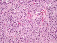
Malignant extrarenal rhabdoid tumor 1 patient population and location? 2 histo? 3 prognosis? 4 IHC/molecular alteration?
|
1 infants and kids, deep soft tissue of trunk, abdomen, pelvis, retroperitoneum (anywhere really) 2 sheets of discohesive round ro polygonal celsl with exccentric nuclei and prominent nucleoli with eoinophilic cytoplasm containing globular cytoplasmic inclusions near nucleus 3 really horrible, very aggressive 4 loss of INI1, called SMARCB1 |
|
|
1 List 4 variants of myxoma with alternate names 2. All have a pretty good prognosis but which two have a higher recurrence rate and what is it? |
1 intramuscular, digital, juxta-articular (digital mucous cyst), cutaneous (superficial angiomyxoma) 2. juxta-articular and superficial angiomyxomas have 30% local recurrence rates |
|
|
|
intramuscular myxoma 1. patient population and location? 2. Histology ? molecular alteration and %? 3. gross appearance, location and patient population for digital myxoma? other name? |
1. W>M 50-70 in large skeletal muscles thigh, buttock, upper ext. 2. few bland spindled to stellate fibroblast cells with scant eosinophilic tapering cytoplasm in myxoid stroma; infiltrative at edges of lesion but no atypia or mits; 61 % have GNAS1 mutations 3. 2x as common in women a dome shaped nodule on finger usually solitary and pinful with a verucous surface; same histology; superficial acral fibromyxoma |
|

|
Juxtarticular myxoma 1. patient population and location? 2. Describe patient populatoin and location for cutaneous myxoma? what is associated with multiple, pathognomonic? 3. Histology of cuteous mysomas? |
1. 4-6th decares M>W, 90% around knee, 25% meniscus of large joint shoulder elbow hip ankle 2. M=W 4-6 decades, usually sucuranous on trunc, genitalia, H&N; multiple associated with Carney complex while an external ear lesion is pathognomonic for Carney 3. dermal pfoliferation of bland plunp spindled or stellate cells in multilobulated pattern, no atypia or mits bug may entrap epithelial or adpiocytic elements |
|
|
1. Which two myxomas have syndrome associations? 2. List and describe each? 3. Which myxoma is NOT associated with any syndroms? |
1. intramuscular and cutenous myxomas 2. INTRAMUSCULAR myxomas: McCune albright: mono or poly ostotic fibrous dysplasia, melanotic pigmentation of skin, endocrine abnormalities (precocious puberty) GI polyps, hypophosphatemic osteomalacia and bone tumors (b9 and malignant) Mazabrauds: fibrous dysplasia CUTANEOUS myxomas: Carney syndrome: cardiac and breast myxomas, spotty pigmentation, endocrine tumors (adrenal or pituitary), large cell calcifying sertoli cell tumor of testis, psammomatous melanotic schwanomma 3. Juxta-articular, its all on its own :) |
|
|
|
Ossifying fibromyxoid tumor 1. patient population and location? 2. histology? 3. Features suggesting an typical or malignant? 4. What positive stain suggests cell of origin and what is it? 5. Prognosis? |
1. m=w over 50yrs with 70% on the extremities> trunk and H&N 2. Nests of uniform round to polygonal cells with pale to clear eosinophilic cytoplasm ill defined cell borders and uniform nuclei in abundant fibromyoid collagneous stroma; stoma may have thrombosed vessels, 80% incomplete shell of lamellar bone 3. central rather than peripheral osteoid, increased cellularity and high mitotic rate 4. 75-80% are s100 positive suggesting schwannian origin 5. Benign!! |
|
|
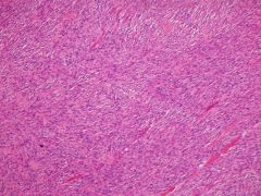
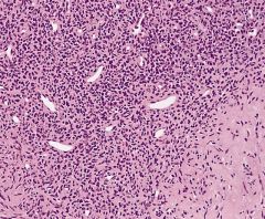
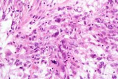
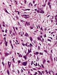
Synovial sarcoma 1. Patient population and location ? 2. Two types and histology? which is more common? 3. List positive stains? which one is actually useful/specific? 4. Molecular alteration? 5. List features portending a better and worse prognosis? |
1. younger patients (15-40 yrs) M>F; usually deep seated mass 80% around knee or ankle! 2. Monophasic and biphasic (more common); spindled cells are plump wiht scant cytoplasm adn oval hyperchromatic nuclei that are arranged in sheets and/or herringbone fascicles with alternating hyper and hypocellular areas; in biphasic an epithelial component is added and makes nests cords glandular structures or papillae 3. cytokeratins EMA, CD99 and BCL2 are all positive but TLE1 is the bomb stain for SSs! 4. t(X;18) translocates SYT with SSX1 or SSX2 4. worse: extensive necrosis, rhabdoid cells, high mits, high brade nuclei, monophasic better: biphasic wiht >50% epithelial component, calcs, osseous metaplasia, youger (<15yrs), distal ext lesions, small lesions, low mitotic rate and low necrosis |
|
|
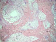
1. List 4 presentations of angiosarcoma and describe each? |
1. cuteaneous w/o lymphedema often on scalp or forhead looking like a bruise, 50% multifocal Cutenous WITH longstanding lymphedema, 90% after mastectomy called Stewart Treves syndrome Breast primary, rapidly enlarging with blue discoloration after radiation therapy Radiation induced Deep soft tissue of ext or abdominal cavity with extensive hemorrhage and necrosis |
|
|
|
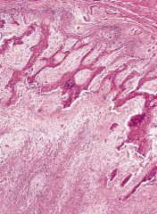
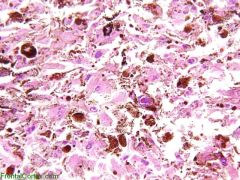
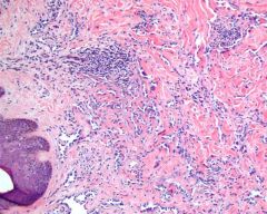
angiosarcoma 1. Histology? 2. Liver angiosarcomas assocated with? |
1. ill defined infiltrative with wide range of differentiation, irregular anastomosing vascular channels that dissect tissue along facial planes and collagen bundles with atypical endothelial cells that may be tufted or form signet ring like single cell vascular channels 2. Thorotrast (thorium dioxide), vinyl cloride (PVC), silver oxide containing insectisides, arsenic poisoning, androgenic anabolic steroids |
|
|
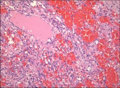
Glomus tumor 1. patient population and location? 2. Histology? List and describe variants? 3. Positive IHC? |
1. equal sex distribution exept for subungal which is more common in women (and the most common site), 3-5 decades and usually a solitary dermal or subcutaneous mass, usually painful and sensitive to cold and touch 2. classic: nests or sheets of round uniform cells with pink cytoplasm and hyprchromatic nuclei surrounding capillaries and peripherally encircled by rim of collagen glomangioma: cavernous hemangioma like arrangement of clusters of glomus cells around gaping blood vessels, +/- secondary thrombosis Glomangiomyoma: cells transitioning between glomus and smooth muscle cells Symplastic: marked cytologic atypia but no other bad features Malignant glomus tumor (glomangiosarcoma) can be spindled or round cell but must be >2cm subfascial or visceral and have lots of atypical mites or marked nuclear atypia 3. Smooth muscle actin |
|
|
1. List variants (5)? 2. What is a hemagioendothelioma? |
1. Epithelioid, retiform, spindle cell, malignant endovascular papillary angioendothelioma (dabska tumor) , kaposiform 2. A vascular tumor with intermediate behavior between angiosarc and hemangioma |
|
|
|
epithelioid HE 1. Patient population and location? Caveat? 2. Histology? 3. Unique treatment for what location? 4. Location with worst prognosis? |
1. M=W, distal ext of young adults, 50% arising in wall of vein; women more likely to have parenchymal (lung liver bone) lesions that are multifocal 2. epithelioid or histiocyte like tumor cells with abundant eosinophilic cytoplasm arranged in short strands or nests with bland nuclei and intracytoplasmic lumina (vacuoles) containing rbs, tumor cells may arise from vessel (50%) extending out into soft tissue, and has either myxoid or hyalinized stroma f variable content 3. Liver primary can be treated with transplant, 75% 5 year survival 4. pulmonary primary has 65% mortality rate, liver 35% |
|
|
Retiform HE 1. Location and patient population? 2. Histology? 3. This tumor is closely related to ? 4. IHC positive?
|
1. plaque like nodule of skin and subcutaneous tissue of distal extremities of young people 2. diffuse dermal involvment charactherized by elongated narrow arborizing vascular channels (looks like rete testis) lined by hyperchromatic endothelial cells hobnailing into lumina, half have prominent stromal and pervascular lymphocytic infiltrate 3. Dabska tumor 4. CD31, CD34, von willebrand factor |
|
|
spindle cell HE 1. Location and patient population? 2. Histology? 3. IHC positive? shared with? 4. Prognosis? 5. Associated with 2 syndromes and one congenital condition? |
1. distal extremities (especially hand) of young M>F (50% <25yrs) 2. Combo of cavernous hemangioma and kaposi sarcoma features; bland spindled tumor cells in multinodular pattern with peripheral gaping vascular channels at periphery and scattered itractyoplasmic vaculones like epithelioid HE 3. Factor VII positive (spp not in the spindled part though but he vascular spaces and epithelioid areas) as is epithelioid HE, and dabscka tumor (factor VII NOT positive retiform and not kaposiform) 4. 60% local recurrence rate 5. Maffucci syndrome, Klippel Trenaunay syndrome and congenital lymphedema |
|
|
Malignant endovascular papillary angioendothelioma 1. AKA? patient population and location? 2. Histology? 3. Prognosis?
|
1. dabska tumor, skin and soft tissue of children and infants, RARE in adults! 2. plumph endothelial cells forming papillay tufts within dilated well formed vascular spaces (glomeruloid) and also hobnailing often with intraluminal rbc's lymphocytes and perivascular lymphocytes 3. Good prognosis despite reginal lymph node metastases |
|
|
Kaposiform hemangioendothelioma 1. Patient population and location? 2. Histology? 3. Associations? (one syndrome to explain) |
1. exclusively in kids and adolescents in superficial (75% skin) or deep soft tissue (20% reroperitoneum) 2. combines features of capillary hemangioma and kaposi sarcoma; intersecting fascicles of bland spindle cells mixed with capilaries; fasicles may be compact or have slit like or cresent vascular lumens and scattered glomeruloid nests are seen 3. lymphangiomatosis, Kasabach Merrit syndrome (consumptive coagulopathy and thrombocytopenia especially with deep tumors like retroperitoneum)
|
|
|
1. List 16 types of hemangiomas with alternate names? |
1. capillary, acquired tufted angioma, hobnail, verrucous, cherry angioma (senile angioma), cavernous, arteriovenous, venous, spindle cell, epithelioid (angiolymphoid hyperplasia with eosinophilia), pyogenic granuloma (granulation tissue type capillary), intramuscular, synovial, perineural (hemangioma of peripheral nerve), diffuse (angiomatosis), papillary enodthelial hyperplasia (intravascular HE of Masson) |
|
|
|
(lobular) Capillary hemangioma 1. list 3 variants of capillary hemangioma with most common first? 2. Patient population, location and natural history? tx? 3. Histology? 4. If congenital or perinatal these are all considered inflantile hemangiomas and are positive for ? |
1. Cellular hemangioma of infancy, pyogenic granuloma (aka lobular capillary hemangioma) and epithelioid hemangioma 2. children especially H&N locations, appears shortly after birth and grows rapidly but will spontaneously regress; corticosteroids or interferon alpha 3. Plump to flat endothelial cells with variable pericytes arranged in multilobular pattern with branching small vascular channels, fibrotic stroma increases with age of lesions (as they involute) 3. GLUT 1 |
|
|
Targetoid hemosiderotic hemangioma 1. aka? 2. patient population and histology? |
1. hobnail hemangioma 2. Exophytic skin lesion young adults, with superficial dilated vessels lined by hobnnail endothelial cells with slit like capillaries at deeper portion |
|
|
Cherry angioma 1. aka? 2. Patient population, gross appearance and histology? |
1. Senile angioma 2. older people, most common angioma that increases with age, has red papule with pale halo on trunk and extremities of adults, made of thin walled dilated capillaries covered wtih atrophic epidermis |
|
|
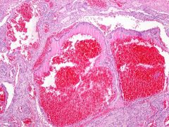
Cavernous hemangioma 1. patient population and location? 2. histology? 3. Treatment? 4. Associations? describe?
|
1. kids upper body, larger, deeper less well circumscribed than capillary hemangioma, may destroy other structures 2. medium to large blood vessels with flattened endothelium arranged in lobular or diffuse pattern 3. recombinant interferon alpha2a and pentoxifylline 4. kasabach merrit syndrome (consumptive coagulopathy) blue rubber bleb nevus syndrome (AD, cutaneous and GI hemangiomas) Maffuci syndrome (enchondromas multiple hemangiomas, cavernous and or spindle) |
|
|
Spindle cell hemangioendothelioma 1. gross appearance? 2. Histology? 3. Associated with what? 4. Prognosis? |
1. often multiple noduels in same anatomic area 2. thin walled cavernous vessels lined by flat endothelium separated by spindled areas containing epithelioid cells with cytoplasmic vacuoles/lumens 3. Mafucci syndrome (multiple enchondromas and soft tissue hemangiomas, spindled or cavernous) Klippel Trenaunay syndrome early onset varicosities, congenital lymphedema and epithelioid hemangioendothelioma 4. 60% recurrence rate |
|
|
Epithelioid hemangioma 1. aka? 2. patient population and location? 3. histology? 4. treatment and prognosis? |
1. angiolymphoid hyperplasia with eosinophilia 2. 30-50 y/o women in superficial H&N region often periauricular 3. epithelioid endothelial cells with vacuolated eosinophilic cytoplasm in multilobular pattern ususally with large central vessel surrounded by smaller vessels with a mixed inflammatory infiltrate with eos 4. superficial radiotherapy gives 80% response rate |
|
|
Papillary endothelial hyperplasia 1. aka? 2. what is it? location? 3. histology? |
1. Intravascular hemangioendothelioma of Masson 2. an organizing thrombus in a blood vessel wiht occasional soft tissue extension, often in association with b9 vascular lesions; often H&N location fingers or trunk (hemorrhoids) 3. well defined interatastomosing channels and papillary structures line by benign endothelium often associated with residual orgnaizing thrombus |
|
|
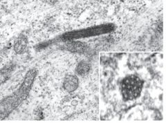
Weibel palade body 1. What is it? |
1. rod shaped storage granules composed of parallel tubules typically found in endothelial cells that contain P-selectin and von Willebrand Factor |
|
|
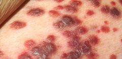
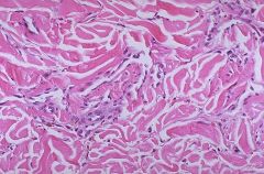
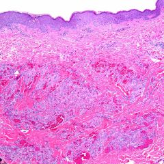
Kaposi sarcoma 1. List and describe 4 types? |
1. classic: indolent, 90% in elderly men of Mediterranean/ east european/ Jewish descent, indolent corse with death in 8-10 yreas, 25-30% have secondary malignancy of which half are heme origin, red cell aplasia, autoimmune hemolitic anemia lymphadenpathic: African, LAD in children or LE dz in men, kids have rapid progression due to internal organ involvement, not so for adults transplant associated: 1% incidene, 100% death rate unless immunosupression tx is decreased by at least 50% which --> marked response rate of 100%, balancing game, associated with Castlemans dz and angioimmunoblastic LAD AIDS related: 30% develop, highest incidence in homosexual men, widespread skin and oral lesions, 50% LN involvement, 30% GI involvement, mortality rate of 40%, |
|
|
Kaposi sarcoma patch 1. Histology? 2. Charactheristic "sign"? |
1. earliest stage, composed of subtle small thin walled vessels dissecting though collagen 2. Promontory sign: when existing vessels and adnexal structures are trapped and protrude into the neoplastic luimina 3. HHV-8 |
|
|
Kaposi sarcoma plaque 1. histology? 2. Characteristic hitologic finding? |
1. more prominent anastomosing irregular vessels + spindled cell component and appearance of intra/extra cytoplasmic hyaline globules and extravasated RBCs 2. autolumination: paranuclear vacuoles containing RBCs |
|
|
Kaposi sarcoma nodule 1. Histology? 2. buzzword? 3. Staining of globules? |
1. same as plaque only more prominent everything especially consolidation of spindled areas into curvilinear fascicles resembling fibrosarcoma and clusters of hyaline globules 2. Seive-like pattern 3. PAS positive diasetase resistant |
|

|
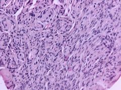
Myopericytoma 1. patient population and location? 2. Histology? 3. pertinent negative stain? |
1. subcutaneous nodule on distal extremities of middle aged adults with = sex predilection 2. well circumscribed nonencapsulated lesion composed of monomophic spindle to oval eosinophlic or amphophilic cells growing in multilayered concentric fashion around lesional blood vessels and occasional whorled or fasicular with myxoid stroma often with subendothelial proliferation (making luminal mass) 4. desmin negative despite myoid appearance |