![]()
![]()
![]()
Use LEFT and RIGHT arrow keys to navigate between flashcards;
Use UP and DOWN arrow keys to flip the card;
H to show hint;
A reads text to speech;
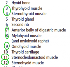
231 Cards in this Set
- Front
- Back
|
Brain Case vs. the Facial Skeleton |

Braincase (neurocranium) protects and supports the brain. Facial skeleton (viscerocranium) protects and supports the airways,food passages, and organs of special sense (sight, smell, and taste) |
|
|
SCALP layers |
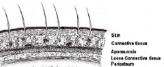
- Aponeurosis is also known as the epicranial aponeurosis - Connective tissue comes off Periosteum easily |
|
|
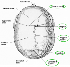
What are the bregma and lambda sutures remnants of? |
- Bregma was the anterior fontanelle - Lambda was the posterior fontanelle
|
|

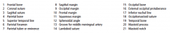
Identify the sutures. |
|
|
|
What vessel crosses the pterion? |
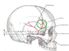
The middle menigeal artery |
|
|
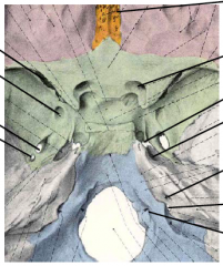
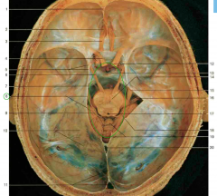
What parts of the brain do the following fossas support? 1. Anterior cranial fossa 2. Middle cranial fossa 3. Posterior cranial fossa |
Anterior cranial fossa = i. frontal lobes Middle cranial fossa = i. the temporal lobes ii. diecephalon iii. pituitary gland Posterior cranial fossa = i. brain stem ii. cerebellum |
|
Identify. |
|
|
Identify. |
|
|
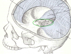
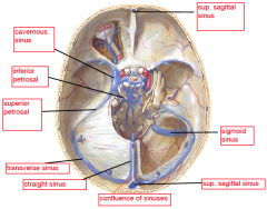
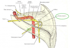
Name the dural folds and the green encircled area. |
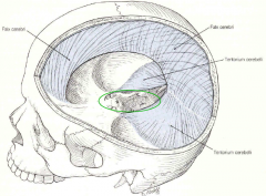
area between the tentorium cerebelli = tentorial notch |
|
|
What parts of the skull are separated by the tentorium cerebelli? |
|
|
Identify. |
|
|
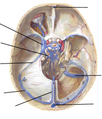
Identify. |
|
|
|
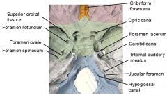
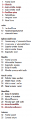
Name Cranial nerves I-V and mention where they go into the skull. |
I – Olfactory – Into the cribriform plate of the ethmoid
II – Optic – Into the optic canal III – Occulomotor – Into the superior orbital fissure IV – Trochlear – Into the superior orbital fissure V – Trigeminal -- V1 - Ophthalmic division – Into the superior orbital fissure -- V2 – Maxillary division – Into the foramen rotundum -- V3 – Mandibular division – Into the foramen ovale |
|
|
Name Cranial nerves VI-XII and mention where they go into the skull. |
VI – Abducens – Into the superior orbital fissureVII – Facial – Into the internal auditory meatus VIII – Vestibulocochlear – Into the internal auditory meatus IX – Glossopharyngeal – Into the jugular foramen X – Vagus - Into the jugular foramen XI – Accessory – Into the jugular foramen XII – Hypoglossal – Into the hypoglossal foramen |
|
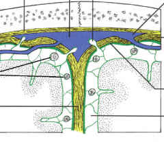
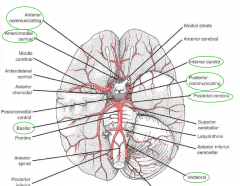
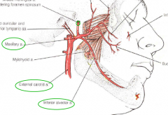
Identify the 4 arteries. |
|
|
|
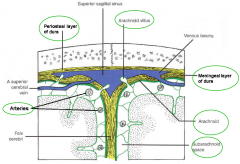
What is the function of the arachnoid villi? |
Clear the cerebral spinal fluid into the sinuses |
|
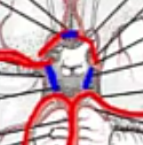
What vessels make up the circle of Willis?
|
The arteries arising from this circle are all end arteries. No anastamosing! |
|
|
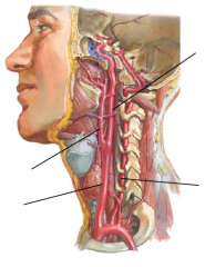
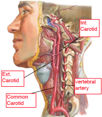
1. What vessel enters the skull through the carotid canal?
2. What vessel enters the skull through the foramen magnum? |
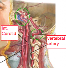
1. Internal carotid arteries 2. Vertebral arteries |
|
|
What forms the zygomatic arch?
|
Temporal process of zygomatic bone meets zygomatic process of temporal bone to form zygomatic arch |
|
|
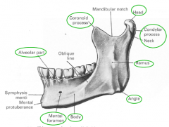
What part of the skull holds the teeth in? |
Alveolar process of the maxilla and the alveolar process of the mandible
|
|
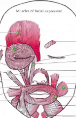
Name all these 5 muscles + the nerve that innervates them. |
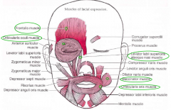
Innervated by Cranial Nerve VII |
|
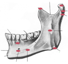
Identify the red dots. |
|
|
|
What are the Muscles of Mastication and what nerve are they innervated with? |
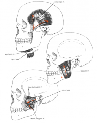
1. Temporalis 2. Masseter 3. Lateral pterygoid 4. Medial pterygoid - Innervated by the mandibular division (V3) of cranial nerve V- the trigeminal |
|
|
Which accessory muscles of mastication act as depressors of the mandible? What innervates them? |
1. Digastric 2. Mylohyoid V3 innervation for ant. belly of digastric. VII for posterior belly |
|
|
What gland is innervated Parasympathetically by cranial nerve IX and what muscle does this gland's duct cross over?
|
Parotid gland. The duct crosses over the masseter muscle and enters the mouth. |
|
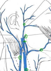
Identify the veins. |
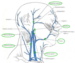
- Superficial temporal vein - Maxillary vein - Retromandibular vein - External jugular vein - Facial vein
|
|
|
Which vein drains into the facial vein? |
Opthalmic |
|
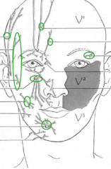
Sensory Innervation of the Face |
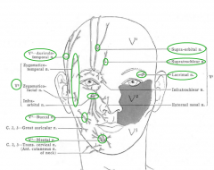
Branches of the Trigeminal: V1 Ophthalmic – supraobital supratrochlear, lacrimal V2 Maxillary – Zygomatic this and that, and infraorbital V3 Mandibular – auriculotemporal, buccal, and mental branch of the inferior alveolar |
|
|
What are the boundaries of the infratemporal fossa? |
- Inferior to the zygomatic arch - Between the ramus of the mandible and the lateral pterygoid plate of the sphenoidbone |
|
|
Pterygomaxillary fissure leads from _________ into the __________, a space between the maxilla and the pterygoid plates of the sphenoid bone. |
Pterygomaxillary fissure leads from the infratemporal fossa into the pterygopalatinefossa, a space between the maxilla and the pterygoid plates of the sphenoid bone. |
|
|
What other parts of the skull does the pterygopalatine fossa connect with and through what fissures/foramena? |
- the orbit (through the infraorbital fissure) - the nose (through the sphenopalatine foramen) - the mouth (through the greater palatine canal and the greater and lesser palatine foramena) - the middle cranial fossa (through the foramen rotundum and the pterygoid canal) |
|
|
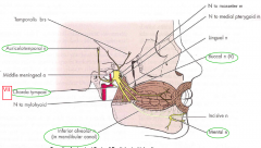
What are the contents of the infratemporal fossa? |
1. The medial and lateral pterygoid muscles. 2. The maxillary artery and some of its branches. 3. Mandibular Division of the Trigeminal (V3) 4. Chorda Tympani Branch of the Facial (VII) 5. The Otic Ganglion |
|
|
Which of the cranial nerves are sensory, which are motor and which are both? |
Some say marry money but matan berson says big brains matter more S = sensory M = motor B = both |
|
|
Mnemonic to remember all the cranial nerves. |
Ooh, ooh, ooh to touch and feel avoluptuous girl's vagina. Soo hot! |
|
|
What are Trigeminal V3 motor branches innervating? |
Motor branches to muscles of mastication:
1. temporalis 2. masseter 3. the pterygoids 4. mylohyoid 5. the anterior belly of the digastric |
|
|
What are Trigeminal V3 sensory branches innervating? |
1. Auriculotemporal (External ear, auditory canal and tympanic membrane)
2. Buccal (skin over buccinator) 3. Inferior alveolar– enters the mandibular foramen, and exits the mental foramen as the mentalnerve. Gives off the inferior dental nerves: teeth in lower jaw, skin of lower lip and jaw 4. The lingual nerve– general sensory (pain, touch, pressure and temperature) to the mucosa of the floor of the mouth and to the anterior 2/3 of the tongue. |
|
|
What nerve enters the infratemporal fossa through the foramen ovale? |
Mandibular Division of the Trigeminal (V3) |
|
|
Which nerve leaves the skull through the squamotympanic/petrotympanic fissure and joins the lingual nerve in the infratemporal fossa? |
Chorda Tympani Branch of the Facial (VII) |
|
|
How are the submandibular and sublingual salivary glands innervated? |
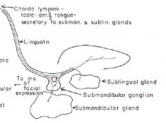
Preganglionic parasympathetic fibres of VII. - synapse on postganglionic neurons in the submandibular ganglion located on the lingual nerve. - They are secretomotor to the glands. |
|
|
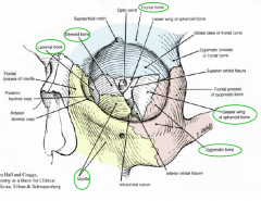
What bones make up the orbital skeleton? |
|
|
|
What parts of the skull is the orbit communicating with and through what structures?
|
Communicating with the middle cranial fossa through the optic canal and superiororbital fissure Communicating with the pterygopalatine fossa through the inferior orbital fissure Communicating with the nose through the nasolacrimal canal. |
|
|
Proptosis |

If something is in the orbit and is pushing the eye forward
|
|
|
Orbital Vs. Visual Axes |
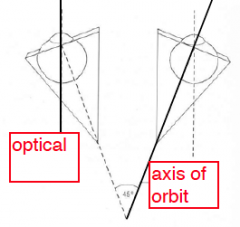
The two axes are not congruent. Muscles attach on teh axis of orbit |
|
|
The Ocular Bulb |
Aka the eyeball = Cornea, sclera, optic nerve |
|
|
The Bulbar Fascia (Bulbar Sheath) |
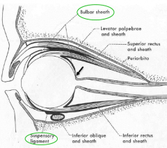
- Forms a fascial socket for the eye, attaching to the sclera where the optic nerve entersthe bulb and extending forwards to attach anteriorly at the corneoscleral junction. - Pierced by the eye muscles - Creates a sling to support the eye – the suspensory ligament. |
|
|
What are the functions of these eye muscles? Medial rectus Lateral rectus Superior rectus Inferior rectus Inferior oblique Superior oblique |
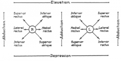
Medial rectus – adduction Lateral rectus – abduction Superior rectus – elevation of the abducted eye Inferior rectus – depression of the abducted eye Inferior oblique – elevation of the adducted eye Superior oblique – depression of the adducted eye |
|
|
Which muscles torque the eye when the head rotates? |
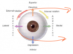
Inf. and Sup. Obliques |
|
|
Innervation of the Extrinsic Muscles of the Eye |
- LR(VI) - SO(IV) - All others by III |
|
|
What makes up the conjunctival sac?
|
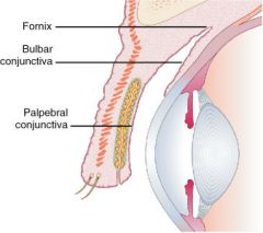
A sac made up of Palpebral and bulbar conjunctiva when the eye lids close. |
|
|
What innervates the Levator palpebrae superioris? |
Voluntary skeletal muscle innervated by somatic motor fibres carried in CN III Smooth muscle innervated by postganglionic sympathetic fibres from the carotid nerve plexus. (Horner's syndrome = failure of sympathetic innervation) |
|
|
What is the Lacrimal Apparatus made up of? |
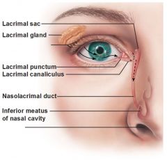
1. Lacrimal gland 2. Superior and inferior lacrimal puncta 3. Lacrimal canaliculi 4. Lacrimal sac and nasolacrimal duct 5. Lacis lacrimalis. |
|
|
Describe the blood supply to the eye |
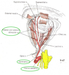
Ophthalmic artery – from the internal carotid, then through the optic canal. Gives offthe central artery of the retina, branches to the orbital tissues and the anterior andposterior ethmoidal arteries to the ethmoidal air cells and lateral nasal wall. |
|
|
How is blood drained from the eye? |
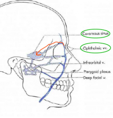
- Ophthalmic veins drain to the middle cranial fossa into the cavernous sinus, posterior to the orbit.
- Facial vein |
|
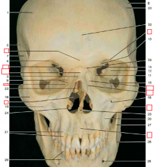
Identify everything but the red boxes. |
|
|

Identify everything but the red boxes. |

|
|
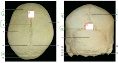
Identify all the sutures, as well as A + B |
A = bregma
B = lambda |
|
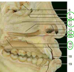
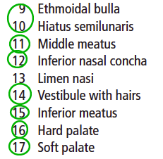
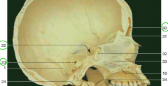
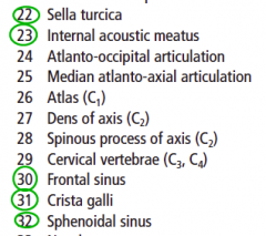
Identify #8 |
|
|
|
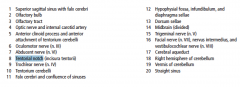
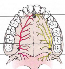
What does V2 split into after it goes through the Foramen rotundum? |
1. infraorbital – through the inferior orbital fissure, groove and infraorbital foramento the cheek, upper lip, and lower lid
2. Superior alveolar nerves – Through the lateral walls of the maxillary air sinus tothe upper teeth and gingivae 3. Zygomatic through the inferior orbital fissure, across the orbital floor to thelateral cheek 4. Greater and lesser palatine nerves – through the greater palatine canals to the hardand soft palates. 5. Lateral Nasal Branches – through the sphenopalatine foramen to the lateral nasalmucosa 6. Nasopalatine nerve – through the sphenopalatine |
|
|
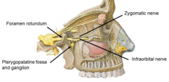
What does V1 split into after it goes through the Superior orbital fissure? |
1. Lacrimal nerve 2. Frontal - then splits to supraorbital and supratrochlear branches 3. Nasociliary - then splits to long ciliary, anterior and posterior ethmoidal and infratrochlear branches |
|
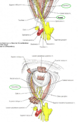
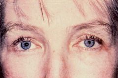
What could have caused the left eye position and pupil dilation? |
- occulomotor nerve (III) Palsy - lesion to the occulomotor nerve --> Eye is dilating (PSy innervates the constrictor pupil muscle occulomotor (III)) --> innervates Levator palpebrae superioris muscle + All Other muscles (aside from the two below). - Lateral Rectus (LR6) is now unopposed - Superior Oblique (SO4) is also unopposed |
|
|
How the Dillator Pupillae innervated sympathetically? |
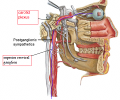
- Postganglionic fibres from the superior cervical ganglion follow the internal carotidinto the skull. - Some of these fibres join the nasociliary nerve and enter the eye through the long ciliary branch - others form a sympathetic root to the ciliary ganglion and pass through it without synapsing to enter the eye with the short ciliary branches |
|
|
What nerves are to be found in the ciliary ganglion and nerves? |
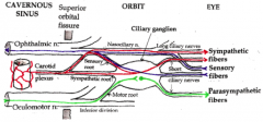
1. Sympathetic fibers
2. Sensory fibers 3. PSy fibers synapse here |
|
|
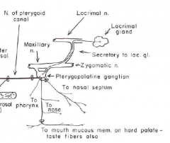
Parasympathetic Innervation of the Lacrimal Gland |
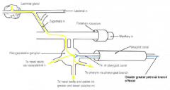
Preganglionic fibers: - Facial Nerve (VII) --> Greater Petrosal nerve --- Pterygopalatine canal --> Pterygopalatine fossa - synapse at the Pterygopalatine ganglionPostganglionic fibers: - follow zygomatic branch (V2) - leave in a communicating branch - lacrimal branch (V1) |
|
|
Vestibule VS. Oral cavity proper |
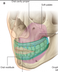
Vestibule: space between lips and teeth/gums Oral Cavity Proper: area surrounded by teeth and gums containing the tounge |
|
|
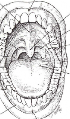
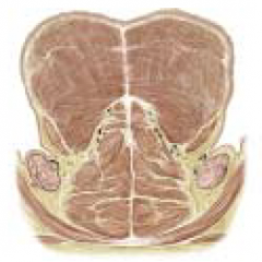
What makes up the Roof of the Mouth? What is the sensory and blood supply? |
Hard Palate:
- palatal processes of maxillae - horizontal plates of palatine Soft Palate: - palatal aponeurosis - Tensor veli palatini (V3-Trigeminal) - leVator veli palatini (X-Vagus) Sensory: - greater and lesser palatine branches - nasopalatine branch (V2) Blood: - greater and lesser palatine branches (maxillary art.) |
|
Identify. |
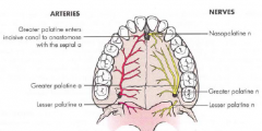
All nerves are from V2
|
|
Identify. |
|
|
|
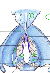
Muscles that Support the Floor of the Mouth |
Mylohyoid and geniohyoid |
|
|
What are the intrinsic and extrinsic muscles of the tongue and what innervates them? What separates the left and right pairs of muscles in the tongue? |
Intrinsic – transverse, vertical and longitudinal fibres. - shape the tongue (Innervated by XII) Extrinsic – Position the tongue.
1.Genioglossus (XII) 2. Hyoglossus (XII) 3. Styloglossus (XII) 4. Palatoglossus (X) Bilateral and separated by the fibrous septum of the tongue |
|
|
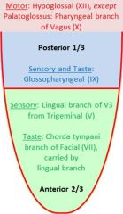
Sensory Innervation of the tongue |
|
|
|
Blood supply and drainage of the tongue |
The lingual artery a branch of the external carotid The lingual vein a tributary of the internal jugular. The vessels lie medial to the hyoglossus, while the lingual and hypoglossal nerves are lateral to the hyoglossus |
|
|
What are the 3 pairs of salivary glands |
1) Parotid 2) Submandibular 3) Sublingual |
|
|
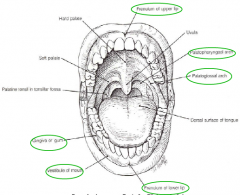
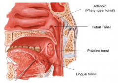
What do the Palatine Tonsils lie in between? |
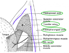
In the fossa between the palatopharyngeal and palatoglossal folds. Bed of the tonsil contains the glossopharyngeal nerve and the tonsillar branch of thefacial artery. |
|
|
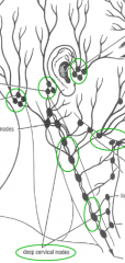
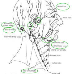
Where do the regional groups of lymphatic nodes drain into? |

The regional groups drain to the deep cervical group of nodes which lies alongthe internal jugular vein, deep to the sternomastoid muscle. |
|
Identify. |
|
|
|
What makes up the nasal septum? |
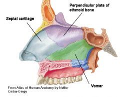
Perpendicular plate of the ethmoid, vomer, septal cartilage |
|
|
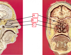
What do the two meningeal layers of the dura create? |
falx cerebri |
|
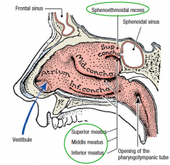
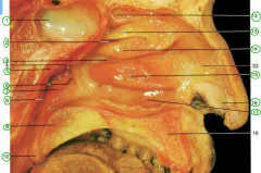
Identify. What is their function? |
Superior, middle and inferior conchae - warm air - filter air |
|
|
1. What is the space above the superior conchae? 2. What is the space between the conchae? |
|
|
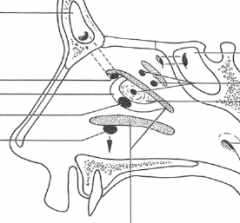
What are the openings into the sphenoethmoidal recess and the 3 meatus?
|
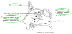
- Sphenoethmoidal recess receives the sphenoidal air sinus - Superior meatus receives ethmoid air sinuses - Middle meatus receives ethmoidal air sinuses on the bulla, the maxillary and frontal air sinus into the hiatus semilunaris. - Inferior meatus receives the nasolacrimal duct lying in the nasolacrimal canal |
|
|
1. What must the maxillary artery pass through in order to get into the pterygopalatine fossa?
2. What must the maxillary nerve pass through in order to get into the pterygopalatine fossa? |
The maxillary artery enters from the infratemporal fossa by passing through the pterygomaxillary fissure The maxillary nerve enters from the middle cranial fossa by passing through the foramen rotundum |
|
|
What bones are the 3 conchae made of? |
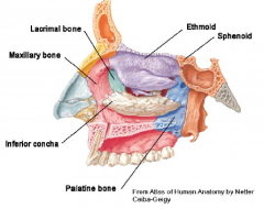
Superior and middle: ethmoid Inferior conchae is its own bone. |
|
|
What is the blood supply to the lateral wall of the nose? |
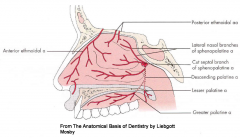
- From the lateral nasal branches of facial - From the anterior and posterior ethmoidal branches of opthalmic - From the lateral nasal branches of the sphenopalatine artery; |
|
|
What is the blood supply to the nasal septum of the nose? |
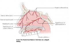
- Septal branch of the sphenopalatine - From the labial branches of the facial - From the anterior and posterior ethmoidal branches of the ophthalmic - From the greater palatine artery - Kisselbach’s area |
|
|
What is the innervation to the lateral wall of the nose?
|
- Olfactory nerves enter through the cribriform plate - general sensory: -- lateral nasal branches (V2) -- anterior ethmoidal (nasociliary, V1) |
|
|
What is the innervation to the nasal septum of the nose? |
- nasopalatine branch (V2) - anterior ethmoidal (nasociliary, V1) |
|
|
What artery enters the nose through the sphenopalatine foramen? |
sphenopalatine artery; a branch of the maxillaryartery given off in the pterygopalatine fossa. - septal branch irrigates nasal septum - lateral nasal branches irrigate lateral wall of nose |
|
|
Name the 3 parts of the pharynx |
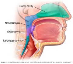
Oropharynx, Nasopharynx, laryngopharynx.
Retropheryngeal space between the pharynx and vertebral column. |
|
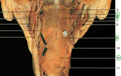
Identify circled numbers. What innervates muscle #5? |
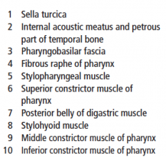
The stylopharyngeus is innervated by IX: glossopharyngeal |
|
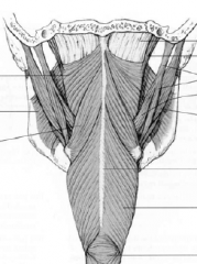
Identify. What innervates the muscular wall of the pharynx? |
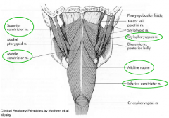
X: Vagus nerve.
Stylopharyngeus is innservated by IX. |
|
|
What is found in the laryngopharynx? |
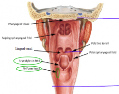
1. Piriform fossae 2. The aryepiglottic folds |
|
|
What is the sensory innervation of the pharynx mucosa? |
Glossopharyngeal nerve (IX) Some overlap with V2 in the nasopharynx Some overlap with the vagus in the laryngopharynx |
|
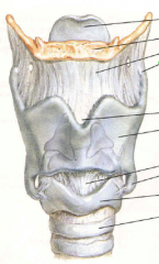
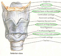
Identify. |
|
|
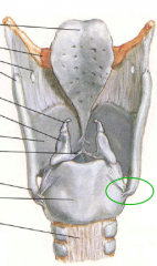
Identify. |
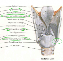
Inferior horn of thyroid cartilage |
|
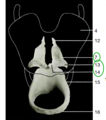
Identify circled structures. |
7 = arytenoid cartilage 13 = muscular process of arytenoid cartilage 14 = vocal process of arytenoid cartilage |
|
|
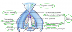
1. What muscle abducts the vocal chords and open the rima glottidis? 2. What muscle tilts the thyroid cartilage to tighten the vocal chord? |
1. Posterior cricoarytenoid 2. Cricothyroid |
|
Identify.
|
|
|
Identify. |
|
|
Identify. What communicates with 6? |
The auditory tube communicates with the middle ear |
|
Identify.
|
|
|
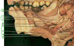
Identify. |
Hypoglossal nerve is slightly more medial and posterior than the lingual nerve |
|
Identify. |
|
|
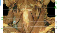
What innervates each of these muscles? |
7 = V3 8 = V3 Strap muscles (3,4,9,12) = cervical spinal nerves 11 = Spinal accessory (XI) |
|
|
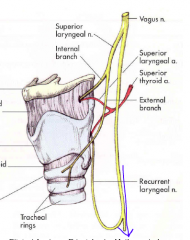
1. The intrinsic muscles of the pharynx (except for cricothyroid) are innervated by? 2. Describe the branching of this nerve 3. Cricothyroid is innervated by? |
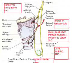
1. the recurrent branches of the vagus nerves. 2. The left recurrent laryngeal nerve recurs around the right subclavian artery; the left recurs under the arch of the aorta. Ascend to the larynx in the groove between the trachea and esophagus. 3. external laryngeal branch of the superior laryngeal nerve |
|
|
1. Sensation from the laryngeal lining above the vocal folds is carried by the 2. Sensation from below the level of the vocal folds is carried by |

1. internal laryngeal branch of the superior laryngeal nerve (a branch of the vagus) 2. the recurrent laryngeal nerves. |
|
|
What is the blood supply and drainage to and from the pharynx and larynx? |
Arterial supply from branches of the superior and inferior thyroid arteries. Venous drainage to the superior and middle thyroid veins, tributaries of the internal jugular veins. The inferior thyroid veins drain into the brachiocephalic veins. |
|
|
1. Names of glands in the external auditory meatus? 2. What fraction of the meatus is cartilagenous/bony? |
1. Ceruminous glands 2. 1/3 cartilagenous, 2/3 bony |
|
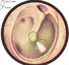
1. What are the two circled structures?
2. What innervates this membrane? |
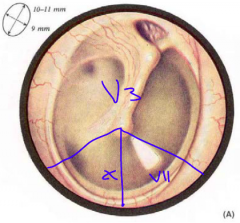
1. Umbo (tip of the malleus) and cone of light of the tympanic membrane 2. external surface = V3, VII, X internal surface = IX |
|
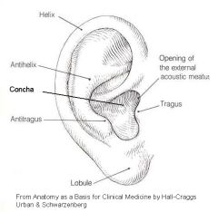
What innervates the auricle of elastic cartilage covered with skin? |
Mostly auricotemporal (V3), some C1, VII, X |
|
|
What is the tunnel the middle ear communicates with the mastoid air cells through? What is the tube the middle ear communicates with the nasopharynx? What does the middle ear communicate with the internal ear though? |

antrum (posterior to middle ear) auditory (eustacian or pharyngotympanic) tube oval and round windows |
|
|
What is the bony floor of the middle ear? What separates the middle ear from the the middle cranial fossa? |
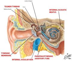
temporal bone (between jugular foramen and opening to carotid canal) tegmen tympani |
|
|
Function of the ossicles? |
Convert vibration of tympanic membrane to mechanical oscillations, which stimulate the organ of Corti (Spiral organ) in the cochlear duct of the inner ear
|
|
|
What are the articulation points between the malleus, incus and stapes? What are the muscles that attach to them and reflexively dampen the oscillations? |
Malleus: - handle: tympanic membrane (umbo) - head: into epitympanic recess > Tensor tympani muscle (V3) Incus: - articulates with malleus and incus heads Stapes: - base of stapes: oval window > Stapedius muscle (VII) |
|
|
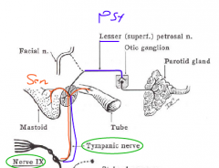
Describe the route the glossopharyngeal (IX) nerve takes to innervate the parotid gland.
|

- leaves jugular foramen - gives off tympanic branch (PSy and Sensory) - this branch enters glossopharyngeal canaliculus to middle ear - forms tympanic plexus - preganglionic PSy enter middle cranial fossa as lesser petrosal nerve - petrosal nerve synapses at the otic ganglion on V3 - postganglionic PSy follow auriculotemporal branch of V3 to parotid gland |
|
|
What do the sensory fibres in the tympanic plexus innervate? |
- lining of the middle ear - mastoid air sinuses - auditory tube - internal aspect of tympanic membrane |
|
|
Describe the route Facial (VII) nerve takes to innervate the lacrimal glands |
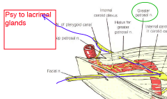
- facial nerve enters the internal auditory meatus - through geniculate ganglion - Greater petrosal nerve (preganglionic PSy) - crosses middle cranial fossa - pterygoid canal - synapse at pterygopalatine ganglion - zygomatic branch (V2) - lacrimal branch (V1) |
|
|
Describe the route Facial (VII) nerve takes to innervate the sublingual and submandibular gland |
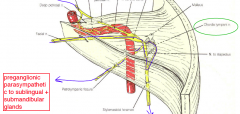
- facial nerve enters the internal auditory meatus
- goes through geniculate ganglion - descends in facial canal (temporal bone) posterior to middle ear - gives off chorda tympani n. (preganglionic PSy) - chorda tympani passes between malleus and incus - re-enter temporal bone via petrotympanic fissure - joins lingual nerve - synapse with post ganglionic PSy at submandibular ganglion - postganglionic fibers innervate the glands |
|
|
Describe the route Facial (VII) nerve takes to innervate the muscles of facial expression |
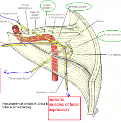
- facial nerve enters the internal auditory meatus - descends in facial canal (temporal bone) posterior to middle ear - exit skull via stylomastoid foramen |
|
|
What cell bodies can be found in the geniculate ganglion?
|
- taste sensory neurons of anterior 2/3 of tongue - general sensory neurons that innervate external auditory meatus and tympanic membrane |
|
|
What is the function of the vestibular apparatus and cochlear duct? What stimulates them? What innervates them? |
- Cochlear duct: hearing - Vestibular apparatus: balance and motion - Stimulated by movement of endolymph - Vestibulocochlear nerve (VIII) |
|
|
1. What are the parts of membranous labyrinth in the temporal bone and what is the labyrinth suspended in? 2. What does the labyrinth contain inside of it? |
1. - vestibule, cochlea, semicircular canals - suspended in perilymph 2. contains endolymph |
|
|
What innervates the platysma muscle? What does the muscle lie on top of? |
- VII
- lies on the inve$ting layer of cervical fascia |
|
|
What surrounds the vertebral compartment of the neck? What can be found inside this compartment? |
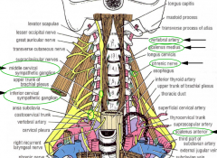
- prevertebral fascia - vertebral arteries (transverse foramina) - phrenic nerve lies on scalenus anterior - cervical part of sympathetic chain (anterior to fascia) - superior, middle and inferior cervical ganglia |
|
|
Post ganglionic fibers can communicate to 3 locations from the sympathetic chain. What are they?
|
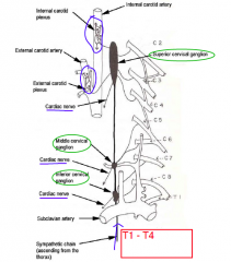
1. Grey rami 2. Cardiac branches---> cardiac and pulmonary plexuses 3. Branches to the carotid plexus (from superior cervical ganglion only) |
|
|
What can be found in the carotid sheath? |
- common and internal carotid arteries - internal jugular vein - deep cervical lymph nodes - vagus nerve |
|
What could be some symptoms of Horner's syndrome? |
Loss of sympathetic to one side can cause:
- lack of sweating - pupils constrict (constrictor pupil (III) is unopposed) - eye lid drops (LPS (III) unopposed) |
|
|
What can be found at the level of bifurcation of common carotid and what innervates ? |
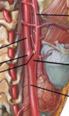
Bifucation @ superior border of thyroid cartilage - Carotid body: chemoreceptor - Carotid Sinus: baroreceptor Innervated by IX and X |
|
|
Name 5 branches off the ext. carotid artery |
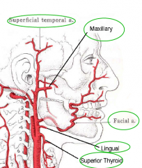
- superior thyroid - lingual - facial - maxillary - superficial temporal a |
|
|
What can be found in the visceral compartment of the neck and what surrounds this compartment? |
- surrounded by the pretracheal fascia 1. Pharynx 2. Larynx 3. Thyroid 4. Parathyroid glands 5. Esophagus 6. Trachea 7. Recurrent laryngeal nerves |
|
|
Where do the branches of the vagus nerve go after the recurrent laryngeal branch off? |
They continue to the cardiac branches to the mediastinum
|
|
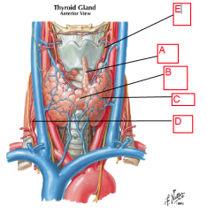
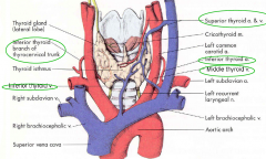
Identify. What artery is not present in this diagram that would be traveling up to irrigate the thyroid. |
A = pyramidal lobe
B = isthmus C = lobes of thyroid D = inferior thyroid branches from thyrocervical branch of the subclavian E = superior thyroid artery of the external carotid - Thyroidea ima from the brachiocephalic artery or from the aortic arch |
|
|
What drains the thyroid and parathyroid? |
- superior and middle thyroid veins which drain to the internal jugular veins - inferior thyroid veins that drain to the brachiocephalic veins |
|
|
What muscles are in the investing layers? |
- Trapezius - Sternocleidomastoid (elevate face/turn head to side) - both innervated by XI |
|
|
Where are the fibers of the Spinal Accessory nerve (XI) coming from? |
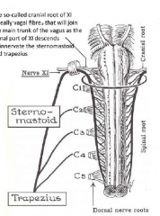
Two roots: spinal and cranial root - Cranial root is vagal fibres that join the spinal root to descend and innervate the SCM and Traps. |
|
|
Once synapsed in the pterygopalatine ganglion, where are 2 locations Facial (VII) post-ganglionic nerve fibres might head to? |
1. zygomatic nerve --> lacrimal nerve --> innervate lacrimal gland 2. follow the nasal and greater palatine branches of the maxillary nerve to innervate glands in the nasal and palatal mucosa. |
|
|
What are two ligaments found in the acromioclavicular joint? What movement does the acriomioclavicular joint permit? What muscles act upon this joint? |
- acromioclavicular lig. - coracoclavicular lig. Minimal gliding Joint: Completing full shoulder elevation requires movement from this joint |
|
|
Name the joint between the clavicle and sternum. What movements are permitted here and what ligaments are found in this joint? What muscles act upon this joint? |
- sternoclavicular joint
Biaxial joint: - glides superior/inferior - glides anterior/posterior - anterior & posterior sternoclavicular lig. - interclavicular lig. |
|
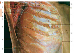
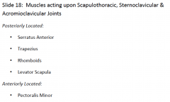
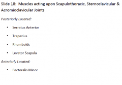
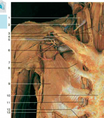
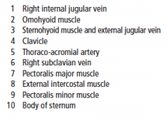
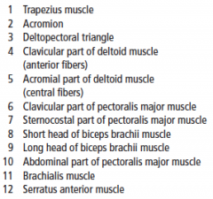
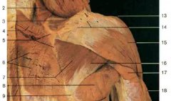
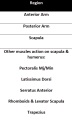
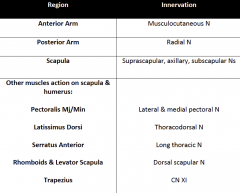
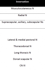
Origin, insertion, innervation and action of #7 |
Origin: ribs 1-9 Insertion: vertebral border of scapula Innervation: long thoracic nerve (C5,C6,C7) Action: abduction (protraction), upward rotation of scapula |
|

Origin, insertion, innervation and action of #3 |
Trapezius Origin: Occiput, Spinous processes of C7-C12 Insertion: spine of scapula, acromion process, clavicle Innervation: Spinal accessory (XI) Action: Elevation (sup.), adduct/retraction (mid.), depression (inf.), upward rotation (sup. & inf. synergistically) |
|

Origin, insertion, innervation and action of #14/15 |
Rhomboids major (15) and minor (14): Origin: Spinous processes of C7-T1 (minor) & T2-T5 (major) Insertion: Vertebral border of scapula Innervation: Dorsal Scapular N. Action: Elevate, Adduct, Downward rotation scapula |
|
Origin, insertion, innervation and action of #12 |
Levator Scapula Origin: Transverse processes C1-C3 Insertion: Vertebral border of scapula Innervation: Dorsal Scapular N. (C5) Action: Elevate scapula |
|
|
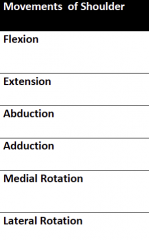
Intrinsic and extrinsic ligaments of the shoulder joint |
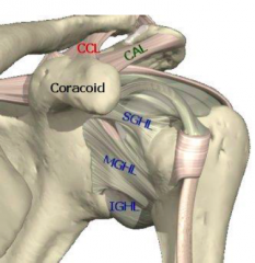
Intrinsic:
- Superior Glenohumeral lig. - Middle Gelnohumeral lig. - Inferior Glenohumeral lig. Extrinsic: - Coracoclavicular lig: limits abduction and flexion - Coracoacromial lig. limits humerus from moving superiorly |
|
|
What movements are allowed in the shoulder joint and what are 3 features found with in this joint? |
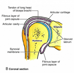
Multiaxial Joint: Movement in all planes + circumduction - Glenoid labrum: fibrocartilagenous ring to deepen socket (increasing joint congruency & support) - Long head tendon of biceps passing through (intra-articular) - Subdeltoid bursae |
|
Origin, insertion, innervation and action of #9 |
Origin: Ribs 3-5 Insertion: Coracoid process Innervation: Medial pectoral N. (from medial cord) Blood supply: Thoracoacromial A Action: Abduction, Downward rotation of scapula |
|

Origin, insertion, innervation and action of #6/7 |
Origin: Ribs 1-6, clavicle, sternum Insertion: lateral lip of intertubecular groove Innervation: Medial lateral pectoral N. (from medial and lateral cord) Blood supply: Thoracoacromial A Action: Adduction, flexion, medial rotation of humerus |
|
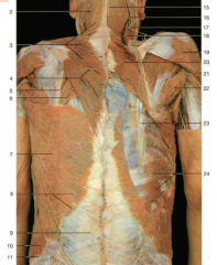
Origin, insertion, innervation, action and blood supply of #7 |
Latissimus Dorsi Origin: Spinous process C7-L5, iliac crest, sacrum Insertion: intertubecular groove Innervation: Thoraco-Dorsal N (off the Posterior cord) Action: Adduction, extension, medial rotation of humerus Blood supply: Thoraco-Dorsal (off subscapular branch) |
|

Origin, insertion, innervation and action of #3 |

Origin: Supraspinous fossa Insertion: Greater tuberosity of humerus Innervation: Suprascapular N. (from upper trunk) Action: abduction, Lateral rotation of humerus |
|

Origin, insertion, innervation and action of #7 |
Origin: Infraspinous fossa Insertion: Greater tuberosity Innervation: Suprascapular N. (from upper trunk) Action: Lateral rotation |
|
Origin, insertion, innervation and action of #8 |

Origin: Inferior angle of scapula (#9) Insertion: Medial lip of intertubercle groove Innervation: Lower subscapular N. (from posterior cord) Action: Adduction, Extension, Medial rotation |
|

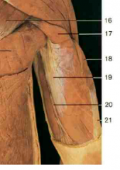
Origin, insertion, innervation and action of #16 |

Origin: Lateral border of scapula Insertion: Greater tubercle Innervation: Axillary N. Action: Lateral rotation |
|
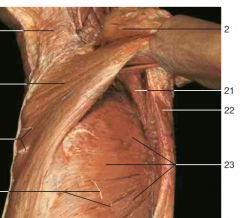
Origin, insertion, innervation and action of #21 |
Subscapularis Muscle Origin: Subscapular fossa Insertion: Lesser tuberosity Innervation: Upper and Lower subscapular N. (off posterior cord) Action: Medial rotation |
|
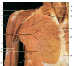
Origin, insertion, innervation, blood supply and action of #4/5 |
Deltoids: Origin: Clavicle, Spine of scapular Insertion: Deltoid tuberosity Innervation: Axillary N. Blood supply: Thoracoacromial A Action: Abduction |
|
|
What are the Axilla Region's borders? |
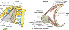
Medial wall: Ribs, serratus anterior Lateral wall: intertubercular groove Anterior wall: Pectoralis Mj. and Mn. Posterior wall: Scapula and muscles (subscapularis, LD, Teres Mj.) |
|
Origin, insertion, innervation and action of #8 |
Biceps Brachii Origin: Sup. Glenoid (long head) and Coracoid process (short head) Insertion: Radial tuberosity Innervation: Muscolocutaneous N. Action: Supination of arm, flex shoulder joint, flex elbow joint |
|

Origin, insertion, innervation and action of #9 |
Brachialis Origin: Humerus Insertion: Ulnar tuberosity Innervation: Muscolocutaneous N. Action: Flexes forearm at elbow joint |
|
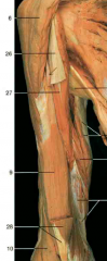
Origin, insertion, innervation and action of #27 |
Coracobrachialis Origin: Coracoid process Insertion: Humerus Innervation: Musculocutaneous N. Action: Flex shoulder joint |
|
Origin, insertion, innervation and action of #18/19 |
Triceps brachii: Origin: Superior Glenoid (long head), humerus (lateral and medial heads) Insertion: Olecranon process Innervation: Radial N Action: Extend elbow joint, some extension of shoulder joint |
|
|
|
|
|

|
|
|
|
|
|
|
|
|
What 5 joints make up the shoulder region? |
- Glenohumeral joint (true shoulder joint) Additional joints which contribute to shouldermovement: - Acromioclavicular (Minimally Gliding Joint) - Coracoclavicular (Minimally Gliding Joint) - Sternoclavicular (Biaxial Joint) Pseudojoint: - Scapulothoracic |
|
|
What is the true elbow joint? What is the true wrist joint? |
- Ulnohumeral
- Radiocarpal joint |
|
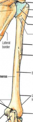
Identify. |
|
|
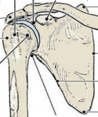
Identify. |
|
|
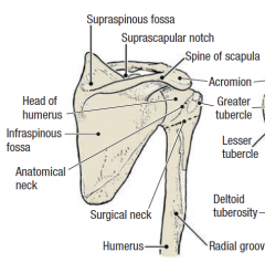
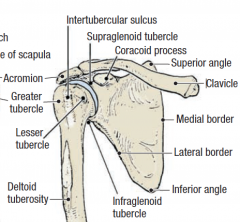
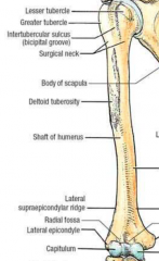
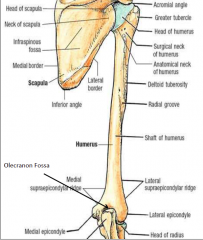
Identify the structures on the humerus. |
|
|
Identify the structures on the humerus. |
|
|
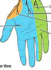
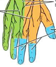
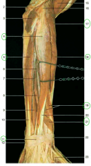
What innervates the different colored portion? |
Orange: Radial Blue: Median Green: Ulnar |
|
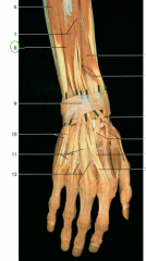
What innervates the different colored portion?
|
Orange: Radian Blue: Median Green: Ulnar |
|
|
The musculocutaneous nerve can be found sandwiched between which two muscles?
|
Biceps brachii and Brachialis
|
|
|
What does the Ulnar nerve innervate? |
- ½ FDP - FCU -hypothenar muscles -thumb adductor & interossei -medial1/2 of lumbricals |
|
|
What does the Median nerve innervate? |
-ANTERIOR forearm (except ½ FDP & FCU) -thumb (thenar muscles) -lateral 1/2 of lumbricals |
|
|
What do the Radial and Axillary nerve innervate? |
Radial: all POSTERIOR muscles of arm & forearm Axillary: Teres Min. and Deltoid |
|
|
Starting with the brachiocephalic artery, describe the breakdown by landmarks of the main artery of the arm. |
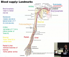
|
|
|
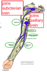
Describe the blood drainage of the arm. |
|
|
|
Humero-ulnar joint allows for what movements?
|
Uniaxial joint, hinge – permits flexion/extension |
|
|
Carrying Angle |
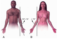
Long axis of a fully extended humerus makes an angle with ulna of approx 170 degrees. - Angled away from hips, more for women then men. Created by obliquity of articulation between humerus & ulna. |
|
|
Medial Collateral Ligament: |
Connects ulna to humerus (also called ulnar collateral) - Prevents excessive valgus forces |
|
|
Lateral Collateral Ligament: |
Connects radius to humerus (also called radial collateral) Prevents excessive varus forces
|
|
|
Anular Ligament |
Anular Ligament: Connects on anterior & posterior aspect of ulna around radial neck & head Maintains radial head’s position and permits pivot |
|
|
What lies in the radial groove? |
- Radial nerve - Deep brachial artery |
|
|
Cubital Fossa is bounded by? |

- Superior: Line joining medial & lateral epicondyles - Lateral: edge of pronator teres - Medial: edge of brachioradialis |
|
|
Cubital Fossa contains? |
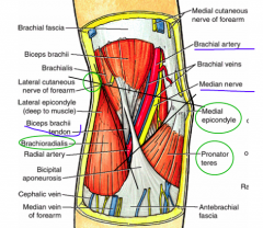
- Biceps tendon - Brachial artery - Median nerve - ‘TAN’ from lateral to medial |
|
|
What are the two joints between the ulna and the radius? What type of movement is possible at these joints? |
Superior Radio-ulnar Joints: - Radial head articulates with radial notch in ulna Joint between radius & ulna Inferior Radio-ulnar Joints: - Ulnar head articulates with ulnar notch in radius Two joints combine to permit pronation &supination |
|
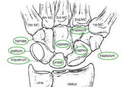
Identify all.
|
|
|
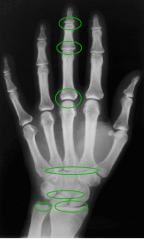
Identify the joints. |
Distal Interphalangeal Joint (DIP): between distal &middle phalanges Proximal Interphalangeal Joint (PIP): between middle &proximal phalanges Metacarpophalangeal Joint (MCP): between metacarpals& phalanges Carpometacarpal Joint (CMC): between distal carpal row& bases of metacarpals Midcarpal Joint: joint between proximal & distal carpalrows Radiocarpal Joint: between proximal carpal row & distalradius; no direct articulation between carpals and ulna(there a disc interposed between ulna and carpal bones) Inferior Radio-ulnar Joint: between the distal ends ofradius and ulna |
|
|
What type of joint is the Carpometacarpal Joints? - what type of movements are permitted here? |
- Planar Joints - Gliding movements permitted - Exception: 1st CMC which is a saddle joint - Permits flexion,extension, adduction,abduction & opposition |
|
|
What types of joints are the Metacarpophalangeal Joints? - What types of movements are possible here? |
- Condyloid Joints - Biaxial Movements: flexion/extension,abduction/ adduction - Exception is 1st MCP –only flexion/extension |
|
|
What type of joints are Interphalangeal Joints? What movements are possible in this joint? |
Interphalangeal Joints: - Hinge Joints - Uniaxial Movements:flexion/extension |
|
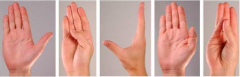
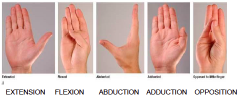
Name the thumb movements.
|
|
|
|
What separates the 2 compartments of forearm? |
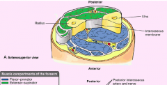
Separated by two bones of forearm and the connecting interosseous membrane |
|
|
Anatomical Snuffbox |
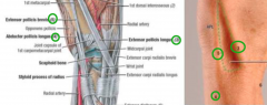
Formed between the tendons of the EPL, APL & EPB - Floor of snuffbox is scaphoid - Crossing through this is branch of radial A |
|
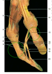
Identify. What is its function? |
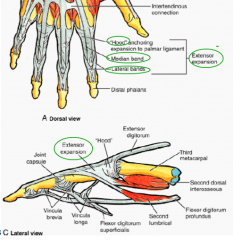
Extensor Expansion. Complex network of fibrous/ligamentous bands which surrounds extensor tendons - Comprised of two lateral bands, a median band and a ‘hood’ Aids in binding tendon down to phalanges: - Provides attachment point for lumbrical, thus allowing synergistic extension from muscles technically on the flexor side |
|
|
|
|
Origin, Insertion, Action, Innervation of #10 and #11 |
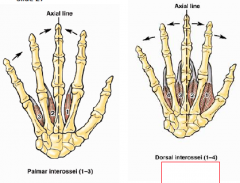
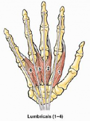
11 = Palmar lumbricals - 3 muscles Attach from metacarpal to phalange Adduction (PAD) 10 = Dorsal lumbricals - 4 muscles Attach from metacarpal to phalange - Abduction (DAB) All innervated by Ulnar N. |
|
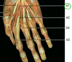
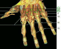
Origin, Insertion, Action, Innervation of #41 |
Lumbricals muscles Origin: Attaches from FDP tendon Insertion: proximal phalanges of 4 fingers Action: flexes MCPs and extends IPs Innervation: Lateral 2 by Median, medial 2 by ulnar |
|
|
1. Tendons on the hand's flexor side are bound down by a series of sheaths called?
2. what are the sheaths that can be found bellow these ones? |
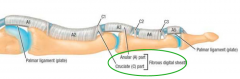
1. Fibrous digital sheaths: - Annular ligaments – thick bands covering tendons - Cruciate ligaments – thin criss-crossed bands close to the joint spaces 2. Synovial sheaths |
|
|
What forms the carpel tunnel and what can be found going through it?
|
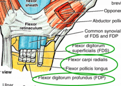
Floor and walls = carpals
Roof = flexor retinaculum
Contents:
- 4 FDS - 4 FDP - FCR - FPL - Median Nerve
|
|
Function of #29? |
Palmar Aponeurosis - Protects tendons |
|
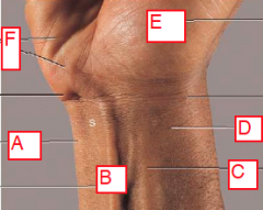
1. Identify. (A and F are on medial side of body) 2. What can be found between A and B? |
A= tendon of ECU B= tendon of PL C= tendon of ECR D= Radial A E= Thenar Eminence F = Hypothenar Eminence 2. Ulnar A |
|
Origin, insertion, action and innervation of #9
|
Adductor pollicis
Origin: 3rd metacarpal Insertion: proximal phalanx of thumb Action: adducts thumb at CMC joint Innervation: Ulnar N. |
|

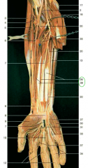
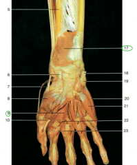
Origin, insertion, action, innervation and blood supply of #4 |
4 = Flexor carpi radialis Origin: Medial epicondyle Insertion: 2nd metacarpal Action: flex wrist, abduct hand Innervation: median N. Blood: Radial A. |
|
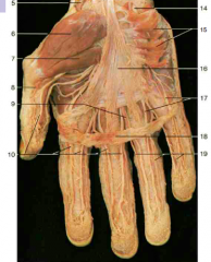
Origin, insertion, action, innervation and blood supply of #6 |
6 = Flexor Digitorum Superficialis Origin: Medial epicondyle Insertion: middle phalanges 2-5 Action: Flex proximal IP, flex MCP, flex wrist Innervation: median N. Blood: Ulnar A. |
|

Origin, insertion, action, innervation and blood supply of #3 |
3 = Brachioradialis Origin: Lateral epicondyle Insertion: Radius Action: Flex elbow of mid-pronated arm Innervation: Radial N. Blood supply: Radial A. |
|

Origin, insertion, action, innervation and blood supply of #17
|
17 = Pronator teres Origin: Medial epicondyle Insertion: Radius Action: Pronation Innervation: Median N. Blood Supply: Ulnar and Radial A. |
|

Origin, insertion, action, innervation and blood supply of #18 |
18 = Palmaris longus Origin: Medial epicondyle Insertion: Palmar fascia Action: Weak wrist flexion Innervation: Median N. Blood Supply: Ulnar A. |
|

Origin, insertion, action, innervation and blood supply of #21 |
21 = Flexor carpi ulnaris Origin: Medial epicondyle Insertion: Hamate and Pisiform Action: Wrist flexion and hand adduction Innervation: Ulnar N. Blood Supply: Ulnar A. |
|
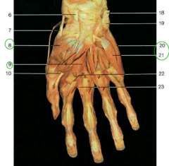
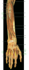
Identify # 5, 7 and 20. |
5 = Radial A 7 = Median N (Anterior forearm except lateral 2 lumbricals, thenar) 20 = Ulnar A |
|

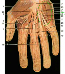
Origin, insertion, action, innervation and blood supply of #36 |
36 = Flexor digitorum profundus Origin: ulna Insertion: distal phalanges 2-5 Action: Flex distal IP, weak wrist flexion Innervation: Median N (lateral half), Ulnar N (medial half) Blood Supply: Anterior Interosseous A. |
|
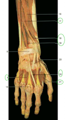
Origin, insertion, action, innervation and blood supply of #39 Identify # 8 |
39 = Flexor pollicis longus Origin: radius Insertion: distal phalanx of thumb Action: flex thumb (MCP, IP) Innervation: Median N Blood Supply: Anterior Interosseous A. 8 = Flexor retinaculum |
|
Origin, insertion, action, innervation and blood supply of #17 |
17 = Pronator Quadratus Origin: Ulna Insertion: Radius Action: Pronation Innervation: Median N. Blood Supply: Anterior Interosseous A. |
|
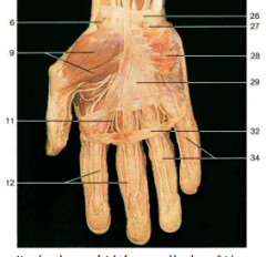
Action, innervation and blood supply of #6 |
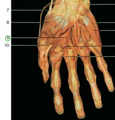
Abductor pollicis brevis Action: abduct thumb Innervation: Recurrent branch of Median N. Blood Supply: Superficial palmar arch |
|

Action, innervation and blood supply of #7 |
Flexor pollicis brevis
Action: flex thumb (MCP) Innervation: Recurrent branch of Median N. Blood Supply: Superficial palmar arch |
|
Action, innervation and blood supply of #8 |
8 = Opponens pollicis Action: Opposition of thumb Innervation: Recurrent branch of Median N. |
|

Action, innervation and blood supply of #21 |
21 = Flexor digiti minimi Action: flex little finger Innervation: Ulnar N. Blood Supply: Ulnar A. |
|

Action, innervation and blood supply of #20 |
20 = Abductor digiti minimi Action: abduct little finger Innervation: Ulnar N. Blood Supply: Ulnar A. |
|
Action, innervation and blood supply of #25 Identify #30 |
25 = opponens digiti minimi Action: opposition of little finger Innervation: Ulnar N. Blood Supply: Ulnar A. 30 = fibrous digital sheaths |
|

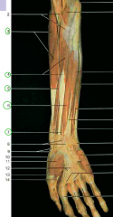
Origin, insertion, action, innervation and blood supply of #4 |
4 = Extensor Digitorum Longus Origin: Lateral epicondyle Insertion: 4 fingers via extensor expansion Action: finger and wrist expansion Innervation: Radial N. Bloody Supply: Posterior Interosseous A. |
|
Origin, insertion, action, innervation and blood supply of #17 |
17 = Extensor carpi radialis longus Origin:Lateral epicondyle Insertion: 2nd metacarpel Action: wrist abduction, wrist extension Innervation: Radial N Bloody Supply: Radial A. |
|

Origin, insertion, action, innervation and blood supply of #18 |
18 = extensor carpi radialis brevis Origin:Lateral epicondyle Insertion: 3rd metacarpel Action: wrist abduction, wrist extension Innervation: Radial N Bloody Supply: Radial A. |
|
Identify #7. Origin, insertion, action, innervation and blood supply of #8 |
7 = Extensor digitorum 8 = Extensor digiti minimi Origin: Lateral epicondyle Insertion: little finger via extensor expansion (#10 are the tendons) Action: Extend little finger Innervation: Radial N. Bloody Supply: Posterior Interosseous A. |
|
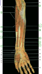
Origin, insertion, action, innervation and blood supply of #7 |
7 = Extensor Carpi Ulnaris Origin: Lateral epicondyle Insertion: 5th metacarpal Action: Adduct wrist, extend wrist Innervation: Radial N. Bloody Supply: Ulnar A. |
|
Origin, insertion, action, innervation and blood supply of #17
|
17 = Extensor pollicis longus Origin: ulna and radius Insertion: distal phalanx of thumb Action: extend thumb Innervation: Radial N. Bloody Supply: Posterior Interosseus A. |
|

Origin, insertion, action, innervation and blood supply of #18 |
18 = Extensor pollicis brevis Origin: radius Insertion: proximal phalanx of thumb Action: Extend thumb Innervation: Radial N. Bloody Supply: Posterior Interosseus A. |
|

Origin, insertion, action, innervation and blood supply of #16 |
16 = abductor pollicis longus Origin: ulna and radius Insertion: 1st metacarpal Action: Abduct thumb Innervation: Radial N. Bloody Supply: Posterior Interosseus A. |
|

Origin, insertion, action, innervation and blood supply of #19 |
19 = extensor indicis Origin: ulna Insertion: index finger via extensor expansion Action: extend index finger Innervation: Radial N. Bloody Supply: Posterior Interosseus A. |
|
|
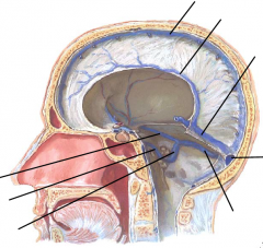
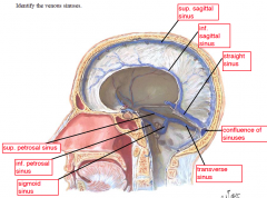
Where do the inf. petrosal and sigmoid sinuses drain into?
|
Internal jugular vein |
|
|
Name the 4 tonsils |
|
|
|
1. Which ligament limits abduction and flexion of the humerus? 2. Which ligament limits the upward movement of the humerus in the glenohumeral joint? |
1. Coracoclavicular lig. 2. Coracoacromial lig. |
|
|
How are the ciliary muscles innervated? |
Occulomotor (CN III) - pre-ganglionic parasympathetic synapse at the ciliary ganglion - post-ganglionic travel to eye through short ciliary |
|
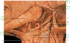
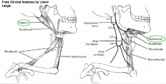
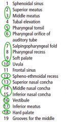
Identify 1,2,9,10 What innervates each of them? |
Suprahyoid muscles: 1 = Anterior belly Digastric (V3) 2 = Mylohyoid (V3) 9 = Stylohyoid (VII) 10 = Posterior belly of Digastric (VII) |