![]()
![]()
![]()
Use LEFT and RIGHT arrow keys to navigate between flashcards;
Use UP and DOWN arrow keys to flip the card;
H to show hint;
A reads text to speech;
22 Cards in this Set
- Front
- Back
|
Frontal lobe |
Motor cortex (further maths) Emotion/ Personality (gage became ^ aggressive) Reasoning (abnormality in OCD) |
|
|
Parietal lobe |
Somatosensory cortex - touch, smell, ect... (people say) Recognition (Recognising a smell > linked to a memory e.g. Sidni & hollister perfume holiday) Movement & ability to orientate (touch > surrounded by things = ability to orientate)
|
|
|
Occipital lobe |
Visual cortex (open van) Shape recognition Sense of perspective |
|
|
Temporal lobe |
Auditory (take away) - hearing, speech, sound Recognition (> as sound) |
|
|
Medulla oblongata |
• automatically controls breathing & heart rate |
|
|
Cerebellum |
• has a folded cortex • important for coordinating movement & balance |
|
|
Hypothalamus |
• homeostasis e.g. thermoregulation • produces hormones that control the pituitary gland |
|
|
Cerebrum |
• largest part of brain • 2 halves called cerebral hemispheres • thin outer layer called cerebral cortex > ^ SA (highly folded to fit into skull) • involves in in vision, learning thinking, emotions & movements • dif parts > dif functions |
|
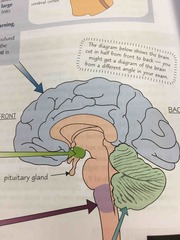
Name the area in dark green (near pituitary gland) |
Hypothalamus |
|
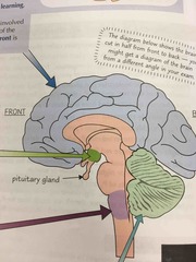
Name the area in light purple |
Medulla oblongata (in brain stem) |
|

Name the area in light green |
Cerebellum (underneath cerebrum) |
|

Name the area in light blue |
Cerebrum/ cerebral cortex |
|
|
What is the purpose of using brain scanning techniques? |
• To investigate the structure and function of the brain • To diagnose medical conditions |
|
|
4 types of scan |
Computed Tomography (CT) Magnetic Resonance Imaging (MRI) Functional Magnetic Resonance Imaging (fMRI) Positron Emission Tomography (PET) |
|
|
fMRI scan |
• ^ oxygenated blood flows to active areas • Deoxyhaemoglobin absorbs radiowaves whilst oxyhemoglobin does not absorb radio waves • structure & function carried out in scanner = brain ^ active in particular region • diagnosis > abnormal activity > damaged area e.g. seizures |
|
|
CT scans |
• uses x rays > dense areas absorb ^ radiation > show ‘dark’ colour • shows major structures of the brain damaged/diseased > able to determine function of that area • diagnosis > blood = lower density than brain tissue so lighter colour > extent of bleeding |
|
|
PET scan (outline) |
• Uses x rays • highlight active areas of the brain > detects the radioactivity of the tracer • radioactive tracer incorporated into compounds (02, water, glucose, ect...) • structure & function > shows structure of areas & activity/inactivity indicates function • diagnosis > areas active/inactive detected > study disorders changing brain activity (e.g. Parkinson’s= reduction in function of the motor cortex) |
|
|
MRI scans |
• use really strong magnetic fields & radio waves • investigating brain structure > able to differentiate between abnormal & normal tissue > dif tissues respond dif to magnetic field > damaged area used to work out function • diagnosis > damaged/diseased brain region > e.g. brain tumour cells respond dif to mag field - lighter colour > treatment |
|
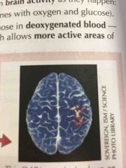
What type of brain scan? |
fMRI scan |
|
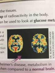
What type of brain scan? |
PET scan |
|

What type of brain scan? |
MRI scan |
|

What type of brain scan? |
CT scans |