Reading...
![]()
Play button
![]()
Play button
![]()
Use LEFT and RIGHT arrow keys to navigate between flashcards;
Use UP and DOWN arrow keys to flip the card;
H to show hint;
A reads text to speech;
156 Cards in this Set
- Front
- Back
- 3rd side (hint)
|
4 areas that the ear is often divided into for study
|
Outer, Middle, Inner, Higher Auditory Pathways
|
|
|
|
These are considered part of the outer ear
|
Auricle/Pinna
Ear Canal/External Auditory Meatus |
|
|
|
These are considered part of the middle ear
|
Auditory tube
Hollow space in temporal bone |
|
|
|
These are considered part of the inner ear
|
cochlea, semicircular canals (sensory)
8th nerve and Higher Auditory Pathways (Neural) |
|
|
|
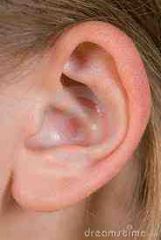
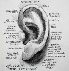
Describe characteristics of the auricle (pinna)
|
-Mainly cartilage covered with skin (except fatty lobe)
-Cutaneous lining (skin) and cartilaginous core extend into temporal bone and make up part of ear canal. -Not a lot of nerves or vascular; susceptible to cold -Not very important acoustically -Helps slightly with sound localization in the front and back |
|
|
|
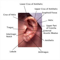
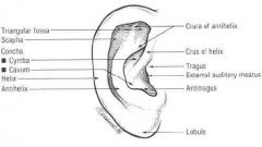
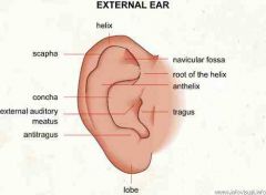
Where is the Helix
|
|
|
|

Where is the Helix?
|

|
|
|

Where is the antihelix?
|

|
|
|
Where is the Crus?
|
|
|
|
|
Where is the Scaphoid Fossa?
|
|
|
|

Where is the Scaphoid Fossa?
|

|
|
|

Where is the Triangular Fossa?
|

|
|
|

Where is the Concha Cymba?
|

|
|
|

Where is the Concha Cavum
|
|
|
|

Where is the Tragus?
|

|
|
|

Where is the AntiTragus?
|
|
|
|

Where is the Intertragical incisure
|

|
|
|

Where is Darwin's Tubercle?
|
|
|
|
|
What has many convolutions and depressions?
|
Auricle/Pinna
|
|
|
|
The lateral end of this is slightly lower than the inside and medial part next the ear drum is also lower
|
Ear Canal/EAM
|
|
|
|
Lateral wall is cartilage, but the medial wall is bones
|
Ear Canal/EAM
|
|
|
|
What is the length of an average ear canal?
|
25-35 mm
|
|
|
|
The lateral portion of the ear canal (1/3-1/2) is made of what?
|
Cartilidge
|
|
|
|
The medial part/wall of the ear canal (2/3-1/2) is made of what?
|
Bone
|
|
|
|
What is the Isthmus?
|
Separation between cartilage and bone in the ear canal
|
|
|
|
Describe the shape and diameter of an average adult ear canal opening?
|
oval and 6-8 mm
|
|
|
|
What are the Cerumen Glands?
|
Produce ear wax
|
|
|
|
What is the shape of the entire ear canal?
|
S shaped
|
|
|
|
Describe the pathway of Pars Externa
|
Medial-Anterior-Superior
|
|
|
|
Describe the pathway of the Pars Media
|
Medial-Posterior
|
|
|
|
Describe the pathway of the Pars Interna
|
Medial-Anterior-Inferior
|
|
|
|
What is the primary function of the EAM?
|
Direct Sound energy to the tympanic membrance
|
|
|
|
What other mechanism is the EAM thought to serve as because of the essential anatomy of the EAM recessed into the boney structures of the skull?
|
Protective mechanism
|
|
|
|
What does the canal act as?
|
Closed tube resonator
|
|
|
|
What HZ does the EAM resonate incoming sounds
|
2500 Hz to 5000 Hz
|
|
|
|
What is the intensity of resonance of the EAM?
|
17 dB
|
|
|
|
What is the resonant frequency of the ear canal alone?
|
2500 Hz
|
|
|
|
What is the resonant frequency of the concha alone?
|
5000 Hz
|
|
|
|
What is the combined resonant frequency of all the structures of the inner ear?
|
3800 Hz
|
|
|
|
Where is the TM located?
|
At the termination of the EAM
|
|
|
|
What does the TM form?
|
Lateral wall of the middle ear cavity
|
|
|
|
Describe the appearance of the TM
|
Smooth, pearl gray, translucent, Concave/cone shaped, Small mass 14 mm
|
|
|
|
What angle is the TM situated in the ear canal at the superior margin?
|
140 degrees
|
|
|
|
What holds the TM in place?
|
An aunulus, or anular ring. A cartiliganious ring that surrounds the TM. The top of the ring is open and the bottom is thickened
|
|
|
|
What is the name for the opening at the top of the aunulus
|
Node of Rivinus
|
|
|
|
What is the typmanic sulcus?
|
Depression that houses the aunular ring
|
|
|
|
What the 3 layers of the TM?
|
Cutaneous, Mucous, Fiberous
|
|
|
|
Describe the Cutaneous Layer of the TM
|
skin layer that is continuous across the face of the tympanic membrane and lining of the EAM
|
|
|
|
Describe the Mucous Layer of the TM.
|
Forms the inner most layer of the TM and is continuous with the lining of the Middle Ear
|
|
|
|
Describe the Fibrous layer of the TM
|
Situated between cutaneous and mucous layers. Gives resiliency to the TM. It is actually made of two sublayers of radial and circular fibers.
|
|
|
|
How do the radial and circular sublayers of the Fibrous layer complement each other?
|
Radial is lateral.
Circular is medial. When superimposed looks like a spiderweb. Gives resilience to TM |
|
|
|
What are the two parts of the ear drum/ TM?
|
Pars Flacid and Pars Tensa
|
|
|
|
Describe the Pars Flacide
|
Superior portion, floppy
|
|
|
|
Describe the Pars Tensa
|
Center portion, tense
|
|
|
|
How does the malleus connect to the Tympanic Membrane?
|
The manubrium of the malleus is firmly attached to the medial surface of the membrane as far as its center, which it draws toward the tympanic cavity; the lateral surface of the membrane is thus concave, and the most depressed part of this concavity is named the umbo.
|
|
|
|
What is the cone of light?
|
When examining the tympanic membrane with an otoscope, a bright cone of light is seen in the anterior-inferior part of the membrane. This light is known as the "cone of light" or "light reflex".
|
|
|
|
What is the umbo?
|
A small anatomical projection on a surface, such as that on the inner surface of the tympanic membrane at the end of the manubrium of the malleus, corresponding to the most depressed point of the membrane.
|
|
|
|
Explain the 4 quadrants of the tympanic membrane.
|
The tympanic membrane is divisible into 4 quadrants by drawing and imaginary line extending down from the handle of the malleus and another imaginary line at right angles to the first line at the umbo. These two lines divide the pars tensa into the anterior-superior, anterior-inferior, posterior-inferior, and posterior superior
|
|
|
|
Explain the 4 quadrants of the TM
|
The tympanic membrane is divisible into 4 quadrants by drawing and imaginary line extending down from the handle of the malleus and another imaginary line at right angles to the first line at the umbo. These two lines divide the pars tensa into the anterior-superior, anterior-inferior, posterior-inferior, and posterior superior
|
|
|
|
How can you tell from looking at the tympanic membrane which ear it belongs to?
|
In the left ear, the handle of the malleus is at 11 o'clock. In the right ear, the handle of the malleus is at 1 o'clock.
|
|
|
|
Point where tympanic membrane connects to the inferior part of the malleus
|
Umbo
|
|
|
|
Latin word for ornamental knob at the center of a warrior’s shield
|
Umbo
|
|
|
|
Located in the petrous portion of the temporal bone
|
Middle ear
|
|
|
|
Air filled cavity that allows air in via auditory tube
|
Middle ear cavity
|
|
|
|
Describe the dimensions of the middle ear from the side view
|
15mm from superior to inferior
15 mm from anterior to posterior (box shape) |
|
|
|
Describe the dimensions of the middle ear from front view
|
Hourglass shape:
Superior portion across the top is 6mm, narrows to 2 mm, base widens to 4mm |
|
|
|
Ceiling of Tympanic Cavity
|
Epitympanic Process
|
|
|
|
thin bony roof of tympanic cavity.
|
Tegmen Tympani
|
|
|
|
Tegmen Tympani
|
Thin sheet of bone in Epitympanic recess (space superior to the tympanic membrane in the middle ear) . So thin that light will transverse. Brain is right behind it; close proximity to cortex
|
|
|
|
Mastoid Antrum
|
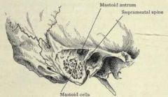
An air-filled space between the upper portion of the middle ear and the mastoid cells.
A cavity in the petrous portion of the temporal bone that communicates with the mastoid sinuses and epitympanic recess of the middle ear. Also called tympanic antrum. |
|
|
|
Opening to Mastoid Antrum
|
Aditus to Antrum
|
|
|
|
Superior wall of middle ear cavity
|
Tegmental; made up of tegman tympani (thin roof)
|
|
|
|
Inferior wall of middle ear cavity
|
Jugular; jugular vein below; prominance into middle ear cavity caused by fossa of jugular vein
|
|
|
|
Posterior wall of middle ear cavity
|
Mastoid; mastoid cavity behind; includes aditus to antrum; pyramidal eminence
|
|
|
|
Anterior wall of middle ear cavity
|
Carotid; divides the tympanic cavity and the carotid canal. Contains the Cochlear Form Process, canal for tensor tympani muscle and an opening for the auditory tube.
|
|
|
|
Lateral wall of middle ear cavity
|
Membranous; houses TM
|
|
|
|
Has a hole in the middle that the stapedial muscle sends a tendon through into the middle ear
|
Pyramidal eminence
|
|
|
|
Superior Wall of Middle Ear Cavity
|
The Tegmental Wall or Roof is formed by a thin plate of bone, the tegmen tympani, which separates the cranial and tympanic cavities.
|
|
|
|
Inferior Wall of Middle Ear Cavity
|
The Jugular Wall or Floor (paries jugularis) is narrow, and consists of a thin plate of bone (fundus tympani) which separates the tympanic cavity from the jugular fossa.
|
|
|
|
Posterior Wall of Middle Ear Cavity
|
The mastoid or posterior wall (paries mastoidea) is wider above than below, and presents for examination the entrance to the tympanic or mastoid antrum, the pyramidal eminence, and the fossa incudis.
|
|
|
|
What are the two parts of the tympanic cavity?
|
The tympanic cavity consists of two parts: the tympanic cavity proper, opposite the tympanic membrane, and the attic or epitympanic recess, above the level of the membrane; the latter contains the upper half of the malleus and the greater part of the incus.
|
|
|
|
Lateral wall of Middle Ear Cavity
|
The Membranous or Lateral Wall (paries membranacea; outer wall) is formed mainly by the tympanic membrane
|
|
|
|
Medial Wall of Middle Ear Cavity
|
The Labyrinthic or Medial Wall (paries labyrinthica; inner wall) is vertical in direction, and presents for examination the fenestræ vestibuli and fenestræ cochleæ, the promontory, and the prominence of the facial canal.
|
|
|
|
pyramidal eminence
|
The pyramidal eminence (eminentia pyramidalis; pyramid) is situated immediately behind the fenestra vestibuli, and in front of the vertical portion of the facial canal; it is hollow, and contains the Stapedius muscle; its summit projects forward toward the fenestra vestibuli, and is pierced by a small aperture which transmits the tendon of the muscle. The cavity in the pyramidal eminence is prolonged downward and backward in front of the facial canal, and communicates with it by a minute aperture which transmits a twig from the facial nerve to the Stapedius muscle.
|
|
|
|
fossa incudis
|
The fossa incudis is a small depression in the lower and back part of the epitympanic recess; it lodges the short crus of the incus.
|
|
|
|
Chorda Tympani
|
Branch of the 7th Cranial Nerve (Facial Nerve)
|
|
|
|
Anterior Wall of the Middle Ear Cavity
|
The Carotid or Anterior Wall (paries carotica) is wider above than below; it corresponds with the carotid canal, from which it is separated by a thin plate of bone perforated by the tympanic branch of the internal carotid artery, and by the deep petrosal nerve which connects the sympathetic plexus on the internal carotid artery with the tympanic plexus on the promontory. At the upper part of the anterior wall are the orifice of the semicanal for the Tensor tympani muscle and the tympanic orifice of the auditory tube, separated from each other by a thin horizontal plate of bone, the septum canalis musculotubarii.
|
|
|
|
Cochlear Form Process
|
canal for tensor tympani muscle
|
|
|
|
What are the ossicles?
|
A chain of 3 small bones that bridge the gap of the middle ear space from the tympanic membrane to the oval window.
|
|
|
|
Name the ossicles.
|
Malleus (hammer), Incus (anvil), Stapes (stirrup)
|
|
|
|
Which is the largest of the ossicles?
|
Malleus
|
|
|
|
What two parts does the malleus have?
|
Head and Neck
|
|
|
|
What are the 3 processes of the malleus?
|
Lateral, Anterior, Manubrium
|
|
|
|
The head of what occupies 1/2 of the epitympanic recess (attic of the middle ear)?
|
Malleus
|
|
|
|
What accounts for the concave nature of the T.M.?
|
Manubrium attached to the middle connective tissue (that fibrous membrane) of the T.M.
|
|
|
|
What contains an articular facet for
attachment to the incus? |
Posterior surface of the head of the Malleus.
|
|
|
|
Where is their a small boney projection for attachment of the tensor tympani tendon?
|
Where the manubrium joins the neck of the malleus, there is a small boney projection for attachment of the tensor tympani tendon
|
|
|
|
Name the parts of the Incus
|
Body, Long process, Short process
|
|
|
|
The body of this occupies 1/2 of the epitympanic recess.
|
Incus
|
|
|
|
On what ossicle do 2 processes arise from the body at nearly right angles?
|
Incus
|
|
|
|
Describe the short process of the Incus
|
The short process (5 mm) runs horizontally backward (posteriorly) and occupies the fossa incudis ( a depression along the back wall of the middle ear).
|
|
|
|
Describe the long process of the Incus
|
The long process (7 mm) runs vertically, parallel to the manubrium.
|
|
|
|
Describe the inferior end of the Incus
|
The inferior end sharply medially and terminates at the lenticular process.
|
|
|
|
What is the lenticular process?
|
A knob at the tip of the long limb of the incus of the ear, articulating with the stapes.
|
|
|
|
Name the parts of the Stapes
|
Head (Concave articular facet , Small boney spine) , Neck, Footplate, Anterior Crus, Posterior Crus, Obturator foramen
|
|
|
|
What is the concave articular facet on the stapes for?
|
Concave articular facet for reception of the lenticular process.
|
|
|
|
What is the purpose of the small boney spine on the head-neck of the stapes?
|
– Small boney spine on the head-neck for attachment of the stapedial tendon.
|
|
|
|
How many Crura are on the stapes, and where do they originate
|
2, Originate near the inferior margin of the footplate – they are connected to the footplate about 3⁄4 of the way down.
|
|
|
|
Which Crus of the stapes is more curved, longer and fatter?
|
Posterior crus. ANTERIOR CRUS = more slender, shorter, and less curved than the posterior crus.
|
|
|
|
What part of what bone of the inner ear is part osseous and part cartilaginous.
|
The footplate of the stapes is Part osseous and part cartilaginous – a bone covered with cartilage
|
|
|
|
What occupies the oval window?
|
Stapes-Footplate
|
|
|
|
What part of an ossicle is held in place by an annular ring?
|
Stapes-Footplate
|
|
|
|
What is the OBTURATOR FORAMEN?
|
(one that closes) = the opening in the stapes (where the cowboy boot goes) --an open air-filled place in the stapes.
|
|
|
|
Who was Eustaschius?
|
16th century anatomist who the Eustachian tube is named for
|
|
|
|
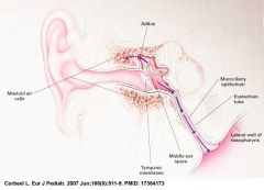
What is the auditory tube (AT) aka eustacian tube?
|
Canal which establishes communication between the middle ear and the nasopharynx
|
|
|
|
What is the auditory tube (AT) aka eustacian tube?
|
Canal which establishes communication between the middle ear and the nasopharynx
|
|
|
|
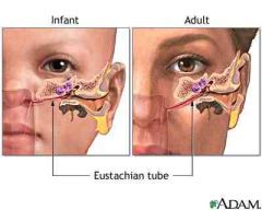
How long is the Aud. Tube in adults?
|
Usually 35-38 mm
|
|
|
|
Describe the path the Aud Tube takes (slope/direction) into the nasopharynx.
|
Directed downward (slope as it drops down into the nasopharynx is about 45 degrees); Forward (angle of about 35 degrees); and medially.
|
|
|
|
What are the 4 sections of the Aud. Tube?
|
Osseus, Cartilaginous, Membraneous, Isthmus
|
|
|
|
What is the Lumen?
|
Opening to the Aud. Tube FROM THE MIDDLE EAR
|
|
|
|
What are the two openings to the Aud tube and how are they different?
|
Lumen is in osseus part of tube, 3-6mm, usually open and is 3 mm above floor of middle ear. Cartilaginous opening is between the lateral and medial cartilage and is the entry into the nasopharynx. It usually closed
|
|
|
|
What part of the aud tube is the narrowest?
|
Isthmus
|
|
|
|
How long is the ossoeus part of the aud tube? Where does the osseous part begin?
|
12-14 mm; @ carotid wall just beneath the septum canalis musculotubari
|
|
|
|
What is the septum canalis musculotubari?
|
Thin sheet of bone that separates encasement for tensor tympani muscle and auditory tube
|
|
|
|
Why does the placement of the middle ear lumen problematic?
|
3 mm above floor of middle ear, which can cause drainage problems
|
|
|
|
Describe the transition through the aud tube from the osseus to cartilaginous
|
Leave osseus portion proceed through the isthmus then enter cartilaginous portion where cartilage starts to appear as a prominence which increases till the end of the tube in the nasopharynx
|
|
|
|
Name the two portions of the cartilage of the aud tube
|
Lateral and Medial
|
|
|
|
What two muscles are associated with the auditory tube
|
Tensor Palatini and Levator Palatine
|
|
|
|
In terms of the aud tube what do the Tensor Palatine and Levator Palatini muscles do?
|
Work in combo to pull lateral cartilage away from medial cartilage to open the lumen of the aud tube.
|
|
|
|
Explain the auditory reflex arc.
|
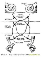
The path of an acoustic stimulus that ascends from the outer ear to the brainstem and then descends via the facial nerves on both sides of the head to innervate the stapedial muscles in both middle ears.
|
A reflex occurs when a signal is transmitted along a sensory neuron to an interneuron to a motor neuron causing a contraction of the muscle tissue innervated by the motor neuron. This is below the level of cognitive control; in other words, patients do not have to think about it. This is why one pulls a hand quickly away from a hot pot without thinking first, “Wow, my hand is burning” (that comes later) and this is why the stapedius muscle contracts in response to a loud sound, even though the patient does not consciously control the action. When a loud sound enters a normal ear, the stapedius muscle will contract on both sides regardless of which ear is stimulated. Therefore, the (acoustic reflex) is a bilateral (“two side”) reflex.
Imagine first a normal right ear and trace the pathway of a loud signal. The signal enters the right ear, travels through the outer, middle (ME) and inner ear (IE), along the VIII nerve, to the brainstem. When the signal reaches the brainstem, the signal arrives first at the cochlear nucleus (CN). From here, the signal travels to both right and left superior olivary complexes and both right and left facial nerve (VII) nuclei. The signal is sent from both facial nerve nuclei to both facial (VII) nerves, which results in a contraction of both stapedius muscles. Thus, both stapes bones are pulled outward and downward, in a direction away from the inner ear. This action makes it harder for energy to travel through the middle ear (increase in impedance/decrease in admittance). |
|
|
What are the 2 muscles associated with the auditory tube? What do they do?
|
Tensor Palatini and Levator Palatini; work in combo to pull lateral cartilage away from medial cartilage to open the lumen of the aud. tube
|
|
|
|
How many times per day does the aud. tube open and under what conditions usually?
|
1000 x per day mainly under reflexive conditions to circulate air
|
|
|
|
What is the Pharyngeal ostium?
|
Entrance into hollow organ or cavity where auditory tube enters nasopharynx
|
|
|
|
What is the Torus Tubaris?
|
Prominence where the aud tube comes into the nasopharynx; The base of the cartilaginous portion of the Eustachian tube lies directly under the mucous membrane of the nasal part of the pharynx, where it forms an elevation
|
|
|
|
What is the function of the Aud Tube?
|
Equalize air pressure of middle ear; drainage of normal and diseased secretions of middle ear
|
|
|
|
Origin and attachment for Superior Malleolar
|
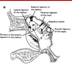
Head of Malleus to Tegmen Tympani
|
|
|
|
Origin and Attachment for Lateral Malleolar Ligament
|

Neck of Malleus to Boney Wall Near Notch of Rivinus
|
|
|
|
Origin and attachment for the Anterior Malleolar Ligament
|

Anterior process to the carotid wall
|
|
|
|
Origin and attachment for the Posterior Ligament of the Incus
|

Tip of short process to Fossa Incudis
|
|
|
|
Origin and attachment for the Annular Ligament of the Stapes
|

Ring like around Foot plate of the stapes secures into oval window
|
|
|
|
Name the 5 ligaments
|
Malleolar (Superior, Lateral, Anterior); Posterior Ligament of the Incus; Annular Ligament of the Stapes
|
|
|
|
What are the two tympanic muscles
|
Tensor Tympani and the Stapedial
|
|
|
|
3 characteristics of the tympanic muscles
|
smallest striated muscles; penate; encased in bone and it is only the tendon that enters the middle ear
|
|
|
|
Who discovered the tensor tympani and when
|
Eustachius and in 1564
|
|
|
|
Length and area characteristics of the Tensor Tympani
|
Length=25 mm
Cross section=5.85 mm2 |
|
|
|
Tensor Tympani Innervation and location in relation to auditory tube
|
Trigeminal/5
Lies superior and parallel to the aud. tube |
|
|
|
Origin and attachment of the Tensor Tympani
|
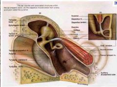
O-Cartilagenous portion of the aud. tube, just above the boney portion of the aud. tube
I-Malleus Handle |
|
|
|
Describe the contraction of the tensor tympani
|
Draws malleus medially and anteriorly; applies a force at a right angle to the rotation of the ossicles; increases tension of the TM.
|
When the tensor tympani is in a tensed state, it pulls the malleus in a medial position so the tympanic membrane is tensed as well. This reduces both the vibration and volume of sound in the middle ear, particularly the sounds created by the act of chewing.
|
|
|
Describe characteristics of stapedial muscle (length and area and innervation)
|
6mm; cross sectional area=5.0mm2
7th/Facial Nerve |
|
|
|
Compare and contrast stapedial and tensor tympani characteristics
|
stapedial: 6mm; area 5.0mm2; 7th facial nerve
Tensor Tympani: Trigeminal 5th nerve, length=25mm and area=5.85mm2 |
|
|
|
Origination and Insertion and Action of Stapedial Muscle
|
O=Boney canal parallel to the facial nerve canal in the posterior wall of the middle ear
I=Tendon inserts @ neck of stapes (muscle is almost verticle and tendon is almost horizonal at 90 degrees) A=Draws head of stapes posteriorly and exerts force at a right angle with the force of ossicles |
|
|
|
describe how the stapedial and tensor tympani work together
|
exert force in opposite directions perpendicular to the rotational axis of the ossicular chain
|
|
|
|
describe important features about tympanic muscle action
|
elicited by sound energy, reflexive in nature (intense sound 85dB and up triggers); elicited by either ipsilateral or contralateral stimulation
|
|
|
|
What is the major player in the accoustic reflex
|
stapedial muscle
|
|
|
|
Motor response initiated then descends through the facial nerves into and back to the middle ear and muscles...The name of this process is...
|
Acoustic Reflex Arc
|
|
|

Look at the pic and name the walls of the tympanic cavity
|
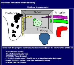
Ceiling=Tegumental
Inferior=Jugular Posterior=Mastoid Anterior=Carotid Lateral=Tympanic Membrane Medial=Labrynthian |
|