![]()
![]()
![]()
Use LEFT and RIGHT arrow keys to navigate between flashcards;
Use UP and DOWN arrow keys to flip the card;
H to show hint;
A reads text to speech;
154 Cards in this Set
- Front
- Back
|
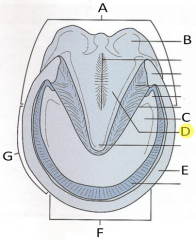
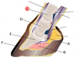
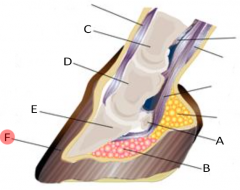
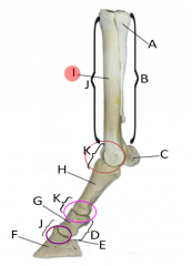
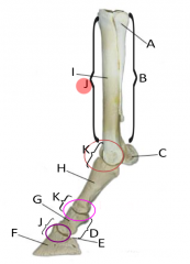
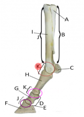
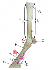
What is A?
|

Carpal pad |
|
|
What is B?
|

Digital pads |
|
|
What is C?
|
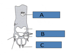
Claw |
|
|
What are dewclaws?
|
Remnants of first digit in small animals. Actual bone found in dewclaws of forelimbs.
|
|
|
Which hooves on ruminants are weight bearing?
|
Rear (3rd and 4th)
|
|
|
How many hooves do ruminants have per foot?
|
Four (including dewclaws)
|
|
|
How many hooves do horses have per foot?
|
One
|
|
|
What are the bones in a hoof?
|
•Coffin
•Navicular |
|
|
What are the sections in the wall of a hoof?
|
•External portion
•Laminae •Regions - Heel, Quarter, Toe |
|
|
Are horns solid or hollow?
|
Hollow in adults, communicates with sinuses
|
|
|
Where is the origin of horns?
|
Frontal bone
|
|
|
What is the main method of dehorning?
|
Hot iron dehorning
|
|
|
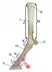
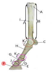
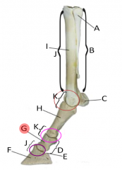
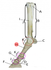
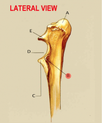
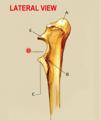
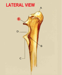
Where would you find a condyle?
|
•distal aspect of the femur and humerus along with the occipital condyles
•where the skull joins the spinal column |
|
|
Facet
|
flat articular surface
|
|
|
Process
|
•projections on the bone
•Usually sites where muscles attach |
|
|
What are the three types of complete fractures?
|
•Transverse
•Oblique •Comminuted |
|
|
What type of bones do birds have?
|
Pneumatic bones
|
|
|
Irregular bones
|
•Vertebrae
•Sesamoids •Patella |
|
|
What is the only joint in the skull?
|
temporomandibular joint
|
|
|
What does the axial skeleton consist of?
|
•Bones of the skull
•Vertebral column •Ribs •Sternum •Hyoid apparatus (cartilages) |
|
|
What does the appendicular skeleton consist of?
|
•Scapula/Clavicle
•Bones of the pelvis •Forelimbs •Hindlimbs |
|
|
Bones of the vertebral column
|
•Cervical
•Thoracic •Lumbar •Sacral •Coccygeal |
|
|
Intervertebral disk
|
in between vertebrae to provide cushioning and allow for increased movement
|
|
|
Rectus abdominis
|
straight muscles on either side of the linea alba
|
|
|
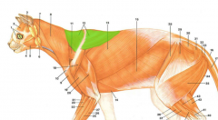
Cutaneous trunci
|
thin broad sheet of skeletal muscle just beneath the skin that some animals twitch
|
|
|
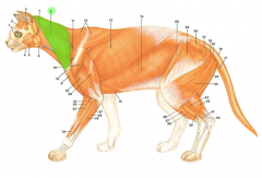
cleidobrachialis
|
Brachiocephalic muscle
|
|
|
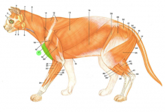
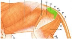
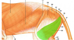
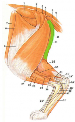
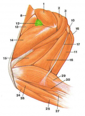
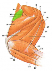
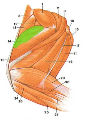
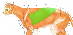
Superficial muscles of the shoulder area
|
•latissimus dorsi
•pectoral muscles •deltoid muscles |
|
|
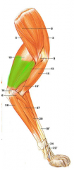
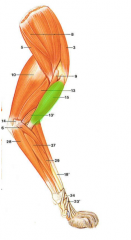
Muscles of the upper arm region
|
•biceps brachii
•triceps brachii |
|
|
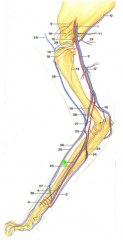
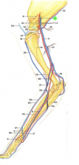
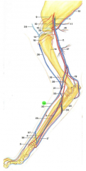
Radial nerve
|
•Largest nerve in front leg
•Extends elbow, carpus, and digits •provides sensation to the cranial lateral surface of the front leg and dorsal surface of the paw |
|
|
Sarcoplasmic reticulum
|
organelle that stores calcium
|
|
|
Main muscles of respiratory system
|
•Diaphragm
•external intercostal muscles |
|
|
Main expiratory muscles
|
•internal intercostal muscles
•abdominal muscles |
|
|
Abdominal muscles
|
•External abdominal oblique
•Internal abdominal oblique •Rectus abdominis •Transverse abdominis |
|
|
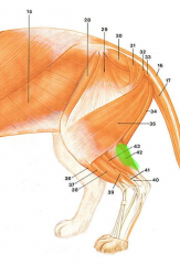
Pelvic limb muscles
|
•Sartorius/Gracilis
•Gluteal muscles •Quadriceps •Biceps femoris •Semimembranosus/semitendinosus •Gastrocnemius/calcanealtendon |
|
|
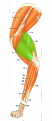
Quadricep muscles
|
•Vastus medialis
•Rectus femoris •Vastus lateralis |
|
|
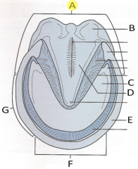
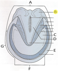
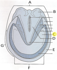
Heel |
|
|
Bulbs of heel |
|
|
Sole |
|
|
Frog |
|

|
Wall |
|
|
Toe |
|
|
Quarters |
|

#1 |
Coronet |
|

#2 |
Wall |
|

#3 |
Toe |
|

#4 |
Quarter |
|

#5 |
Heel |
|

#6 |
Bulb |
|
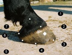
#7 |
P2 (Small pastern) |
|

#2 |
Bulb
|
|

#3 |
Frog |
|
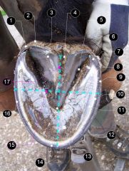
#6 |
Heel |
|

#7 |
Bar |
|

#11 |
White line |
|

#12 |
Frog |
|

#13 |
Sole |
|

#14 |
Toe |
|

#16 |
Quarter |
|

|
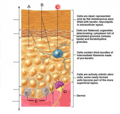
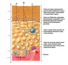
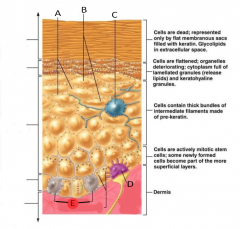
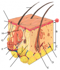
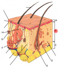
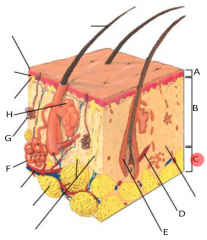
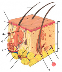
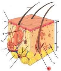
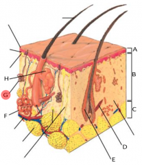
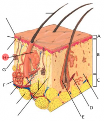
Stratum corneum |
|

|
Stratum granulosum |
|

|
Stratum spinosum |
|

|
Stratum basale |
|
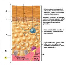
|
Dermis |
|
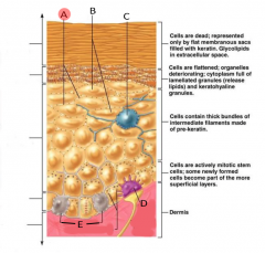
|
Desmosomes |
|
|
Keratinocytes |
|
|
Langerhans' Cells |
|
|
Merkel cell |
|
|
Melanocytes |
|
|
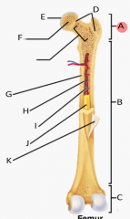
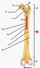
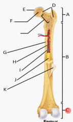
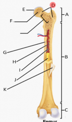
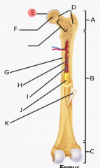
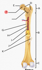
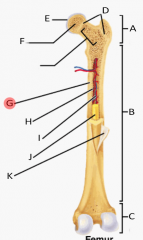
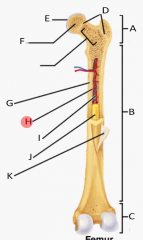
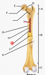
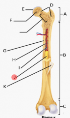
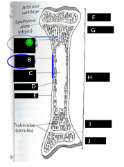
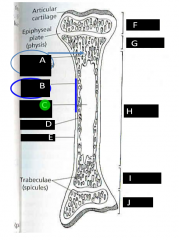
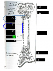
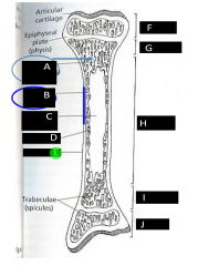
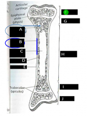
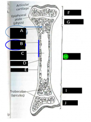
Parts of bone: Proximal epiphysis |
|
|
Parts of bone: Diaphysis |
|
|
Parts of bone: Distal epiphysis |
|
|
Parts of bone: Epipheseal disks |
|
|
Parts of bone: Articular cartilage |
|
|
Parts of bone: Spongy bone |
|
|
Parts of bone: Endosteum |
|
|
Parts of bone: Compact bone |
|
|
Parts of bone: Medullary cavity |
|
|
Parts of bone: Yellow marrow |
|
|
Parts of bone: Periosteum |
|
|
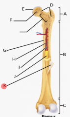
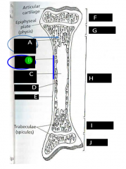
Parts of bone: Spongy (cancellous bone) |
|
|
Parts of bone: Compact bone |
|
|
Parts of bone: Medullary cavity |
|
|
Parts of bone: Endosteum |
|
|
Parts of bone: Periosteum |
|
|
Parts of bone: Epiphysis |
|
|
Parts of bone: Diaphysis |
|

|
Navicular bone |
|

|
Digital cushion |
|
|
Proximal phalanx (P1) |
|
|
Middle phalanx (P2) |
|
|
Distal phalanx (P3- Coffin bone) |
|
|
Hoof wall |
|
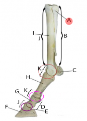
|
2nd and 4th metacarpals |
|
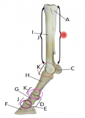
|
Splint bones |
|
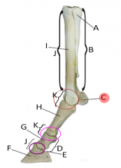
|
Proximal sesamoids |
|

|
Short pastern |
|
|
Distal sesamoid (Not visible due to distal phalanx) (Navicular bone) |
|
|
Distal phalanx (Coffin bone) |
|
|
Middle phalanx |
|
|
Proximal phalanx |
|
|
Third metacarpal |
|
|
Cannon bone |
|
|
Fetlock joint |
|
|
Pastern joint |
|
|
Epidermis |
|
|
Dermis |
|
|
Sub Q |
|
|
Arrector pili muscle |
|
|
Hair follicle |
|

|
Apocrine sweat gland |
|
|
Eccrine sweat gland |
|
|
Sebaceous gland |
|
|
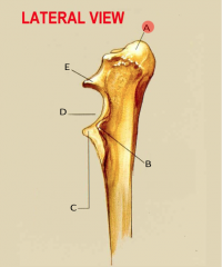
Olecranon |
|
|
Lateral coronoid process |
|

|
Medial coronoid process |
|
|
Trochlear notch |
|
|
Anconeal process |
|
|
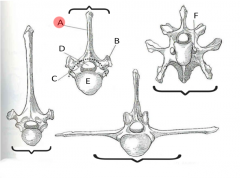
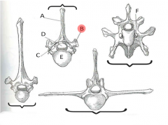
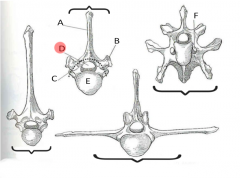
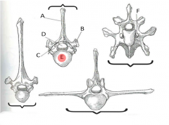
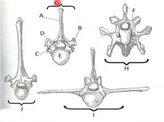
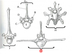
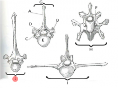
Spinous process |
|
|
Transverse process |
|
|
Vertebral foramen |
|
|
Body |
|

|
Transverse foramen |
|
|
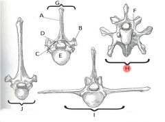
Typical vertebra |
|
|
Cervical vertebra |
|
|
Lumbar vertebra |
|
|
Thoracic vertebra |
|
|
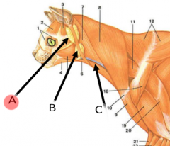
Parotid salivary gland |
|
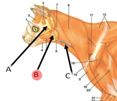
|
Mandibular salivary gland |
|
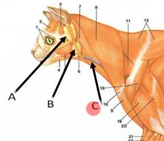
|
External jugular vein |
|
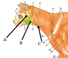
|
Masseter muscle |
|

|
Pectoral muscle |
|
|
Medial saphenous vein |
|
|
Cleidocervicalis |
|
|
Cleidobrachilas |
|
|
Trapezius |
|
|
Heads of the triceps brachii |
|
|
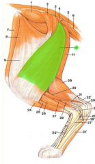
Biceps femoris |
|
|
Gracilis |
|
|
Sartorius |
|
|
External abdominal oblique |
|
|
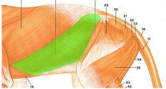
Gluteal muscles |
|
|
Biceps femoris |
|
|
Heads of triceps brachii |
|
|
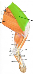
Gastrocnemius |
|
|
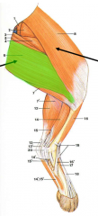
Semimembranosus |
|
|
Brachial artery |
|
|
Axillary artery |
|

|
Median artery |
|
|
Cephalic vein |
|
|
Vastus medialis |
|
|
Vastus femoris |
|
|
Vastus lateralis |
|
|
Latissimus dorsi |
|
|
Biceps brachii |