![]()
![]()
![]()
Use LEFT and RIGHT arrow keys to navigate between flashcards;
Use UP and DOWN arrow keys to flip the card;
H to show hint;
A reads text to speech;
49 Cards in this Set
- Front
- Back
|
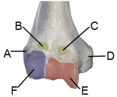
Name the three bones at the elbow joint |
Radius Humerus Ulna |
|
|
A: lateral epicondyle B: radial fossa C: coronoid fossa D: medial epicondyle E: trochlea F: capitulum |
|
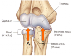
Name the joints |
A: humero-radial joint B: humero-ulna joint |
|
|
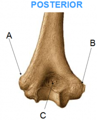
A: lateral epicondyle B: medial epicondyle C: olecranon fossa |
|
|
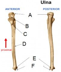
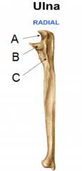
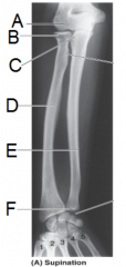
A: olecranon process B: coronoid process C: ulnar tuberosity D: interosseous crest E: head F: styloid process |
|
|
A: trochlear notch B: radial notch C: supinator crest |
|
|
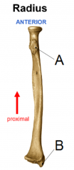
A: radial (bicipital) tuberosity B: styloid process |
|
|
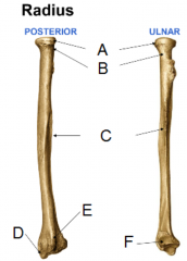
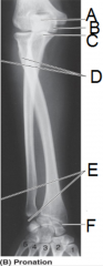
A: head B: neck C: interosseous crest D: styloid process E: dorsal (Lister's) tubercle F: ulnar notch |
|
|
What types of movement are allowed at the elbow joint? |
flexion/extension and the humero-radial and humero-ulna joint pronation/supination at the proximal radio-ulnar joint |
|
|
What feature allows some people to hyper extend their arm? |
Some people have an olecranon foramen instead of an olecranon fossa. This allows part of the olecranon to enter the foramen, allowing hyperextension. |
|
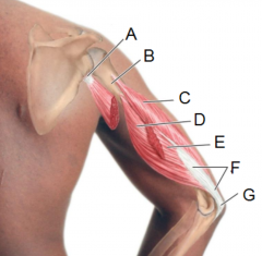
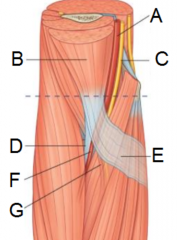
ANTERIOR |
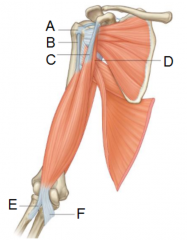
A: transverse humeral ligament B: long head of biceps brachii C: short head of biceps brachii D: coracobrachialis E: tendon of biceps brachii F: bicipital aponeurosis |
|
|
What are the three flexor muscles of the arm? Which of the three is considered to be an accessory flexor? |
Brachialis, biceps brachii and brachioradialis. Brachioradialis is an accessory flexor - it helps flexion when the forearm is mid-pronated. |
|
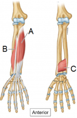
ANTERIOR |
A: brachialis muscle B: bicipital aponeurosis (cut) C: tuberosity of ulna D: radial tuberosity E: brachioradialis |
|
|
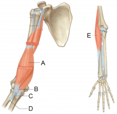
Which muscle(s) allow extension of the elbow? |
triceps brachii: long, lateral and medial head |
|
|
A: attachment of long head of triceps to infraglenoid tubercle of scapula B: shaft of humerus C: lateral head D: medial head E: long head (cut) F: triceps tendon G: attachment at olecranon process of ulna |
|
|
Where is the common extensor origin and common flexor origin, respectively. |
Common extensor origin - lateral epicondyle Common flexor origin - medial epicondyle |
|
|
What attaches to the medial and lateral epicondyles, respectively |
Lateral epicondyle - superficial extensor muscles Medial epicondyle - superficial flexor muscles |
|
|
What is the correct term for tennis elbow, and what causes it? |
lateral epicondylitis resisting/limiting wrist extension (eccentric) |
|
|
What is the correct term for golfer's elbow, and what causes it?
|
medial epicondylitis resisting/limiting wrist flexion (eccentric) |
|
|
What is the common treatment for epicondylitis? |
Rest and injection of corticosteroids if severe |
|
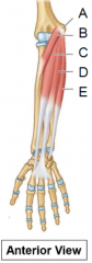
superficial/intermediate layers |
A: common flexor tendon B: pronator teres C: flexor carpi radialis D: palmaris longus E: flexor carpi ulnaris |
|
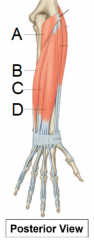
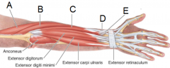
superficial |
A: anconeus B: extensor carpi ulnaris C: extensor digiti minimi D: extensor digitorum |
|
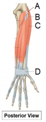
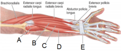
superficial |
A: common extensor tendon B: extensor carpi radialis longus C: extensor carpi radialis brevis D: extensor retinaculum |
|
|
What do the following words correspond to; A: opponens B: carpi C: digitorum D: pollicis E: digiti minimi |
A: rotates bone along longitudinal axis B: wrist C: fingers D: thumb E: little finger |
|
|
What do the following words correspond to;
A: indicis B: profundus C: palmaris D: brevis E: teres |
A: index finger B: deep C: palm D: shorter than something E: rounded or cylindrical |
|
|
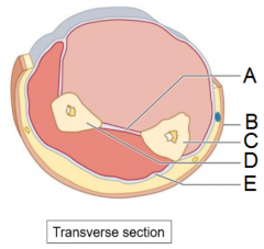
The interosseous membrane divides the forearm into what two compartments? |
anterior/flexor compartment posterior/extensor compartment |
|
|
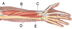
A: interosseous membrane B: skin C: shaft of ulna D: shaft of radius E: antebrachial fascia |
|
|
Which nerve innervates the muscles of the superficial and intermediate muscle layers of the anterior compartment? |
All muscles are innervated by the musculotaneous nerve, except for flexor carpi ulnaris, which is innervated by the ulnar nerve. |
|
intermediate (anterior) |
A: humero-ulnar joint B: radial head C: flexor digitorum superficialis |
|
|
In the deep muscle layer of the anterior compartment, which muscles are not innervated by the musculocutaneous nerve? |
medial half of the flexor digitorum profundus which is supplied by the ulnar nerve |
|
deep muscles |
A: flexor digitorum profundus B: flexor pollicis longus C: pronator quadratus |
|
|
In the anterior compartment of the forearm, which muscles are not innervated by the musculocutaneous nerve? |
flexor carpi ulnaris (ulnar nerve) medial half of the flexor digitorum profundus (ulnar nerve) |
|
superficial muscles (posterior) |
A: anconeus B: extensor digitorum C: extensor digiti minimi D: extensor carpi ulnaris E: extensor retinaculum |
|
superficial muscles (posterior) |
A: brachioradialis B: extensor carpi radialis longus C: extensor carpi radialis brevis D: abductor pollicis longus E: abductor pollicis brevis |
|
|
Which nerve innervates the superficial and deep muscles of the posterior compartment of the forearm? |
radial nerve |
|
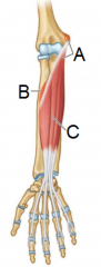
deep, posterior |
A: supinator B: abductor pollicis longus C: extensor pollicis brevis D: extensor pollicis longus E: extensor indicis |
|
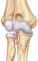
Name the circled joint |
radio-ulnar joint |
|

|
A: radial collateral ligament B: anular ligament of radius C: ulnar collateral ligament D: sacciform recess of synovial membrane |
|
|
What is the function of the anular ligament of the radius? |
1. stabilise the radius 2. allow rotation of the radius for pronation/supination |
|
|
A: capitulum B: head of radius C: proximal radio-ulnar joint D: radius E: ulna F: distal radio-ulna joint |
|
|
A: olecranon B: trochlea C: coronoid process D: radial tuberosity E: site of articular disc of distal radio-ulnar joint F: radial styloid process |
|
|
Which muscles are used for pronation? What nerves innervate these muscles? |
pronator teres and pronator quadratus median nerve |
|
|
Which muscles are used for supination?
What nerves innervate these muscles? |
supinator and biceps brachii radial and musculocutaneous |
|
|
What is the name of the triangular shaped depression anterior to the elbow? |
cubital fossa |
|
|
What muscles mark the lateral and medial borders of the cubital fossa? |
lateral: brachioradialis medial: pronator teres |
|
|
What signifies the floor and roof of the cubital fossa? |
floor: brachialis roof: deep fascia reinforced by the bicipital aponeurosis (broad, flat tendon) |
|
|
A: brachial artery B: biceps brachii C: median nerve D: biceps brachii tendon E: bicipital aponeurosis F: radial artery G: ulnar artery |
|
|
Which tendon, nerves and artery are found in the cubital fossa? |
biceps brachii tendon median nerve and radial nerve brachial artery |
|
|
What are the arteries which enter and leave the cubital fossa? |
In: brachial artery Out: brachial bifurcates into radial and ulnar arteries |