![]()
![]()
![]()
Use LEFT and RIGHT arrow keys to navigate between flashcards;
Use UP and DOWN arrow keys to flip the card;
H to show hint;
A reads text to speech;
64 Cards in this Set
- Front
- Back
|
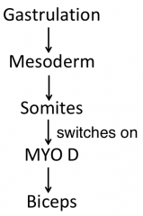
what is a somite |
A somite is a division of the body of an animal or embryo |
|
|
what musculature is derived from somites |
Musculature of the axial skeleton, body wall and limbs are derived from somites |
|
|
in their formation, somites undergo epithelialisation, they are classified as either being part of the upper region or the lower region as part of this, name the components of the upper and lower region |
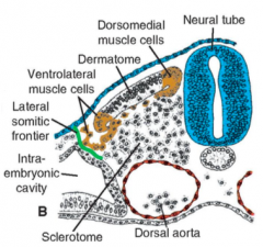
upper region: dermatome dorsomedial muscle cells ventrolateral muscle cells
lower region: vertebrae and ribs |
|
|
the dermatome is directed to become the dermis by what and from what location |
by neutrophin-3 from the dorsal neural tube |
|
|
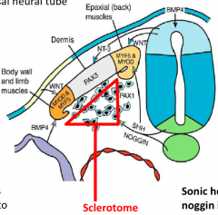
what sequence of events combine for the expression of MYF5 |
WNT (activating) and BMP (inhibitting) combine to form MYOD which creates a group of muscle cell precursors which expresses MYF5 |
|
|
what are MYOD and MYF5 |
they are myogenic regulatory factors, they can: -activate muscle specific genes -convert non-muscle cells to muscle cells |
|
|
what induces sclerotome formation |
sonic hedgehog and noggin |
|
|
where is the scerotome |
|
|
|
what are the two mesodermal domains |
the primaxial and abaxial domain |
|
|
what is the lateral somitic frontier |
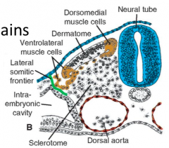
it is a well defined border between each somite and the lateral plate mesoderm |
|
|
what two mesodermal domains are separated by the lateral somatic frontier |

the primatial domain and the abaxial domain |
|
|
what is the primaxial domain |
the region around the neural tube, somite derived mesoderm |
|
|
what is the abaxial domain |
parietal layer of lateral plate mesoderm and some migratory somitic cells |
|
|
the lateral somatic frontier also separates two other things other than the primaxial and abaxial domains, what are these? |
also separates the dermis derived from the dermatome, and the dermis derived from the lateral plate mesoderm in the body wall |
|
|
the signals controlling the development of the primaxial and abaxial domains of the mesoderm come from different places, where does the primaxial domain get its signals from |
Primaxial: many from neural tube and notochord |
|
|
the signals controlling the development of the primaxial and abaxial domains of the mesoderm come from different places, where does the abaxial domain get its signals from |
Abaxial: many from lateral plate mesoderm |
|
|
each spinal nerve divides into two nerve segments, what are these called |
the dorsal primary ramus the ventral primary ramus |
|
|
what are the epaxial muscles |
the true muscles of the back |
|
|
what are the hypaxial muscles |
the limb and body wall |
|
|
are the epaxial (true back muscles) innervated by the dorsal or ventral primary ramus |
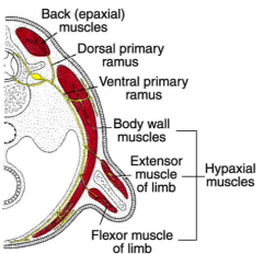
dorsal primary rami |
|
|
are the hypaxial (limbs and body wall) innervated the by the dorsal or ventral primary ramus |

ventral primary ramus |
|
|
Molecular signals for muscle induction arise from tissue which is where in relation to prospective muscle cells |
Molecular signals for muscle induction arise from tissue adjacent to prospective muscle cells |
|
|
what is gastrulation |
Gastrulation is a phase early in the embryonic development of most animals, during which the single-layered blastula is reorganized into a trilaminar ("three-layered") structure known as the gastrula. These three germ layers are known as the ectoderm, mesoderm, and endoderm |
|
|
give a summary of how muscles are made |
|
|
|
in embryonic development, when do the 'limb buds' first become visible |
week 4 |
|
|
when does limb morphogenesis occur |
weeks 4-8 |
|
|
is the lower limb development slightly faster or slower than upper limb development |
sightly slower, however it catches up by the end of the developmental period |
|
|
from which embryonic part is the mesenchyme derived from |
from the dorsolateral cells of the somites |
|
|
what type of tissue dictates the pattern of muscle formation |
connective tissue |
|
|
since connective tissue dictates the pattern of muscle formation, what layer is connective derived from |
parietal layer of lateral plate mesoderm |
|
|
lim buds consists of a mesenchymal core, from where is this core derived |
From parietal layer of lateral plate mesoderm |
|
|
what is the mesenchymal core covered by |
the cuboidal ectoderm |
|
|
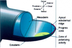
when the ectoderm is growing, what area of it "thickens" |
the distal border thickens |
|
|
what is the distal border of the ectoderm where the ectoderm thickens also known as |
the 'progress zone' or APICAL ECTODERMAL RIDGE |
|
|
does the apical ectodermal ridge ever differentiate? |
no, it always remains undifferentiated |
|
|
as the limb grows, at what places do the cells differentiate and what do they differentiate into |
the cells that are farthest form the Apical ectodermal ridge, differentiate the most and they differentiate into cartilage and muscle |
|
|
what are the 2 main things the Apical Ectodermal Ridge is responsible for |
limb development key signalling centre |
|
|
development of limbs occurs in what direction |
proximodistally |
|
|
what are the 3 components of the proximodistal limb development |
stylopod zeugopod autopod |
|
|
what are the components in the stylopod zeugopod and autopod subsequently |
– Stylopod – humerus and femur – Zeugopod – radius/ulna and tibia/fibia – Autopod – carpels, metacarpals, digits, tarsals/metatarsals |
|
|
at week 6 the terminal portion of the 'buds' become flattened to form what 2 different entities |
handplates footplates |
|
|
how do the fingers form |
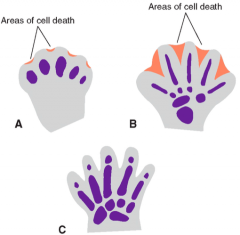
• Cell death in the AER separates ridges into 5 parts – 5 digits grow out under influence of 5 ridge parts • Mesenchyme condenses to form cartilaginous digits • By d56, digit separation is complete |
|
|
roughly how far behind is the development of the lower limbs compared with the upper limbs |
1-2 days |
|
|
when does limb rotation occur in the embryo |
week 7
|
|
|
what direction do the upper and lower limbs rotate in |
upper limbs --> 90o laterally lower limbs --> 90o medially |
|
|
limb outgrowth is regulated by what growth factor and from where is it secreted |
FGF10 secreted by lateral plate mesoderm |
|
|
what is the AER induced by |
BMPs |
|
|
what does the RADICAL FRINGE in the dorsal limb do |
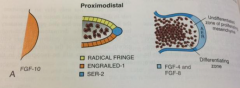
restricts AER to the distal tip and induces SER2 |
|
|
what maintains the undifferentiated zone |

FGF 4 and FGF 8 |
|
|
the positioning of the limbs along the craniocaudal axis is controlled by what gene |
the HOX gene |
|
|
in what sort of way is the HOX gene expressed |
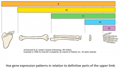
|
|
|
what transcription factors are expressed in the upper limbs |
TBX-5 |
|
|
what transcription factors are expressed in the lower limbs |
TBX-4 and PITX1 |
|
|
from where do limbs arise from |
the lateral mesoderm and overlying ectoderm |
|
|
explain how bone limb development occurs |
• As the external shape is being established, mesenchyme in the buds becomes condensed • Cells differentiate into chondrocytes seen • Areas where chondrogenesis is arrested makes joints – Cell proliferation, increased density, differentiation then cell death – induced by WNT 14 |
|
|
when is the first cartilage laid down in bone development |
week 6 |
|
|
by what week are the primary ossification centres present in all long bones |
Primary centres of ossification are present in all long bones by week 12 |
|
|
list the stages in limb bone development |
Cells in centre of cartilage model proliferate, enlarge, make new kind of matrix – can be calcified – calcified cartilage matrix does not allow diffusion of nutrients, so cartilage cells die –left with spicules of calcified cartilage matrix –acts as a scaffolding on which bone can be deposited Periosteum – vascular connective tissue around the model – blood vessels grow in – bringing progenitor cells Osteoprogenitor cells become osteoblasts, line up on spicules and start producing bone matrix Core of calcified cartilage matrix is removed by osteoclasts Trapped osteoblasts – become osteocytes |
|
|
during childhood in what directions do bones grow |
in both length and width |
|
|
define interstitial growth |
growth throughout the tissue |
|
|
what is appositional growth |
growth at the bone surface |
|
|
cartilage is capable of interstitial growth, is bone? |
nope |
|
|
when do growth plates 'close' and what happens to them |
they close at puberty they are converted to bone |
|
|
during the growth period, what is the remodelling of bones done by? and why |
it is done by osteoclasts to retain overall shape and proportion |