Reading...
![]()
Play button
![]()
Play button
![]()
Use LEFT and RIGHT arrow keys to navigate between flashcards;
Use UP and DOWN arrow keys to flip the card;
H to show hint;
A reads text to speech;
71 Cards in this Set
- Front
- Back
|
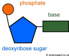
DNA structure
|
|
|
|
What are the four different bases?
|
adenine, thymine, guanine, cytosine
|
|
|
which kind of bond links the bases?
|
hydrogen bond
|
|
|
How are the DNA strands arranged?
|
the two strands have their 3' and 5' terminals at opposite ends - they are anti-parallel
|
|
|
In which direction does DNA replication occur?
|
DNA replication can only occur in a 5' --> 3' direction so a different method is needed for the two Strands.
|
|
|
Adenine and guanine are ...
|
purines
|
|
|
Thymine and cytosine are ...
|
pyrimidines
|
|
|
Adenine links to ...
|
thymine
|
|
|
Guanine links to ...
|
Cytosine
|
|
|
What is a nucleotide?
|
|
|
|
(DNA replication) The cell ...
|
produces many free nucleotides. Each has three phosphate groups (deoxyribonucleoside triphosphate). Two phosphates are removed during replication to release energy.
|
|
|
(DNA replication) Helicase ...
|
uncoils the DNA double helix and splits it into two template strands.
|
|
|
(DNA replication) DNA polymerase III ...
|
adds nucleotides in a 5' to 3' direction. On obe strand it moves in the same direction as the replication fork. On the other template strand it moves in the opposite direction.
|
|
|
(DNA replication) RNA primase ...
|
adds a short length of RNA attached by base pairing to the template strand of DNA. This acts as a primer, allowing DNA polymerase to bind and begin replication.
|
|
|
(DNA replication) DNA polymerase III ...
|
starts replication next to the RNA primer and adds nucletides in 5' to 3' direction. It therefore moves away from the replication fork on this Strand.
|
|
|
(DNA replication) DNA polymerase I ...
|
removes the RNA primer and replaces it with DNA. A nick is left where two nucleotides are still unconnected.
|
|
|
(DNA replication) DNA ligase ...
|
seals up the nick by making another sugar-phosphate bond.
|
|
|
What kind of DNA do prokaryotes have?
|
naked DNA which consists mostly of single copy genes that are transcribed and translated without modification.
|
|
|
What are replication initiation sites?
|
special initiation points where the replication of DNA begins
|
|
|
Where are replication initiation sites?
|
many along each chromosome (eukaryotes)
only one point on their DNA molecule (prokaryotes) |
|
|
nucleosomes?
|
globular structures that contain eight histone proteins, with DNA wrapped around
|
|
|
Functions of nucleosomes? (2)
|
1. they help to package up the DNA during mitriss and meiosis by the process of supercoiling
2. they can be used to mark particular genes |
|
|
repetitive sequences
|
much of the DNA consists of repetitive sequences which are not translated
|
|
|
introns
|
sequences of DNA that are transcribed but not translated
|
|
|
exons
|
sequences of DNA that are transcribed and translated
|
|
|
(Transcription) RNA polymerase ...
|
Splits the DNA into two strands. One of the strands forms the template for transcription. The base sequence of the mRNA is complementary to it.
|
|
|
The Strand that forms the template and is transcribed is called ...
|
the antisense strand
|
|
|
sense strand
|
the strand that has the same base sequence as the mRNA (except for U instead of T)
|
|
|
What is the function of mRNA?
|
it carries the information needed for making polypeptides out from the nucleus to the cytoplasm of eukarytotic cells. The information is in coded form which is decoded in Translation.
|
|
|
Translation
|
the base sequence of the mRNA is translated into the amino acid sequence of a polypeptide.
|
|
|
The genetic code is a ...
|
triplet code
|
|
|
triplet code?
|
three bases code for one amino acid
|
|
|
codon?
|
a group of three bases, there are 64 different codons (4^3)
|
|
|
All tRNA molecules have
|
- sections that become souble stranded by base pairing, creating loops
- a triplet of bases called anticodon, in a loop of seven bases - two other loops - the base sequence CCA and the 3' Terminal, which forms a site for attachement of amino acids |
|
|
tRNA activating enzyme
|
attaches the correct amino acid to the 3' terminal of the tRNA, energy from ATP is needed for the attachment
|
|
|
What are the features of ribosomes? (4)
|
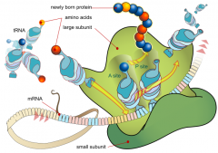
1. consist of proteins and ribosomal RNA molecules
2. two subunits, one large and one small 3. 3 binding sites for tRNA, 2 tRNA can bind at the same time 4. binding site for mRNA |
|
|
free ribosomes
|
in the cytoplasm
|
|
|
bound ribosomes
|
on the endoplasmic reticulum
|
|
|
function of ribosomes?
|
site of polypeptide synthesis
|
|
|
peptide bond
|
linkage between amino acids
|
|
|
polysomes
|
groups of ribosomes moving along the same mRNA, as they simultaneously translate it
|
|
|
polypeptide elongation
|
1. one of the binding sites for tRNA is vakant. The small subunit of the ribosome esures that only a tRNA with the anticodon that is complementary to the next codon binds to it.
2. The large subunit of the ribosome advances over the small subunit and detaches the polypeptide from the tRNA shown on the left. The polypeptide is attached by a peptide linkage to the single amino acid held by the tRNA shown on the right. 3. The small subunit slides across the large subunit. At the same time it moves three nucleotides on along the mRNA in a 5' to 3' direction. Translation always occurs in a 5' to 3' direction along mRNA. 4. The tRNA shown on the left has been displaced into the third binding site, and detaches from the ribosome. It can be used again in Translation after a tRNA activating enzyme has added another amino acid to it. |
|
|
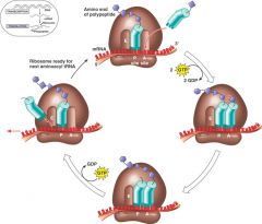
initiation
|
start the process of Translation
1. tRNA with the anticodon complementary to the start codon binds to the small subunit of the ribosome 2. the small subunit, carrying the tRNA binds to the 5' ebd of the mRNA 3. the small subunit slides along the mRNA until it reaches the start codon, which shows where Translation should be started. 4. The large subunit of the ribosome binds to the small subunit. 5. Another tRNA, with the anticodon complementary to the enxt codon on the mRNA, binds to the ribosome. Elongation of a polypeptide can now start. |
|
|
termination
|
stop the process of Translation
1. The ribosome moves along the mRNA in a 5' to 3' direction, translating each codon into an amino acid on the elongating polypeptide. 2. The ribosome reaches a stop codon. No tRNA molecule has the complementary anticodon. 3. The large subunit advances over the small subunit. The polypeptide is released and starts to fold into its final protein shape. 4. The tRNA detaches and the large subunit, small subunit and mRNA all separate. 5. Proteins synthesized by free ribosomes mostly remain and are used in the cytoplasm. Proteins synthesized by ribosomes bound to the ER are mostly excreted from the cell or are used in lysosomes. |
|
|
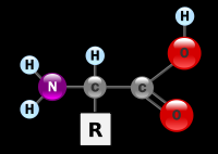
Draw an amino acid!
|
|
|
|
alpha helix
|
|
|
|
beta-pleated sheet
|
|
|
|
ionic bonds
|
form between positively and negatively charged R groups
|
|
|
hydrophobic interactions (weak bonds)
|
can form between R groups that are non-polar
|
|
|
hydrogen bonds
|
form between some R groups
|
|
|
disulfide bridges (strong covalent bonds)
|
form between pairs of cysteines
|
|
|
primary structure
|
the number and sequence of amino acid in a polipeptide, most polypeptides consist of between 50 and 1000 amino acid
|
|
|
secondary structure
|
regular repeating structures including alpha helices and beta-pleated sheets stabilized by hydrogen bonds between groups in the main chain of the polypeptide
|
|
|
tertiary structure
|
the three-dimensional conformation of a polypeptide stabilized by intramolecular bonds that form especially between R groups
|
|
|
quaternary structure
|
linking together of two or more polypeptides to form a single Protein
|
|
|
prosthetic group
|
non-polypeptide structure contained in some Proteins
|
|
|
conjugated Proteins
|
proteins with a prosthetic group
|
|
|
(Functions of Proteins): Structural. Example/Details/Shape
|
Example: Collagen
Details: strengthen bone, tendon and skin Shape: fibrous |
|
|
(Functions of Proteins): Transport. Example/Details/Shape
|
Example: Hemoglobin
Details: transports oxygen to different tissues Shape: Globular |
|
|
(Functions of Proteins): Movement. Example/Details/Shape
|
Example: Myosin
Details: cause contraction in muscle fibres and cause movement in animals Shape: Fibrous |
|
|
(Functions of Proteins): Defence. Example/Details/Shape
|
Example: Immunoglobin
Details: act as antibodies Shape: globular |
|
|
Polar amino acids
|
hydrophilic R groups
|
|
|
nonpolar amino acids
|
hydrophobic R groups
|
|
|
Position of polar amino acids in and out of membranes
|
- on the surface of proteins make the water soluble
- create channels through which hydrophilic substances can diffuse - positively charged R groups allow negatively charged ions through and vice versa - cause parts of the membrane proteins to protrude from the membrane |
|
|
Position of nonpolar amino acids in and out of membranes
|
- in the centre of water-soluble proteins stabilize their structure
- cause proteins to remain embedded in membranes |
|
|
activation energy
|
energy needed to start a chemical reaction
|
|
|
Competitive inhibition
|
- substrate and inhibitor are chemically very similar
- inhibitor binds to the active site of the Enzyme |
|
|
Non-competitive inhibition
|
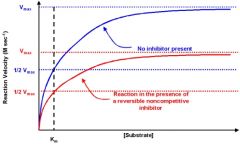
- substrate and inhibitor are not similar
- inhibitor binds to the enzyme at a different site from the active site |
|
|
features of metabolic pathways (6)
|
- many chemical reactions that are carried out in a particular sequence
- an enzyme catalazes each reaction - all reactions occur inside the cell - soma pathways build up organic compounds (anabolic pathways) and some break them down (catabolic pathways) - some metabolic pathways consist of a chain of reactions - some metabolic pathways consist of cycles of reactions |
|
|
end-product inhibition
|
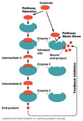
the product of the last reaction in the pathway inhibits the enzyme that catalazes the first reaction
|
|
|
allosteric Enzyme
|
The enzyme that is inhibited by the end products
|