![]()
![]()
![]()
Use LEFT and RIGHT arrow keys to navigate between flashcards;
Use UP and DOWN arrow keys to flip the card;
H to show hint;
A reads text to speech;
41 Cards in this Set
- Front
- Back
|
crown |
the portion of tooth above the gum |
|
|
neck |
the part of took where the root and gum meet |
|
|
root |
the portion of tooth below the gum |
|
|
enamel |
a cell free secretion that covers the tooth before it erupts if it is damaged it must artificially be repaired |
|
|
dentine |
hard yellow tissue that makes up the tooth |
|
|
pulp cavity |
a space occupied with pulp ( connective tissue, blood, lymphatic vessels ) |
|
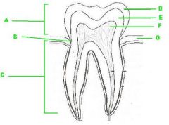
the tooth structure labeling |
A.Crown B.Neck C. Root D. Enamel E. Dentine F. pulp G GuMS |
|
|
4 types of teeth |
1. incisor 2. canine 3. premolar 4. molar |
|
|
incisor |
chisel like cutting teeth , used for biting |
|
|
canine |
more pointed than incisor - puncture and shred food |
|
|
premolar: |
broad surface for crushing and grinding |
|
|
molar: |
broad surface for crushing and grinding |
|
|
Function of the liver |
detoxifies and secretes bile |
|
|
lobule |
the whole unit ( with portal triads on the outside) -- small division of the liver |
|
|
central vein of a liver lobule |
after being processed by the hepatocytes , blood collects here- exits through the hepatic veins |
|
|
portal triad on a liver lobule |
bile duct, hepatic portal venules, hepatic arteries |
|
|
hepatocytes |
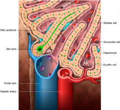
have brush border of microvilli that help regulate the contents of the blood |
|
|
visceral pleura |
serous membrane that covers each lung |
|
|
parietal pleura |
outer covering which is attached to the thoracic cavity |
|
|
what function does the space between the parietal and visceral pleura serve ( pleura cavity ) |
the space = helps reduce friction , create pressure gradient , create compartments |
|
|
trachea - tissue type |
pseudostratisfied columnar ciliated epithelium cells - contain goblet cells for mucus secretion |
|
|
bronchus - tissue type |
ciliated pseudostratisfied columnaar |
|
|
bronchioles |
ciliated cuboidal epithelium |
|
|
4 layers of the digestive system |
1.mucosa 2. sub mucosa 3. muscularis externa 4. serosa |
|
|
folds on internal of stomach |
gastric rague |
|
|
stomach portion closest to the esophagus is known as -- |
cardia region |
|
|
upper portion of the stomach after the cardia |
The fundus region |
|
|
the lesser vs the greater curvature of the stomach |
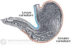
lesser is interior portion greater more exterior portion |
|
|
the pyloric region of the stomach |
the furthest region of the stomach - connects to the dueodenum |
|
|
What are the six parts of the large intestine |
1. cecum ( also where appendix comes off) 2.ascending colon 3.transverse colon 4. descending colon 5. Sigmoid colon 6. Rectum |
|
|
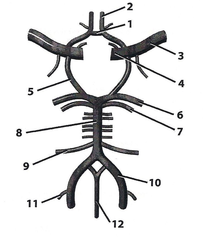
the aorta slits into what at the groin region |
the common illiac artery |
|
|
the illiac artery splits into what two arteries |
the internal illiac artery and external illiac artery |
|
|
from the anterior view the femoral artery is more anterior or posterior |
femoral = anterior looking from posterior view -- the artery will be the one underneath |
|
|
popliteal artery |
found in the knee region -- follow down from femoral artery |
|
|
the political artery splits into what two arteries |
the anterior and posterior tibial artery |
|

1. 3. 4. |
1. anterior communicating artery 3. middle cerebral artery 4. internal carotid artery ` |
|
5. 6. |
5. posterior communicating 6. posterior cerebral |
|
|
what connects the posterior cerebral artery to the vertebral artery |
basilar artery |
|

10. |
10. vertebral artery |
|
|
what are the three parts of the small intestine |
duodenum jejunum ileum |
|
|
the main artery of the abdomin |
celiac trunk |