![]()
![]()
![]()
Use LEFT and RIGHT arrow keys to navigate between flashcards;
Use UP and DOWN arrow keys to flip the card;
H to show hint;
A reads text to speech;
195 Cards in this Set
- Front
- Back
|
It is the job of the respiratory system to |
exchange carbon dioxide and oxygen |
|
|
Pulmonary ventilation |
Movement of air into and out of the lungs which results in exchange of gasses in alveoli of the lungs |
|
|
Pulmonary ventilation is also known as |
breathing |
|
|
External respiration |
Movement of oxygen from the lungs to the blood and carbon dioxide from the blood to the lungs |
|
|
Movement into the blood is called |
loading |
|
|
Movement out of the blood is called |
unloading |
|
|
Both oxygen and carbon dioxide move via |
diffusion |
|
|
External respiration occurs in the |
lungs |
|
|
Transport of respiratory gasses |
The circulatory system carries oxygen to the tissues of the body and carbon dioxide to the lungs for excretion |
|
|
Internal respiration |
Movement of oxygen from the blood into the tissues and carbon dioxide from the tissue into the blood |
|
|
In internal respiration oxygen is ____________ and carbon dioxide is _________________ |
unloaded, loaded |
|
|
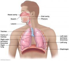
Conduction zone structures |
nasal cavity, nostril, oral cavity, pharynx, larynx, trachea, carina of the trachea, left primary bronchus, right primary bronchus |
|
|
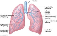
Respiratory zone structures |
superior lobe of the right lung, middle lobe of the right lung, inferior lobe of the right lung, superior lobe of the left lung, inferior lobe of the left lung |
|
|
Conduction zone |
respiratory passageways which provide fairly rigid conduits for air to reach the gas exchange sites |
|
|
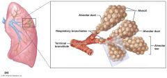
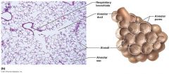
Respiratory zone |
actual site of gas exchange, composed of respiratory bronchioles, alveolar ducts, and alveoli |
|
|
Conduction zone picture |
|
|
|
Respiratory zone picture |
|
|
|
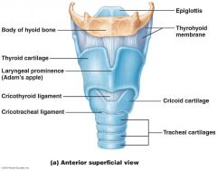
Epiglottis |
keeps food out of the respiratory pathways by covering the trachea and directing it toward the esophagus |
|
|
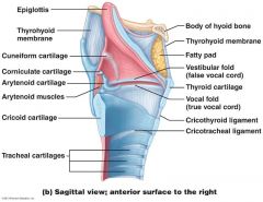
Vocal folds (true) |
vibrates and produces sound/voice as air rushes up from the lungs |
|
|
Glottis |
Opens and closes during vibrations. Functions in speech development and pronunciation |
|
|
Anterior superficial view of larynx |
|
|
|
Saggital view of structure of larynx |
|
|
|
Surface structures of the lungs |
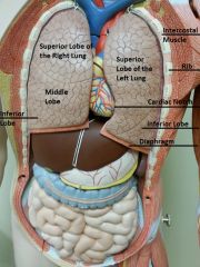
|
|
|
The lung is surrounded by parietal and visceral pleura which form a thin double layered |
serosa |
|
|
Parietal pleura |
outer layer that covers the thoracic wall and superior face of the diaphragm |
|
|
Visceral pleura |
inner layer that covers the external lung surface dipping into and lining its fissures |
|
|
Serous membranes of the lungs |
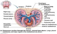
|
|
|
Lobule Structure |
|
|
|
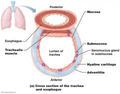
Cross section of the trachea and esophagus |
|
|
|
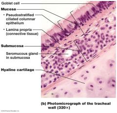
Photomicrograph of tracheal wall |
|
|
|
Histology of the Lung |
|
|
|
Systemic venous blood that is to be oxygenated in the lungs is delivered by the |
pulmonary arteries |
|
|
Pulmonary arteries branch profusely along with the bronchi and feed into the |
pulmonary capillary network |
|
|
Pulmonary circulation |
allow diffusion of oxygen into the blood but doesn't nourish the lungs |
|
|
Bronchial circulation |
bronchial arteries provide oxygenated systemic blood to lung tissue |
|
|
The tissues of the lungs are nourished by |
bronchial circulation |
|
|
Tidal volume |
normal, quiet breathing. 500mL average |
|
|
Vital capacity |
Total amount of exchangeable air (4800mL). Max amount of air that can be exhaled after a max inspiration (sum of TV, IRV, and ERV) |
|
|
Expiratory reserve volume |
amount of air that can be expelled from the lungs after a normal tidal volume expiration (1200mL) |
|
|
Inspiratory reserve volume |
amount of air that can be inspired forcibly beyond the tidal volume (3200 mL) forced inspiration after normal inspiration |
|
|
Minute respiratory volume |
Tidal volume multiplied by respiration per minute |
|
|
Residual volume |
about 1200mL of air that remains in the lungs which helps to keep the alveoli open and prevent lung collapse |
|
|
Bronchial sounds |
produced by air rushing through the large respiratory passageways (trachea and bronchi) |
|
|
Vesicular breathing sounds |
air filling the alveolar sacs and resembles the sound of a rustling or muffled breeze |
|
|
The digestive system |
ingests food then breaks down the food into nutrient molecules |
|
|
Enzymatic hydrolisis |
the addition of a water molecule to break bonds |
|
|
Absorption |
passage through cells lining the GI tract into the blood |
|
|
Digestion |
break down food into smaller fragments either physically or chemically |
|
|
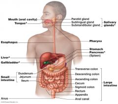
Organs of the digestive tract |
|
|
|
Accessory digestive organs |
teeth, tongue, gallbladder, large digestive glands, liver, and pancreas |
|
|
Order of the digestive tract |
mouth, esophagus, stomach, small intestine, large intestine, rectum, anus |
|
|
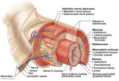
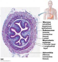
Mucosa |
epithelium, lamina propria, muscularis mucosae |
|
|
Functions of mucosa |
secretion, absorption, protection |
|
|
Submucosa |
dense connective tissue with blood and lymphatic vessels, lymph nodes, and nerve fibers |
|
|
Functions of submucosa |
holds the blood and lymphatic vessels that supply the tissues surrounding it |
|
|
Muscularis externa |
two layers of smooth muscle |
|
|
Functions of muscularis externa |
segmentation and peristalsis, intrinsic control of the digestive tract, includes muscle and myenteric nerve plexus, circular muscles form sphincters in some areas |
|
|
Serosa |
protective, outermost layer |
|
|
Histological structure of the alimentary canal |
|
|
|
Ingestion |
taking in food into the digestive tract |
|
|
Propulsion |
moves the food through the alimentary canal |
|
|
Mechanical breakdown |
increases the surface area of ingested food, physically, preparing it for digestion by enzymes |
|
|
Segmentation |
mixes food with digestive juices and makes absorption more efficient by repeatedly moving different parts of the food mass over the intestinal wall |
|
|
Oral cavity and pharynx view |
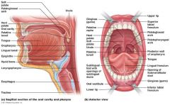
|
|
|
Histological structure of the esophagus |
|
|
|
Stomach |
where mechanical and chemical break down of bolus continues |
|
|
Cardiac region |
surrounds the cardial orifice through which food enters the stomach from the esophagus |
|
|
Fundus |
stomach's dome shape part, tucked beneath the diaphragm |
|
|
Body |
midportion of the stomach |
|
|
Pyloric region |
pyloric antrum, pyloric canal, pyloric sphincter |
|
|
Gastric gland secretions |
hydrochloric acid, and pepsinogen |
|
|
Mucosal gland secretions |
secrete mucus that prevents the stomach from being digested by the gastric gland secretions |
|
|
Stomach diagram |
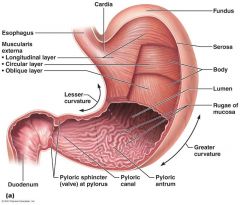
|
|
|
Gastric pits |
lead into gastric glands that produce the stomach secretion called gastric juice |
|
|
Pyloric sphincter |
controls stomach emptying and is located at the end of the stomach entering the small intestine |
|
|
Rugae |
wrinkly folds inside the stomach that appear when the stomach is empty and disappear when the stomach is full |
|
|
In what case might intrinsic factor be absent? |
Gastric bypass surgery |
|
|
Three sections of the small intestine |
duodenum, jejunum, illeum
|
|
|
Small intestine diagram |
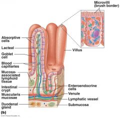
|
|
|
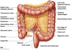
Large intestine functions |
absorb most of the remaining water from indigestible food residues, stores the residues temporarily, and then eliminates them from the body as semisolid feces |
|
|
Large intestine diagram |
|
|
|
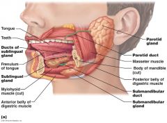
Salivary glands diagram |
|
|
|
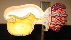
Gallbladder |
green, muscular sac that stores bile that is not immediately needed for digestion and concentrates it by absorbing some of its water and ions |
|
|
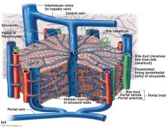
Liver microscopic structure |
|
|
|
Pancreas |
produces enzymes that breakdown all categories of foodstuffs |
|
|
Pancreas exocrine function |
produces pancreatic juice, includes hydrolytis enzymes and an alkaline fluid that neutralizes stomach acid |
|
|
Pancreas endocrine function |
produces the hormone insulin and glucagon |
|
|
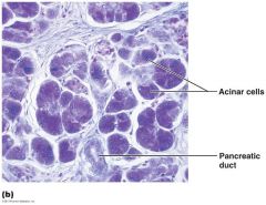
Pancreas microscopic view |
|
|
|
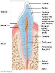
Tooth structure |
|
|
|
Kidney function |
excrete nitrogenous waste, filter the blood, maintain electrolyte balance, maintain fluid balance, maintain pH |
|
|
Kidneys |
produce urine which passes through the ureters to the urinary bladder |
|
|
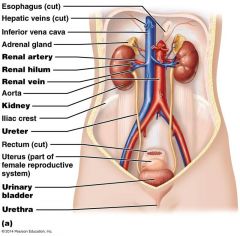
Urinary system diagram |
|
|
|
Position and coverings of kidneys |
lower back covered by fibrous capsule surrounded by perirenal fat capsule outermost covering of kidneys is renal fascia |
|
|
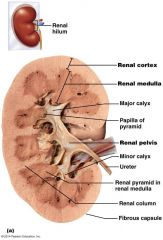
Gross anatomy of kidney |
|
|
|
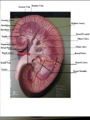
Gross anatomy of sheep kidney |
|
|
|
Nephrons |
structural and functional units of the kidneys and each kidney has over one million of them |
|
|
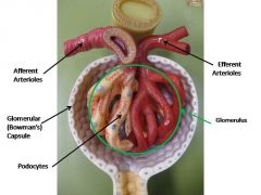
Structure and function of nephrons diagram |
|
|
|
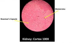
Glomerulus |
ball of fenestrated capillaries |
|
|
Parietal layer of the glomerular capsule |
the outer portion of the capsule forms the impermeable barrier that holds the filtrate |
|
|
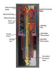
Renal tubule structure and function diagram |
|
|
|
Proximal convoluted tubule |
very coiled tube line with simple cuboidal epithelium with many microvilli to increase surface area |
|
|
Descending limb of Henle |
Continues tubular reabsorption so is also composed of cuboidal epithelium with microvilli |
|
|
Descending thin limb of nephron loop |
This portion of the descending limb of Henle is composed of simple squamous epithelium and is very permeable to water so water is reabsorbed here rather than nutrients |
|
|
Thick ascending limb of the nephron loop |
This portion of the renal tubule is NOT permeable to water |
|
|
Distal convoluted tubule |
Coiled and composed of simple cuboidal epithelium. It's responsible for re-absorption and secretion and conserves bodily fluids |
|
|
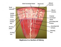
Nephrons in section of kidney diagram |
|
|
|
Urine is formed using the processes of |
glomerular filtration, tubular re-absorption, tubular secretion |
|
|
Blood flow through kidney |
renal artery, segmental arteries, interlobar arteries, arcuate arteries, cortical radiate arteries, afferent arterioles, glomerulus, efferent arterioles, peritubular capillaries, cortical radiate veins, arcuate veins, interlobar veins, renal vein |
|
|
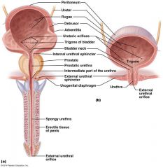
Urinary bladder diagram |
|
|
|
Trigone |
area of the bladder that forms the triangle between the ureter entrances and the urethra exit |
|
|
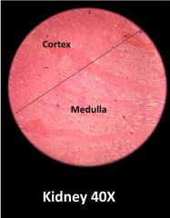
Kidney microscopic 40x |
|
|
|
Kidney microscopic slide 100x |
|
|
|
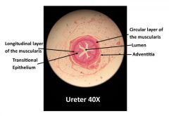
Ureter slide |
|
|
|
pH of fresh urine |
4.5 to 8.0 |
|
|
Normal components of urine |
water, urea, ions, creatinine, uric acid, other ions |
|
|
Nitriuria |
bacteria such as e. coli break nitrates to nitrites |
|
|
Pyuria |
presence of white blood cells or pus in the urine |
|
|
Zygote |
first cell of a new individual, from which all body cells will arise |
|
|
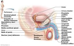
Anatomy of the male reproductive system |
|
|
|
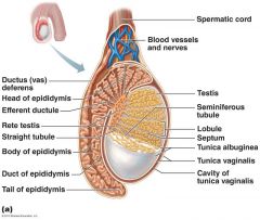
Septum |
divides the testes in the male |
|
|
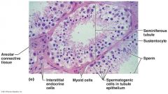
Testes diagram |
|
|
|
Where are immature sperm stored? |
Epididymis |
|
|
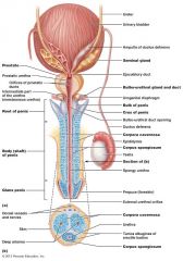
Three portions of the urethra |
prostatic urethra, intermediate part, spongy urethra |
|
|
Urethra diagram |
|
|
|
Prostate |
surrounds urethra just below urinary bladder secretion is milky white and activates sperm |
|
|
Anatomy of male reproductive organs |
|
|
|
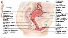
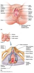
Anatomy of the female reproductive system |
|
|
|
External genitalia diagram |
|
|
|
Labia majora |
homologous to the scrotum of the male enclose two smaller hair free folds |
|
|
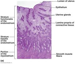
Myometrium |
function is to enlarge during the pregnancy |
|
|
Serosa (perimetrium) |
composed of a thin layer of connective tissue covered by mesothelium |
|
|
Anatomy of selected female reproductive organs |
|
|
|
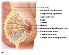
Mammary glands diagram |
|
|
|
Human gametes have |
23 chromosomes |
|
|
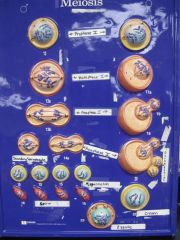
Meiosis 1 |
prophase, metaphase, anaphase, telophase, interkinesis |
|
|
Meiosis 2 |
prophase 2, metaphase 2, anaphase 2, telophase 2 |
|
|
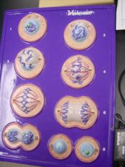
Mitosis diagram |
|
|
|
Meiosis diagram |
|
|
|
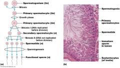
Human spermatogenesis begins at |
puberty |
|
|
Spermatogenesis diagram |
|
|
|
Oogenesis occurs in the |
ovaries |
|
|
Immature ovum develop within a |
follicle |
|
|
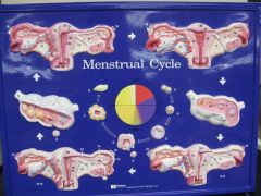
Menses (1-5 days) |
sloughing off the thick layer of the lining of the uterus; accompanied by bleeding |
|
|
Proliferative phase (6-14 days) |
estrogen causes the endometrium to repair, glands and vessels to proliferate, and the endometrium to become thick |
|
|
Secretory phase (15-28 days) |
progesterone causes the vascular supply to the endometrium to increase |
|
|
Menstrual cycle diagram |
|
|
|
Menopause |
cessation of woman's menstrual cycle occurs at ages 45 to 55 |
|
|
Embryology |
Study of the changes that occur in the developing human beginning at conception
|
|
|
3 things to happens to form a zygote |
number and size of cells must increase cells must specialize organ systems must develop |
|
|
From fertilization through week 8, a developing human is called an |
embryo |
|
|
Human ovum diagram |
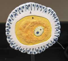
1. Corona radiata 2. zona pellucida 3. egg plasma with yolk 4. female pronucleus 5. spermatozoid |
|
|
Entering the spermatozoid diagram |
1. zona pellucida 2. fertilization membrane 3. Protoplasm 4. female pronucleus |
|
|
Formation of the second polocyte diagram |
1. chromosomes in the female pronucleus 2. spindle 3. male pronucleus 4. first polar body |
|
|
Oocyte pronucleus moves toward the center diagram |
1. female pronucleus 2. male pronucleus 3. first and second polar bodies |
|
|
Nucluear copulation diagram |
1. female pronucleus with maternal chromosomes 2. male pronucleus with paternal chromosomes 3. centrosome 4. first and second polar bodies |
|
|
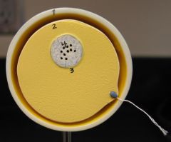
The zygote diagram |
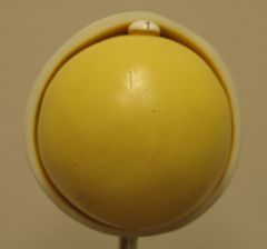
unicellular stage of the embryo the first cell that has the genetic composition of the new person |
|
|
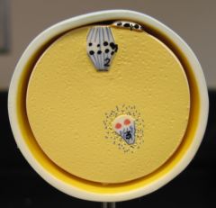
Cleavage diagram: Bi-cellular stage |
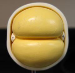
first and second polar bodies |
|
|
Cleavage |
perioid of rapid mitotic divisions of the zygote without growth of the cells |
|
|
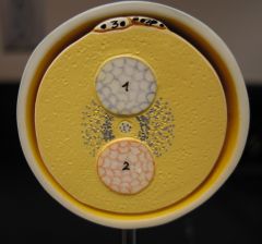
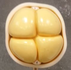
Four cell stage diagram |
|
|
|
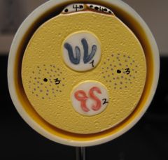
Sixteen cell stage diagram |
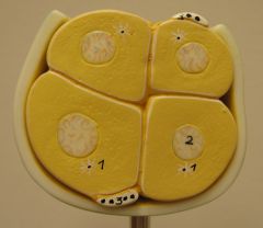
1. centriole 2. interphase nucleus 3. first and second polar bodies |
|
|
The morula |
a solid ball of 32 cells that looks like a blackberry |
|
|
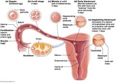
When the morula hollows out, it becomes a |
blastula |
|
|
Blastocyst |
composed of a single layer of trophoblast cells and inner mass cells |
|
|
The embryonic disc develops into the |
embryo |
|
|
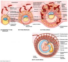
Beginning of implantation (6-7 days after fertilization) diagram |
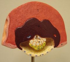
1. primary wall of the blastula 2. cavity of the blastula 3. primary endoderm 4. ectoderm of the germ plate 5. cavity of amnion 6. trophoblast 7. propria of the mucous coat of the uterus 8. uterine epithelium |
|
|
Ovulation diagram |
|
|
|
After the blastula stage, the embryo enters |
gastrulation |
|
|
Gastrulation |
process involving cellular rearrangements and migrations |
|
|
Gastrulation sets the stage for |
organogenesis |
|
|
Embryo diagram |
|
|
|
15th day of human embryo development diagram |
1. ectoderm 2. cavity of amnion 3. endoderm 4. yolk sac 5. clining peduncle 6. mesenchyme 7. exocoel cycts 8. magma reticulate 9. trophoblast 10. female blood sinus 11. sinus like uterine vein 12. coil artery 13. decidua capsularis 14. terminal coagulum |
|
|
Longitudinal section end of third week diagram |
1. nervous plate 2. chorda 3. endoderm 4. mesoderm 5. cavity of amnion 6. vitelline sac 7. rudiment of heart 8. blood islands 9. clining peduncle 10. chorionic villous |
|
|
Amnion |
develops when cells of the epiblast organize themselves into a transparent membranous sac it later becomes amniotic fluid |
|
|
Yolk sac |
human eggs contain very little yolk and used to have nutritive functions |
|
|
Allantosis |
the structural base for the umbilical cord that links the embryo to the placenta |
|
|
Primary germ layers |
ectoderm, endoderm, mesoderm |
|
|
Ectoderm |
forms structures of the nervous system and skin epidermis |
|
|
Endoderm |
forms the digestive and respiratory tracts, and urogenital systems |
|
|
Mesoderm |
forms everything in between: bones, skeletal muscle, kidneys, etc |
|
|
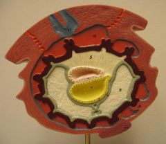
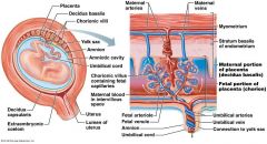
Placenta |
provides nutrients and oxygen to the embryo and carrying away embryonic metabolic wastes |
|
|
Chorion |
cells from inner cell mass give rise to layer of extraembryonic mesoderm that lines the inner surface of the trophoblast |
|
|
Decidua basalis |
part of the endometrium beneath the embryo |
|
|
Placenta and its function diagram |
|
|
|
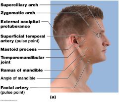
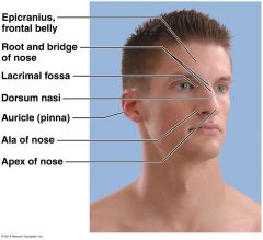
Palpation |
feeling internal structures through the skin with the fingers |
|
|
Landmarks of the head diagram |
|
|
|
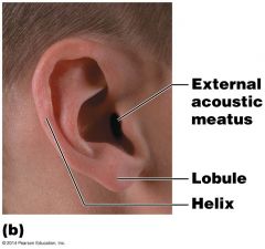
Landmarks of the ear diagram |
|
|
|
Frontal view of landmarks of the head |
|
|
|
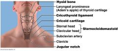
Landmarks of the neck diagram |
|
|
|
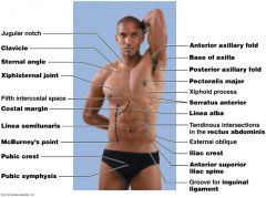
Landmarks of the thorax and abdomen diagram |
|
|
|
Linea alba |
tendon line that runs from the unbilicus superiorly to the xiphoid process |
|
|
Umbilical hernia |
linea alba weakens and coils of the intestine protrude through the opening |
|
|
McBurney's point |
superficial to the appendix common incision point for appendectomy |
|
|
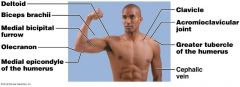
Landmarks of the upper limb |
|
|
|
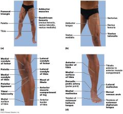
Landmarks of the lower limb |
|
|
|
Injection sites |
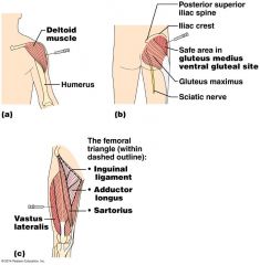
|