![]()
![]()
![]()
Use LEFT and RIGHT arrow keys to navigate between flashcards;
Use UP and DOWN arrow keys to flip the card;
H to show hint;
A reads text to speech;
223 Cards in this Set
- Front
- Back
|
Three Types of Muscle Tissue |
|
|
|
Skeletal Muscle Tissue
|
|
|
Cardiac Muscle Tissue
|
|
|
Smooth Muscle
|
|
|
Skeletal Muscles |
Organs composed of skeletal muscle tissue, plus nerves, blood vessels, and connective tissue |
|
|
Function of Skeletal Muscles |
|
|
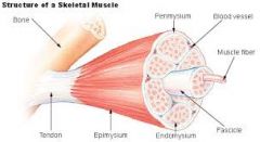
Organization of Skeletal Muscle |
Three Layers of Connective Tissue:
|
|
|
Epimysium |
Surrounds the entire muscle, it is a dense layer of collagen fibers |
|
|
Perimysium |
Divides the muscle into fascicles, contains blood vessels and nerves |
|
|
Endomysium |
Surrounds individual muscle cells (fibers) within the fascicle |
|
|
Skeletal Muscle Fibers = |
Skeletal Muscle Cells |
|
|
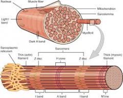
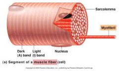
Sarcolemma |
The plasma membrane of a muscle cell
|
|
|
Sarcoplasm |
The cytoplasm of the muscle cell |
|
|
Organization of Muscle Cell |
|
|
|
T- tubules |
Narrow tubes that are continuous with the sarcolemma |
|
|
Myofibrils |
Bundles of protein filaments
|
|
|
Muscle contraction occurs when _________ is released into the sarcoplasm |
Calcium |
|
|
Sarcomeres |
Functional repeating units in the myofibril
|
|
|
The A Band |
Consists of the thick filaments in the center of the sarcomere |
|
|
M Line |
Central portion of the thick filament |
|
|
H Zone |
Light region, only thick filaments |
|
|
Zone of Overlap |
Overlap of thick and thin filaments |
|
|
The I Band |

Contains thin, but not thick filaments |
|
|
Z Line |
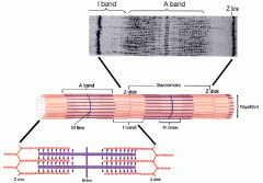
Marks the boundary of the sarcomere |
|
|
Sliding Filaments |
Contain myosin, actin, tropomyosin, and troponin |
|
|
Thin Filaments Contain |
Actin, Tropomyosin, and Troponin |
|
|
Thick Filaments Contain |
Myosin |
|
|
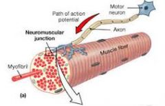
Neuromuscular Junction |
Where communication between the nervous system and muscles occurs |
|
|
Each muscle fiber is controlled by... |
a neuron. |
|
|
Acetyl Choline (ACH) |
A neurotransmitter stored in vesicles |
|
|
Synaptic Cleft |
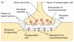
The narrow space that separates the neuron from the (muscle) |
|
|
Motor End Plate |
The sarcolemma surface that faces the synaptic terminal, contains Ach receptors |
|
|
Acetyl Cholinesterase (AChE) |
An enzyme that breaks down ACh |
|
|
Autorhythmic |
Contracts without neural stimulation |
|
|
Pacemaker Cells |
Specialized cardiac cells that determine contraction |
|
|
Intercalated Disks |
MAKE CARDIAC CELLS FUNCTIONS AS ONE ENORMOUS CELL |
|
|
Smooth muscle location: |
Located in many organ systems such as the cardiovascular, respiratory, digestive, and reproductive |
|
|
Thick and Thin Filaments |
Are not organized in sarcomers but are scattered |
|
|
Smooth muscle carries out... |
slow and sustained contractions |
|
|
Two Types of Smooth Muscle |
|
|
|
A typical muscle has an: |
|
|
|
Origin |
Where the fixed end of a muscle attaches -typically proximal to the insertion |
|
|
Insertion |
The site where the movable end attaches
|
|
|
Belly |
The central body |
|
|
Where the muscle contracts it produces... |
an action or movement
|
|
|
Based on their functions, muscles can be described as: |
|
|
|
Agonist |
Prime mover, is a muscle whose contraction is responsible for movement |
|
|
Antagonist |
A muscle that opposes the action of the agonist -Ex: the triceps antagonizes the biceps |
|
|
Synergist |
A muscle that helps the agonist to work more efficiently
|
|
|
# of Muscles in the Body |
Over 700 |
|
* |
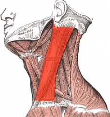
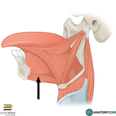
Sternocleidomastoid
|
|
|
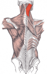
Splenius Capitis
|
|
|
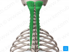
Semispinalis Capitis
|
|
|
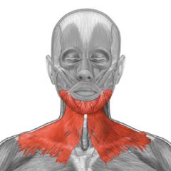
Platysma
|
|
* |
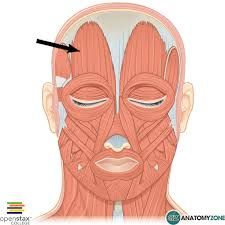
Occipitofrontalis
|
|
|
Rectus Abdominis
|
|
|
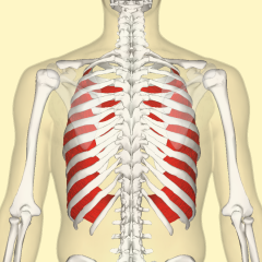
Internal Intercostalis
|
|
* |
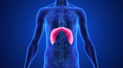
Diaphragm
|
|
|
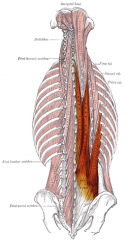
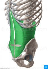
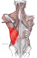
Erector Spinae
|
|
|
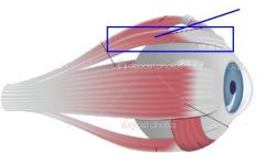
Medial Rectus
|
|
* |
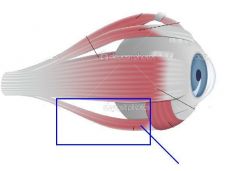
Lateral Rectus
|
|
|
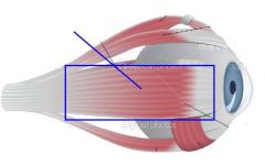
Inferior Rectus
|
|
|
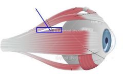
Superior Rectus
|
|
|
Genioglossus
|
|
|
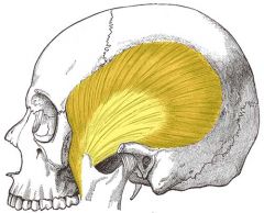
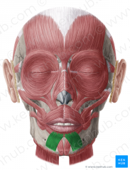
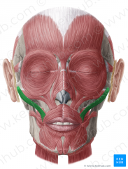
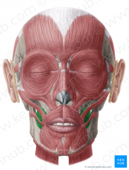
Temporalis
|
|
* |
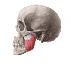
Masseter
|
|
|
Depressor Labii Inferioris
|
|

|
Depressor Anguli Oris
|
|

* |
Zygomaticus Minor
|
|
* |
Zygomaticus Major
|
|
|
Buccinator
|
|
|
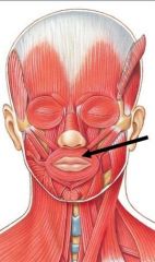
Orbicularis Oris
|
|
|
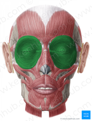
Orbicularis Oculi
|
|
|
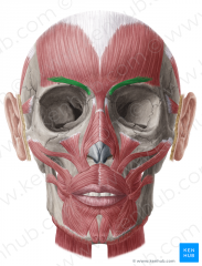
Corrugator Supercilii
|
|
|
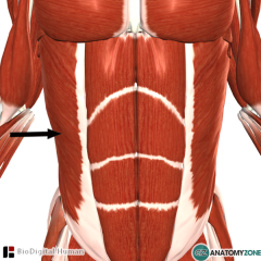
External Abdominal Obliques
|
|
|
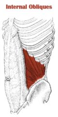
Internal Abdominal Obliques
|
|
|
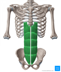
Transverse Abdominis
|
|
|
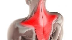
Trapezius
|
|
* |
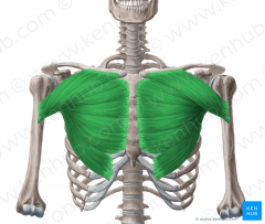
Pectoralis Major
|
|
|
Pectoralis Minor
|
|
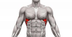
|
Serratus Anterior
|
|
|
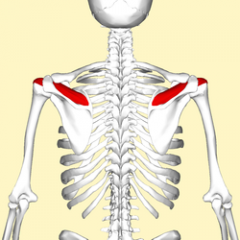
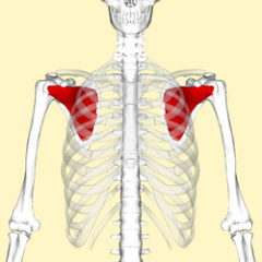
Supraspinatus
|
|
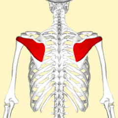
|
Infraspinatus
|
|
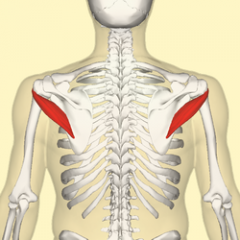
|
Teres Minor
|
|
|
Subscapularis
|
|
|
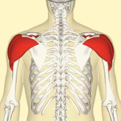
Deltoid
|
|
* |
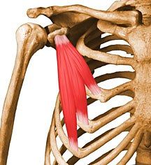
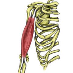
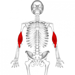
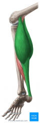
Biceps Brachii
|
|
|
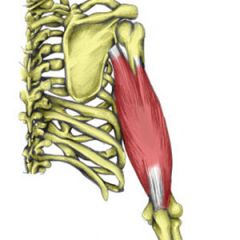
Triceps Brachii
|
|
|
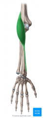
Brachialis
|
|
|
Latissimus Dorsi
|
|
|
Brachioradialis
|
|
|
Flexor Carpi Ulnaris
|
|
|
Flexor Carpi Radialis
|
|
|
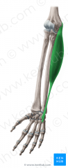
Extensor Digitorum
|
|
|
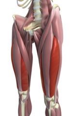
Gluteus Maximus
|
|
* |
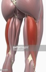
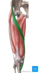
Rectus Femoris
|
|

|
Vastus Medialis
|
|
|
Vastus Lateralis
|
|
|
Biceps Femoris
|
|
|
Sartorius
|
|
* |
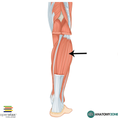
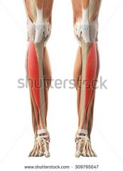
Gastrocnemius
|
|
|
Soleus
|
|
|
Tibialis Anterior
|
|
|
Cranial and spinal nerves are part of the... |
Peripheral Nervous System |
|
|
The peripheral nervous system is composed of... |
Axons and nerve fibers |
|
|
Actions of the Peripheral Nervous System |
|
|
|
The peripheral nervous system is... |
All nervous tissues outside the central nervous system |
|
|
The Central Nervous system is composed of... |
The brain and spinal cord |
|
|
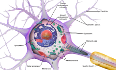
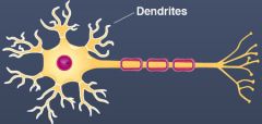
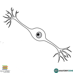
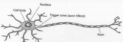
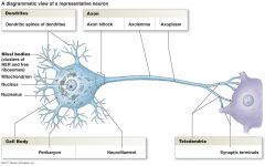
Neuron Cell Body- Nucleus, cytoplasm, organelles (neurotransmitters, ATP, protein synthesis), no centrioles (no cell division except in the nose & hippocampus) |
|
|
Slender extensions from the cell body, highly branched,recieve information
|
|

A |
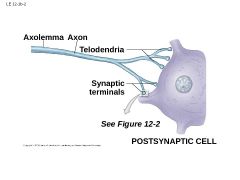
Axon- long cytoplasmic processes capable of propagating electrical impulses, they branch in telodendria |
|
|
Synapse |
A specialized site where the neuron communicates with other neurons |
|
|
Every synapse involves two cells
|
|
|
|
Neurotransmitters |
Chemical messages that mediate communication |
|
|
Synaptic Vesicles
|
Store neurotransmitters |
|
|
Synaptic Cleft |
A tiny opening between neurons |
|
|
A postsynaptic cell can be a... |
neuron or other cell
|
|
|
Synaptic Knob |

The round structure of the synapse |
|
|
Synaptic Cleft |
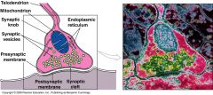
The space between the pre and postsynaptic membranes |
|
|
Anaxonic Neuron |
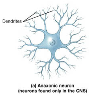
The axon is not distinguished from the dendrites (in brain) |
|
|
Bipolar Neuron |
One axon and one dendrite (in special sense organs) |
|
|
Unipolar Neuron |
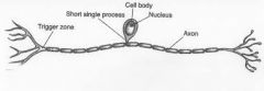
Dendrite and axon are continuous (fused), cell body to the side (sensory neurons of the PNS) |
|
|
Multipolar Neuron
|
Two or more dendrites and one axon (motor neurons) |
|
|
Sensory Neurons |
Afferent, include receptors (10 million) such as:
|
|
|
Exteroreceptors |
Monitor the external environment (touch, temperature) |
|
|
Internoreceptors |
Monitor internal environment (digestive, urinary systems) |
|
|
Proprioreceptors |
Monitor position of skeletal muscles |
|
|
3 Functional Classifications of Neurons |
|
|
|
Six Types of Neuroglia (Four in CNS/ Two in PNS) |
|
|
|
Ependymal Neuroglia |
Line the central canal and produce cerebrospinal fluid |
|
|
Cerebrospinal Fluid |
A fluid that provides cushion, transports nutrients, gases and waste and surrounds the brain |
|
|
Astrocytes |
|
|
|
Oligodendrocytes |
Myelinate axons of CNS -myelin is a membranous material for electrical insulation |
|
|
Microglia |
Remove cell debris, wastes, and pathogens by phagocytosis |
|
|
Satellite Cells |
Surround cell bodies of PNS and regulate the environment around PNS neurons |
|
|
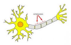
Schwaan Cells |
Myelinate axons in the PNS and help to repair injured neurons (nodes and internodes) |
|
|
Axon Classification- Type A Fibers |
|
|
|
Axon Classification- Type B Fibers |
|
|
|
Axon Classification- Type C Fibers |
|
|
|
Presynaptic cell is... |
a neuron |
|
|
Postsynaptic Cell is... |
a neuron or another cell |
|
|
Two types of Synapses |
|
|
|
Chemical Synapse |
|
|
|
Cholinergic Synapsis |
Synapses that release Ach (neuromuscular junction) |
|
|
Electrical Synapse |
|
|
c |

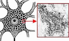
Muscle Fiber |
|

a |
Fascicle- bundle of skeletal muscle fibers surrounded by the perimysium |
|
126 |
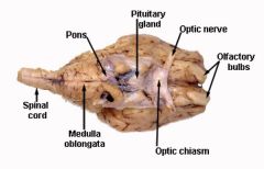
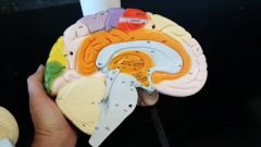
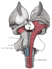
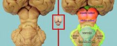
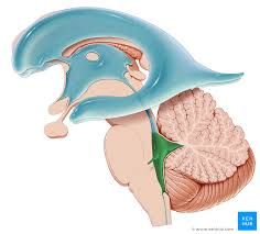
Medulla Oblongata |
|

65 |
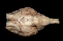
Pons |
|
6 |
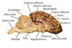
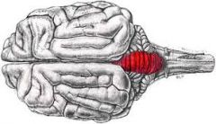
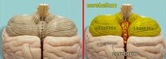
Cerebellum |
|

67 |
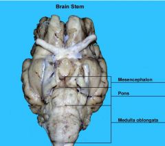
Mesencephalon
|
|

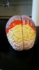
Innermost Orange Layer |
Diencephalon |
|
|
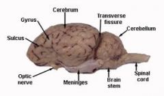
Cerebrum |
|

41-43 |
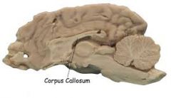
Corpus Callosum |
|
|
Pia Mater |
Innermost meninge of the brain |
|
|
Dura Mater |
Outermost Meninge of the brain |
|
|
Arachnoid Mater |
Webby middle meninge of the brain |
|
|
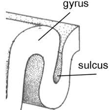
Sulci & Gyri |
|
|
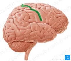
Central line through brain |
Longitudinal fissure |
|
|
Central Sulcus |
|
|
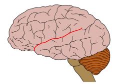
Lateral Sulcus |
|

1 |
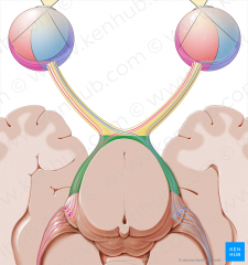
Olfactory Bulbs |
|

75 |
Olfactory Tract |
|
|
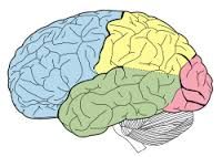
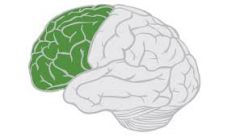
Frontal Lobe |
|
|
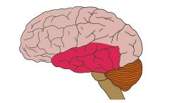
Temporal Lobe |
|
|
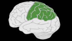
Parietal Lobe |
|
|
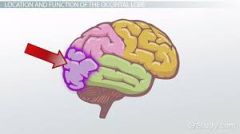
Occipital Lobe |
|

II |
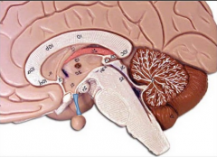
Optic Chiasm |
|
|
Infundibulum |
|
|
Optic Tract |
|
47 or 49 |
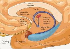
Mammillary body |
|
|
Cerebral Peduncle |
|
|
Corpora Quadrigemina |
|

Bulbous area at 54 |
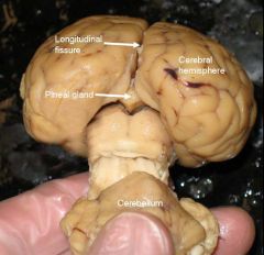
Pineal Gland |
|
|
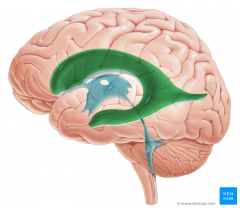
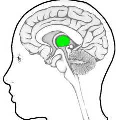
Lateral Ventricle |
|
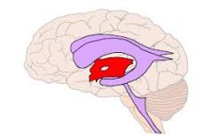
Red area |
Third Ventricle |
|
Green Area |
Fourth Ventricle |
|
|
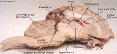
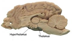
Thalamus |
|

71 & 72 |
Hypothalamus |
|
|
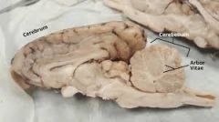
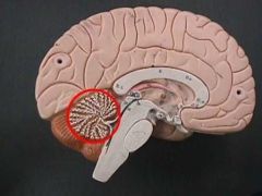
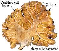
Arbor Vitae |
|
|
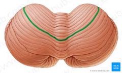
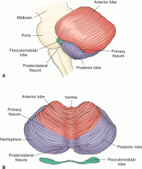
Vermis |
|
|
Folia |
|
|
|
Primary cerebral fissure |
|
|
Lobes of the cerebellum |
|
|
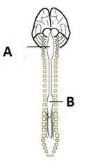
Cauda Equina |
|

A |
Cervical Enlargement of Spinal Cord |
|
B |
Lumbar Enlargement of Spinal Waco |
|
|
Conus Medullaris |
|
|
Filium Terminal |
|
|
Motor Neuron Nissl Bodies |
|
|
Axolemma |
The middle portion of the axon |
|
7 |
Axon Hillock |
|
|
Telodendria |
|
|
|
Internodes |
|
|
|
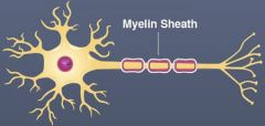
Myelin Sheath |
|
|
|
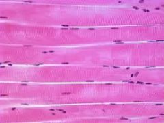
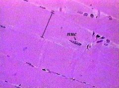
Skeletal Muscle Nuclei |
|
|
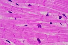
Skeletal Muscle Myofibril |
|
|
|
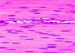
Skeletal Muscle Sarcolemma |

|
|
|
Neuromuscular Junction Muscle Fibers |
|
|
|
Neuromuscular Junction Axon Terminal |

|
|
|
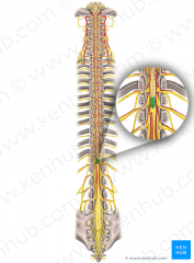
Spinal Dura Matter |
Outer Layer |
|
|
Spinal Pia Mater |
Innermost Layer |
|
|
Spinal Arachnoid Mater |
Middle webb like layer |
|
|
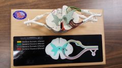
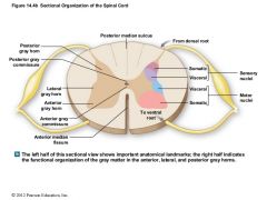
Spinal Cord Gray Matter |
Middle darker section of the spinal cord |
|
|
Spinal Cord White Matter |
Lighter areas surrounding the gray matter of the spinal cord |
|

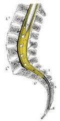
7 |
Spinal Cord- Anterior Horn |
|

6 |
Spinal Cord- Lateral Horn |
|

5 |
Spinal Cord- Posterior (dorsal) Horn |
|

14 |
Anterior (ventral) Median Fissure |
|

10 |
Posterior (dorsal) Median Sulcus |
|

25 |
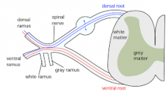
Ventral Root |
|

Small Center Circle |
Central Canal |
|
23 |
Dorsal Root |
|

24 |
Dorsal Root Ganglion |
|

4 |
Anterior Gray Commissure |
|
|
Posterior Gray Commissure |
|
|
|
Spinal Nerve |
|
|
|
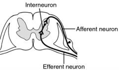
Interneuron |
|
|
|
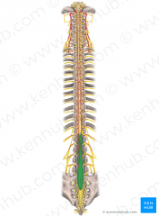
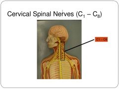
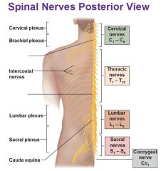
Cervical Spinal Nerves C1-C8 |
|
|
|
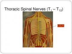
Thoracic Spinal Nerves T1-T12 |
|
|
|
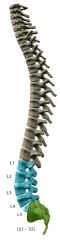
Lumbar Spinal Nerves L1-L5 |
|
|
|
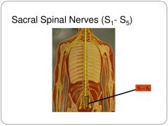
Sacral Spinal Nerves S1-S5 |
|
|
|
Coccygeal Nerve Co1 |
|