Reading...
![]()
Play button
![]()
Play button
![]()
Use LEFT and RIGHT arrow keys to navigate between flashcards;
Use UP and DOWN arrow keys to flip the card;
H to show hint;
A reads text to speech;
34 Cards in this Set
- Front
- Back
|
What are the boundaries of the femoral triangle?
superior, medial, lateral, base, apex, floor, roof |
1. Superior inguinal ligament
2. medially adductor longus 3. laterally sartorius 4. base- inquinal ligament 5. Apex- medial border of sartorius crosses medial border of adductor longus 6. Floor- later to medial by ilipsoas, pectineus, and adductor longus 7. Roof- fascia lata and cribriform fascia |
|
|
What are the contents of the femoral triangle?
|
lateral to medial
NAVEL nerve- femoral artery- femoral empty space- femoral canal lymphatics- deep inguinal |
|
|
What is the role/location of femoral sheath?
|
1. encloses proximal parts of femoral vessels and femoral canal
a. this allows femoral vessels to glide deep to inguinal ligament during hip joint movements |
|
|
What are the three compartments of femoral sheath/
|
1. lateral (femoral artery)
2. Intermediate (femoral vein) 3. Medial (femoral canal) not contain femoral nerve! |
|
|
In a laceration of the femoral artery what happens to circulation if it is ligated?
|
cruciate anastomosis may supply blood to the lower extremity
|
|
|
What is the main role of the femoral canal? What is it a part of?
|
allows femoral vein to expand during increased venous return (excercise)
1/3 compartments of femoral sheath (medial) |
|
|
What is significant to the proximal opening of the femoral canal?
|
femoral ring: a loop of small intestine could petrude into this canal which leads to femoral hernia.
|
|
|
What ligament do femoral hernias occur under and why are women more prone to get them?
|
under inguinal ligament
because of the wider bone structure of the female pelvis |
|
|
Where is location of inguinal hernia? What are the two types and where are they located?
|
1. indirect
2. direct Both located superior to inguinal ligament |
|
|
What are the two groups and subgroups of inguinal lymph nodes?
|
1. superficial inguinal lymph nodes
a. proximal (horizontal) b. Distal (vertical) 2. deep inguinal lymph nodes |
|
|
Where are the two superficial inguinal lymph nodes located?
|
1. proximal- (horizontal)- 1 cm inferior to inguinal ligament
2. Distal (vertical)- along each side of great saphenous vein - both pass deep to inguinal ligament then into external ilac lymph nodes |
|

Label the groups of lymph nodes
|
a. distal (vertical) group/; inferior nodes
b. superolateral of proximal c. superomedial of proximal |
|

Label the deep inguinal lymph nodes and where is it located? Where do they drain into?
|
1-3 along medial side of femoral vein
- inside femoral canal of femoral sheath - drain into external iliac lymph nodes a. external iliac lymph nodes b. femoral nerve c. femoral sheath d. femoral canal e. femoral artery/vein |
|

What 4 muscles does the femoral nerve intervate and what is root?
What is 4 and 5? |
L2-L4
1. illiacus 2. sartorius 3. quadriceps femoris 4. pectineus a. lateral femoral cutaneous nerve b. anterior (intermediate and medial) cutaneous branches of femoral nerve (L2, L3, L4) |
|
|
Where does the femoral nerve start, enter thigh, go to, and end?
What muscles does it innervate? |
Starts- in abdomen wit psoas major
Enters thigh- just lateral to middle of inguinal ligament Supplies- anterior thigh muscles and hip and knee joints ends- turns into saphenous nerve |
|
|
What might an anterior hip dislocation cause?
|
Femoral nerve damage- located under inguinal ligament
1. meralgia paresthetica- compression of lateral femoral cutaneous nerve causes pain along lateral thigh down to the knee |
|
|
Which vein is commonly used for coronary artery bypass surgery?
|
great sephanous
|
|
|
Describe what the saphenous nerve arises from and where...
What does it innervate and when is it most vulnerable? |
from femoral nerve in the femoral triangle...
Innervates skin on medial side of knee, leg, and foot Most vulnerable- on proximal area during surgery to repair varicose veins |
|
|
What is the adductor Canal and where does it end?
|
Starts- apex of femoral triangle and
Ends- adductor hiatus intermuscular passage or fascial tunnel where femoral vessels reach popliteal fossa |
|
|
What is Adductor hiatus and where does it end?
|
in tendon of adductor magnus
- leads femoral vessels to popliteal fossa |
|
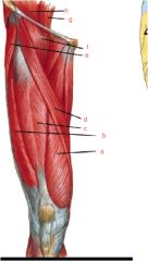
What muscles are this thigh?
|
a. vastus medialis muscle
b. vastus lateralis muscle c. rectus femoris muscle d. sartorius muscle e. tensor fasciae latae muscle f. illipsoas muscle g. psoas major mscle h. illiacus muscle |
|
|
1. What is chief arterial supply to lower limb?
2. What is it a continuation of and where does it start? |
1. femoral artery
2. external iliac artery- starts deep to middle inguinal ligament and lateral to femoral vein |
|
|
What does the femoral artery gives rise to inferiorly in thing?
Name the branches (a and b) |
- genicular artery
a. articular b. saphenous |
|
|
Where is femoral artery palpated?
|
between asis and pubic tubercle
- used for radiographic vizualization of the left side of heart and coronary vessels |
|
|
What is the largest branch of femoral artery?
Where does it descend and give rise to? |
- profunda femoris a.
- descends behinds adductor longus - gives rise to medial and lateral circumflex femoral a. |
|
|
What is the main blood supplier to femoral head and neck? What artery supplies the small proximal part of the neck and head of the femur?
|
medial circumflex femoral artery
obturator artery |
|
|
Where does the medial circumflex femoral artery pass between?
|
iliopsoas and pectineus toward posterior thigh
|
|

Label 1-7
|
1. lateral circumflex femoral a. branches
2. ascending branch 3. transverse branch 4. descending branch 5. lateral circumflex femoral artery 6. deep femoral artery 7. medial circumflex femoral artery |
|

Label...
|
1. deep circumflex iliac artery
2. lateral femoral cutaneous nerve 3. sartorius muscle (cut) 4. ilipsoas muscle 5. tensor fasciae latae muscle (retracted) 6. gluteus medius and minimus muscles 7. femoral nerve 8. rectus femoris muscle 9. ascending, transvers, and descending branches of lateral circumflex femoral artery 10. femoral artery 11. external iliac artery and vein 12. inguinal ligament 13. femoral artery and vein 14. pectineus muscle (cut) 15. obturator canal 16. obturator externus muscle 17. adductor longus muscle (cut) 18. anterior branch of obturator nerve 19. posterior branch of obturator nerve 20. quadratus femoris muscle 21. adductor brevis muscle |
|
|
What muscles does the profunda femoris (deep femoral artery) go through?
What do the perforating arteries supply blood to? |
anterior to pectineus and posterior to adductor longus
- adductor magnus and hamstring muscles |
|
|
If the femoral or external iliac arteries are obstructed what anastomosis bypasses the obstruction?
|
cruciate anastomosis
|
|
|
Where does the femoral vein lie in relation to the femoral artery?
|
medial
|
|
|
Where are emergency blood transfusions performed?
|
anterior to medial malleolus on great saphenous vein and graft can be used for coronary bypass surgery
|
|
|
What might cause medial side foot paralysis?
|
"saphenous cutdown" due to saphenous nerve
|