![]()
![]()
![]()
Use LEFT and RIGHT arrow keys to navigate between flashcards;
Use UP and DOWN arrow keys to flip the card;
H to show hint;
A reads text to speech;
305 Cards in this Set
- Front
- Back
|
3 Types of Joints |
Fibrous: bones joined by dense regular CT Cartilaginous: bones joined cartilage Synovial: most common and most movable type of joint, bones separated by a joint cavity. |
|
|
Most moveable and least moveable types of synovial joints |
Least: Plane Joints Most: Ball and Socket |
|
|
Condylar Joint |
Biaxial Movement ex) metacarpophalangeal joints, wrist joint |
|
|
Saddle Joint |
Biaxial Movement ex) Carpometacarpal joints in thumbs |
|
|
Ball-and-Socket Joint |
Multiaxial Movement ex) should and hip joints |
|
|
Plane Joint |
No axial Movement (gliding only) ex) Intercarpal joints, intertarsal joints, joints between vertebral bodies |
|
|
Hinge Joint |
Uniaxial Joint ex) elbow joints, IP joints |
|
|
Pivot Joint |
Uniaxial Movement ex) proximal radioulnar joints |
|
|
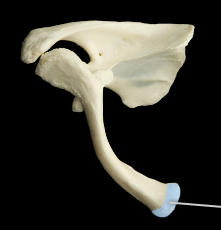
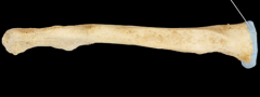
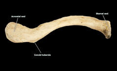
Acromial end of clavicle |
|
|
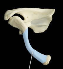
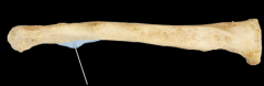
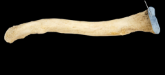
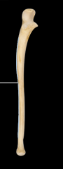
Shaft of clavicle |
|
|
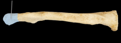
Sternal end of clavicle |
|
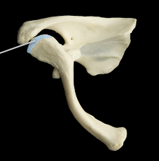
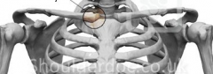
What joint is this? |
Acromiolclavicular joint |
|
|
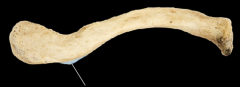
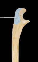
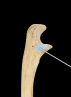
Conoid Tubercle |
|
|
Acromial End of clavicle |
|
|
Sternal End of clavicle |
|
|
Conoid tubercle of clavicle |
|
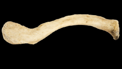
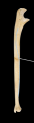
Which end is the acromial and which end is the sternal? |
|
|
|
Sternal End
|
|
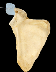
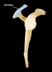
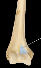
Name the highlighted structure |
Acromion |
|
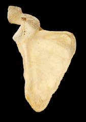
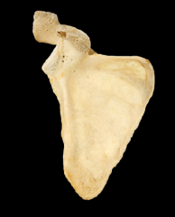
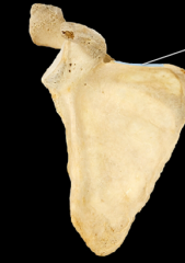
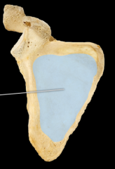
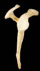
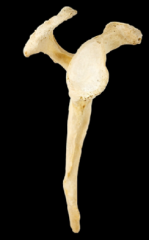
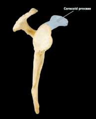
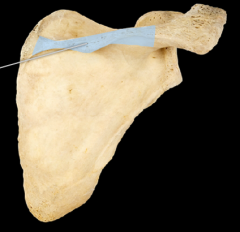
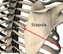
Bone name and orientation in the body (what view?) |
Scapula -- anterior view |
|
|
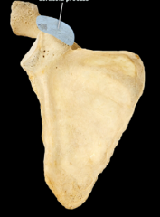
Coracoid Process |
|
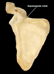
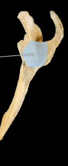
Where is the suprascapular notch located? |
|
|
|
Superior border of the scapula |
|
|
Superior Angle of scapula |
|
|
Medial border of scapula |
|
|
Inferior angle of scapula |
|
|
Subscapular Fossa |
|
|
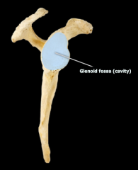
Glenoid Fossa |
|
|
Infraglenoid tubercle |
|
|
Lateral border of scapula |
|
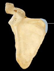
Identify the glenoid fossa |
|
|
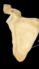
Identify the acromion |
|
|
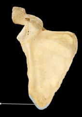
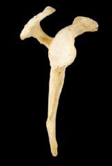
Identify the coracoid process |
|
|
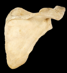
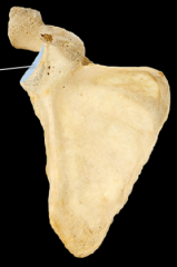
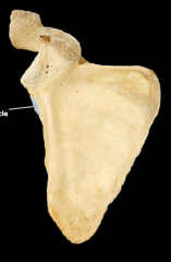
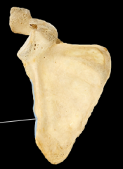
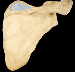
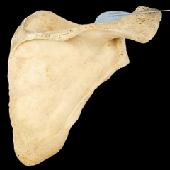
Name the bone and orientation |
Scapula -- posterior view |
|
|
Supraspinous Fossa |
|
|
Where is the Infraspinous Fossa? |

|
|
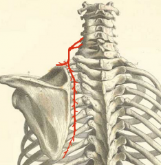
What is the highlighted structure? |
Spine of the scapula |
|
|
Coracoid Process |
|
|
Supraspinous Fossa |
|
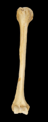
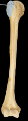
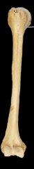
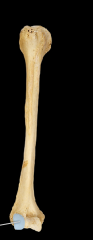
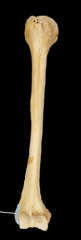
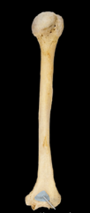
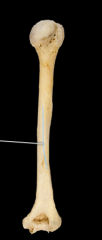
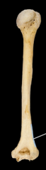
Identify the bone and the orientation |
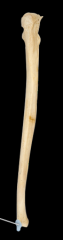
Humerus -- anterior view |
|
|
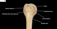
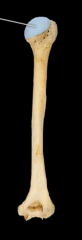
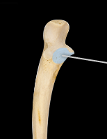
Greater tubercle of the humerus |
|
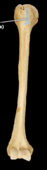
|
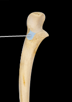
Intertubercular Sulcus of humerus |
|
|
Lesser tubercle of humerus |
|
|
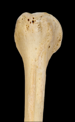
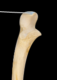
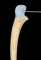
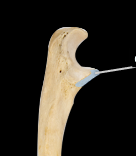
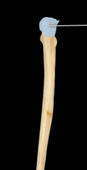
Head of the humerus |
|
|
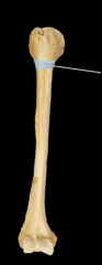
Anatomical neck of the humerus |
|
|
Surgical neck of the humerus |
|
|
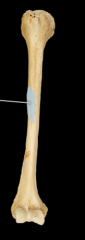
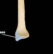
Deltoid Tuberosity |
|
|
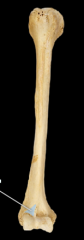
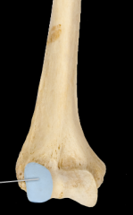
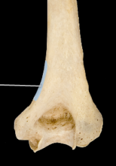
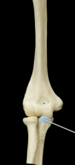
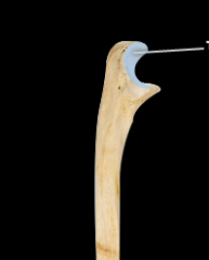
Trochlea |
|
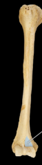
|
Capitulum |
|
|
Radial Fossa |
|
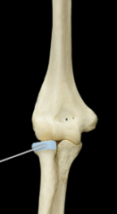
|
Coronoid Fossa |
|
|
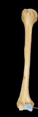
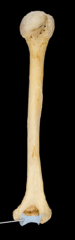
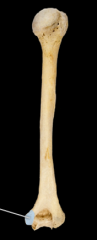
Medial Epicondyle |
|
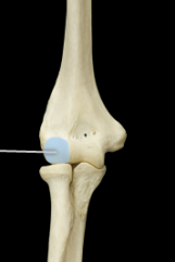
|
Lateral Epicondyle |
|
Identify the: Greater Tubercle Intertubercular sulcus Lesser tubercle |
|
|
|
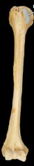
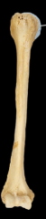
Capitulum |
|
|
Coronoid Fossa |
|
|
Olecranon Fossa |
|
|
Trochlea |
|
|
Medial Epichondyle |
|
|
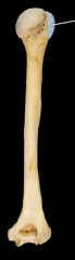
Radial Groove of the humerus |
|
|
Head of the humerus |
|
|
greater tubercle |
|
|
Medial Supracondylar Ridge |
|
|
Lateral Supracondylar Ridge |
|

|
Trochlea of humerus |
|
|
Coronoid Process of the ulna |
|
|
Head of the radius |
|
|
Capitulum of the humerus |
|
|
Coronoid Fossa |
|
|
Radial Tuberosity |
|
|
Olecranon Process of ulna |
|
|
Head of the radius |
|
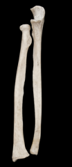
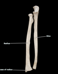
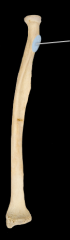
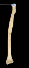
What bone is the radius? Ulna? |
|
|
|
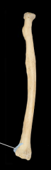
Styloid Process of the ulna |
|
|
Styloid Process of radius |
|
|
Olecranon Process (ulna) |
|
|
Trochlear notch (ulna) |
|
|
Coronoid Process (ulna) |
|
|
Radial notch (ulna) |
|
|
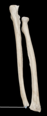
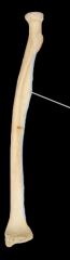
Interosseous border (ulna) |
|
|
Interosseous border (ulna) |
|
|
Olecranon Process (ulna) |
|
|
Radial notch (ulna) |
|
|
Coronoid Process (ulna) |
|
|
Trochlear notch (ulna) |
|
|
Olecranon Process (ulna -posterior view) |
|
|
Styloid process (ulna) - posterior view |
|
|
Radial Tuberosity-- radius |
|
|
Head of the radius |
|
|
Interosseous border (radius) |
|
|
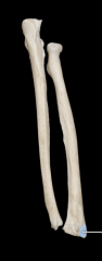
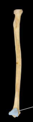
Styloid Process (radius) |
|
|
Ulnar Notch (radius) |
|
|
Ulnar notch (radius) |
|
|
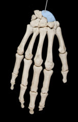
Lunate |
|
|
Scaphoid |
|
|
Triquetrum |
|
|
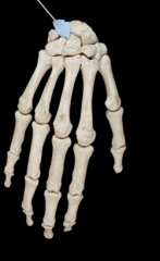
Capitate |
|
|
Trapezoid |
|
|
Trapezium |
|
|
Hamate |
|
|
Scaphoid |
|
|
Capitate |
|
|
Trapezoid |
|
|
Hamate |
|
|
Pisiform |
|
|
Trapezium |
|
|
Metacarpals |
|
|
Proximal phalanges |
|
|
Middle phalanges |
|
|
Distal Phalanges |
|
|
Sternoclavicular Joint -- connects manubrium and clavicle |
|
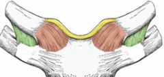
What is labeled red? |
Sternoclavicular Ligament |
|
|
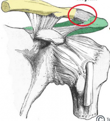
Acromioclavicular Joint |
|
|
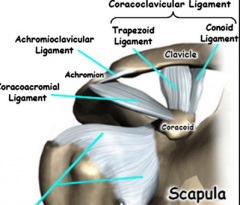
Identify the Acromioclavicular ligament and the Coracoclavicular Ligament |
|
|
|
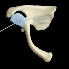
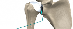
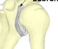
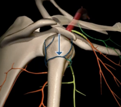
Glenohumeral Joint |
|
|
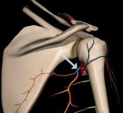
Glenoid Labrum |
|
|
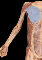
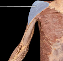
Scapulothoracic Joint |
|
|
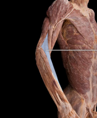
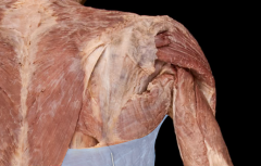
Pectoralis Major |
|
|
Deltoid |
|
|
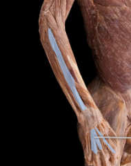
Biceps Brachii |
|
|
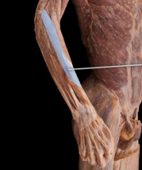
Brachioradialis - |
|
|
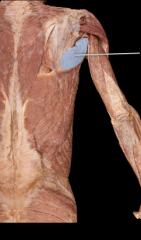
Pectoralis Minor |
|
|
Coracobrachialis |
|
Short or long head of the biceps brachii? |
Long head |
|
|
Latissimus Dorsi |
|
|
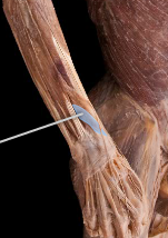
Triceps brachii |
|
|
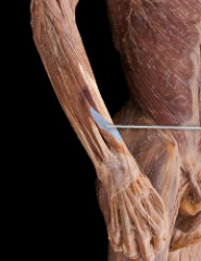
Brachialis |
|
|
Brachioradialis |
|
|
Teres Major |
|
|
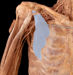
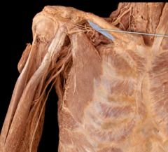
Pectoralis Minor |
|
|
Subclavius |
|
|
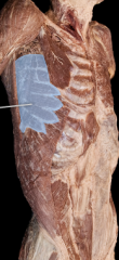
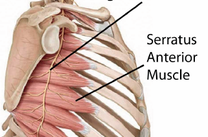
Serratus Anterior (Under pec minor) |
|
|
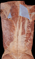
Latissimus Dorsi |
|
|
Serratus Anterior |
|
|
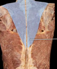
Trapezius |
|
|
Rhomboid Major |
|
|
Supraspinatus |
|
|
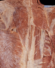
Rhomboid Major |
|
|
Rhomboid Minor |
|
|
Levator Scapulae |
|
|
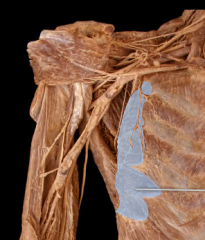
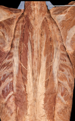
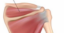
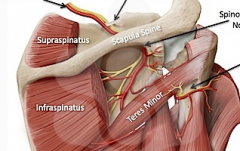
Supraspinatus |
|
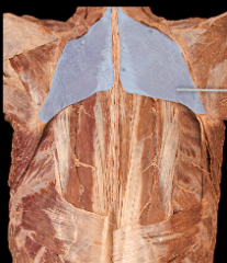
What is the top arrow pointing to? |
Infraspinatus |
|
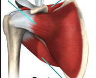
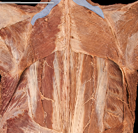
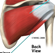
What is the top arrow pointing to? |
Subscapularis |
|
|
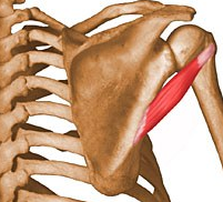
Teres minor |
|
|
Prime Mover |
Muscle causing the action |
|
|
Agonist |
Produces aspecific movement when it contracts-Also called aprime mover |
|
|
Antagonist |
A muscle whoseaction opposes that of an agonist |
|
|
Synergist |
A muscle that assists the agonist in performing itsaction |
|
|
Isometric contraction |
Length is constant; tension is changing |
|
|
Isotonic Contraction |
Tension is constant; length is changing a.Concentric contraction: Muscle isshorteningb.Eccentric contraction: Muscle is lengthening |
|
|
Associated structures of: Acromial end of clavicle |
Articulates with the acromion process of the scapula |
|
|
Associated structures of: Sternal end of clavicle |
Articulates with the sternum |
|
|
Associated structures of: Conoid Tubercle |
Attachment site for the coracoclavicular ligament |
|
|
Associated structures of: Glenoid Fossa |
Articulates with the head of the humerus |
|
|
Associated structures of: Coracoid Process |
Attachment site for the coracobrachilalis, short head of biceps brachii, and pectoralis minor muscle; attachment for the coracoclavicular and coracoacromial ligaments |
|
|
Associated structures of: Acromion Process |
Articulates with the clavicle, attachment for the middle part of the deltoid, acromioclavicular and coracoacromial ligaments |
|
|
Associated structures of: Scapular Spine |
Separates the supraspinatus and infraspinatus fossa, attachment for the deltoid and trapezius muscles |
|
|
Associated structures of: Medial border of the scapula |
Attachment for the levator scapulae, rhomboid major, rhomboid minor, and serratus anterior) |
|
|
Associated structures of: Lateral Border of scapula |
Attachment for teres major and teres minor |
|
|
Associated structures of: Inferior Angle of Scapula |
Attachment site for teres major muscle |
|
|
Associated structures of: Subscapular Fossa |
Fossa on the anterior scapula that holds the subscapularis muscle |
|
|
Associated structures of: Supraspinous Fossa |
Fossa on the posterior surface that holds the supraspinatus |
|
|
Associated structures of: Infraspinous Fossa |
Fossa on the posterior surface that holds the infraspinatus |
|
|
Associated structures of: Infraglenoid Tubercle (scapula) |
Attachment for the long head of the triceps |
|
|
Associated structures of: Suprascapular Notch |
Passageway for the suprascapular nerve |
|
|
Associated structures of: Deltoid Tuberosity |
Attachment for the deltoid muscle |
|
|
Associated structures of: Lesser Tubercle of the humerus |
Attachment for the subscapularis muscle |
|
|
Associated structures of: Greater tubercle |
Attachment for the supraspinatus, infraspinatus, teres minor muscles |
|
|
Associated structures of: Intertubercular sulcus |
Passageway for the tendon of the long head of the biceps; attachment for the latissimus dorsi muscle |
|
|
Associated structures of: Radial Groove of humerus |
The radial nerve travels here on its course around the posterior aspect of the humerus |
|
|
Associated structures of: Medial Epicondyle |
Attachment site for muscle of the anterior forearm (flexors and pronator teres) |
|
|
Associated structures of: Lateral Epicondyle |
Attachment site for the muscles of the posterior forearm (extensors and supinator) |
|
|
Associated structures of: Capitulum |
Articulates with the head of the radius |
|
|
Associated structures of: Trochlea |
Articulates with the ulna |
|
|
Associated structures of: Radial Fossa |
Space for radius during elbow flexion (humerus) |
|
|
Associated structures of: Coronoid Fossa |
Space for the coronoid process of the ulna during elbow flexion |
|
|
Associated structures of: Olecranon process |
Attachment for the triceps brachii (ulna) |
|
|
Associated structures of: Radial notch of ulna |
Articulates with the head of the radius |
|
|
Associated structures of: Styloid process of the ulna |
Attachment for a ligament of the wrist |
|
|
Associated structures of: radial tuberosity |
Attachment for the biceps brachii tendon (on radius) |
|
|
Associated structures of: Styloid Process of the radius |
Attachment site for the brachioradialist muscle and a ligament of the wrist |
|
|
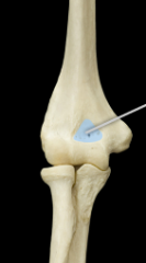
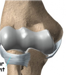
Humeroulnar Joint |
|
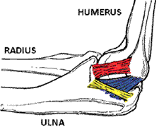
|
Ulnar Collateral Ligament (UCL) |
|
|
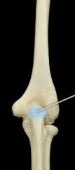
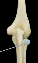
Annular Ligament (humeroradial joint) |
|

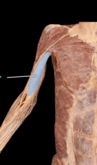
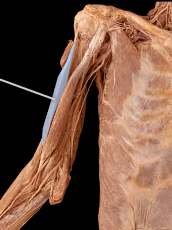
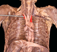
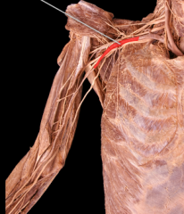
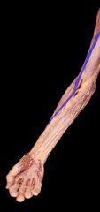
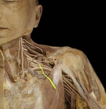
What is this artery and what does it branch into? |
|
|
|
What does the axillary artery arise from? |
The subclavian artery |
|
|
What does the brachial artery arise from? |
From the axillary artery! |
|
|
What is the first artery to branch from the axillary artery? |
Superior Thoracic Artery |
|
|
What arteries branch from the second part (under the pec minor) of the axillary artery? |
Thoracoacromial artery and lateral thoracic artery |
|
|
Anterior Circumflex Artery |
|
|
Posterior Circumflex artery |
|
|
What arteries branch from the third part of the axillary artery? |
Anterior circumflex humeral artery, posterior circumflex humeral artery, and subscapular artery |
|
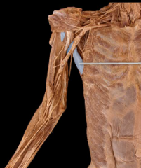
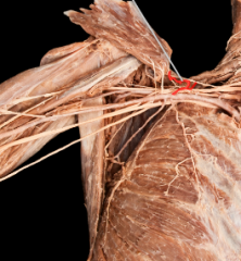
What is this artery and what does it branch into? |
Subscapular artery 1) Thoracodorsal artery 2) Circumflex scapular artery |
|
|
What does the axillary artery turn into? And when does this happen? |
Brachial artery -- at the level of the lower border of the teres major muscle |
|
|
Axillary artery |
|
|
Subclavian Artery |
|
|
Brachial Artery |
|
|
Thoracoacromial artery |
|
|
What spinal segments make up the brachial plexus? |
C5-T1 |
|
|
What forms the M shape of the brachial plexus? |
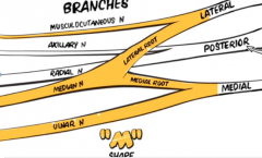
Terminal Branches -- anterior to the axillary artery |
|
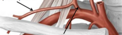
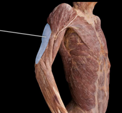
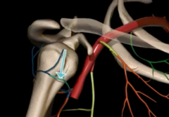
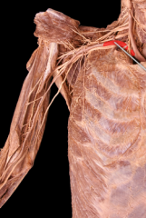
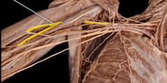
Identify the bottom arrow |
Tyrocervical Trunk - comes off Subclavian artery |
|
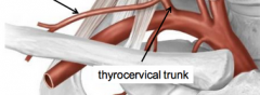
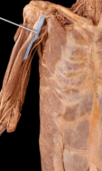
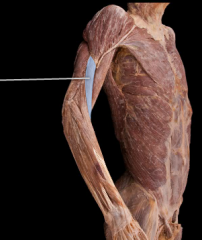
Identify the artery (top left) |
Suprascapular artery - branches from tyrocervical (from subclavian) This artery runs posteriorly towards the scapula with the suprascapular nerve to supply the supraspinatus and infraspinatus. |
|
|
What does the lateral thoracic artery supply? |
This artery branches from the second part of the axillary artery and runs with the long thoracic nerve to supply the lateral wall and the serratus anterior. |
|
|
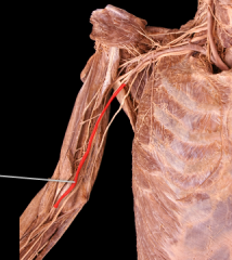
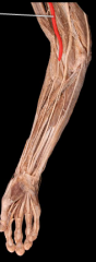
Deep Brachial Artery |
Branches from the brachial artery and runs posteriorly. |
|
|
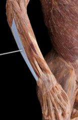
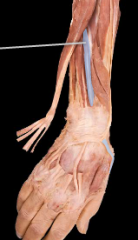
Flexor Retinaculum -- is a fibrous band on the palmar side of the hand near the wrist. It arches over the carpal bones of the hands, covering them and forming the carpal tunnel. |
|
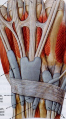
What is the fibrous sheath called? |
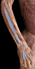
Extensor retinaculum - an anatomical term for the thickened part of the antebrachial fascia that holds the tendons of the extensor muscles in place. |
|
|
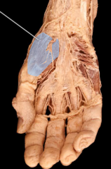
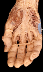
Hypothenar Eminence |
The hypothenar muscles are a group of three muscles of the palm that control the motion of the little finger. The three muscles are: Abductor digiti minimi,Flexor digiti minimi brevis,Opponens digiti minimi |
|
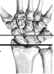
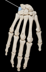
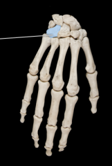
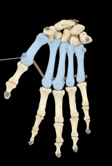
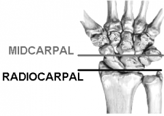
Identify the radiocarpal and midcarpal joints |
|
|
|
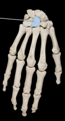
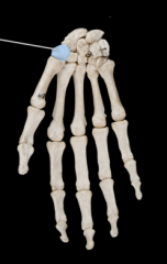
Intercarpal joints |
|
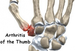
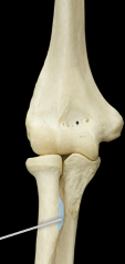
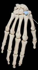
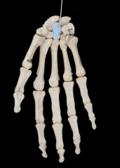
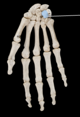
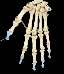
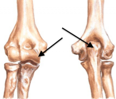
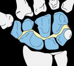
What joint is this arthritis occurring at? |
CMC - Carpometacarpal joint |
|
|
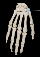
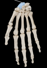
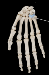
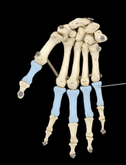
Where are the MP joints located? |
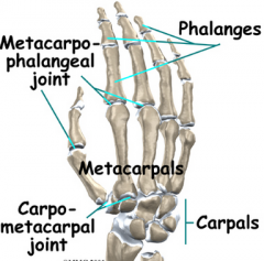
Metacarpophalngeal joints |
|
|
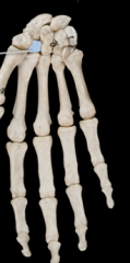
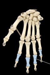
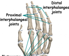
Where are the PIP and DIP joints? What do these abbreviations stand for? |
|
|
|
Trapezius - Actions, Innervation |
Super fibers: Elevate and superiorly rotate scapula Middle fibers: Retract scapula Inferior fibers: Depress scapula Innervated by: Accessory Nerve |
|
|
Levator Scapulae -- Actions, Insertion, Innervation |
Elevates and inferiorly rotates scapula I: Superior medial border of scapula Innervated by Dorsal Scapular nerve |
|
|
Rhomboid Major- Actions, Insertions, Innervation |
Elevates, retracts and inferiorly rotates scapula Insertion: Medial border of scapula Innervated by Dorsal Scapular Nerve |
|
|
Rhomboid Minor - Actions, Insertions, Innervation |
Elevates, retracts, and inferiorly rotates scapula Insertion: Superior medial borer of scapula Innervation: Dorsal Scapular nerve |
|
|
Pectoralis Minor- Actions, Insertions, Innervation |
Protracts and depresses scapula I: Coracoid process of scapula Innervation: Medial Pectoral nerve |
|
|
Serratus Anterior -- Actions, Insertions, Innervation |
Protracts and superiorly rotates scapula; stabilizes scapula Insertion: Anterior medial border of scapula Innervated: Long thoracic nerve |
|
|
Subclavius - Actions, Innervation |
Stabilizes and depresses the clavicle Innervated by nerve to subclavius |
|
|
Latissimus Dorsi - Actions, Insertions, Innervation |
Extends, adducts, and medially rotates GH joint (swimmer's muscle) Insertion: Intertubercular groove of humerus Innervation: Thoracodorsal |
|
|
Pectoralis Major - Actions, Insertions, Innervation |
Arm flexion, adduct, and medially rotates GH joint Insertion: Intertubercular groove of humerus Innervation: Lateral pectoral and medial pectoral nerves |
|
|
Deltoid -- Actions, Insertions, Innervation |
Anterior fibers: flex and medially rotate GH joint Middle fibers: GH abduction Posterior Fibers: Extend and laterally rotates GH Insertion: Deltoid tuberosity of humerus Innervation: Axillary Nerve |
|
|
Coracobrachialis - Actions, Origin, Insertions, Innervation |
A: Adducts and flexes GH joint O: Coracoid process I: Middle medial shaft of humerus Innervation: Musculocutaneous Nerve |
|
|
Teres Major - Actions, Origin, Insertions, Innervation |
A: Entends, adducts, and medially rotates GH joint O: Inferior lateral border and inferior angle of scapula I: Lesser tubercle and intertubercular groove of humerus Innervation: lower subscapular nerve |
|
|
Triceps brachii on arm - Actions, Origin, Insertions, Innervation |
Actions: Extends and adducts GH joint O: Infraglenoid Tubercle I: Olecranon process Innervation: Radial Nerve |
|
|
Biceps Brachii on arm- Actions, Origin, Insertions, Innervation |
Actions: Flexes GH joint O: Supraglenoid Tubercle I: Radial tuberosity Innervation: Musculocutaneous Nerve |
|
|
Subscapularis - Actions, Origin, Insertions, Innervation |
Actions: Medially rotates GH joint, stabilizes the GH joint O: Subscapular fossa I: Lesser tubercle of humerus Innervation: Upper and lower subscapular nerves |
|
|
Supraspinatus - Actions, Origin, Insertions, Innervation |
Action: Abducts GH joint, stabilizies the GH joint O: Supraspinous Fossa I: Greater tubercle of humerus Innervation: Suprascapular nerve |
|
|
Infraspinatus - Actions, Origin, Insertions, Innervation |
Actions: Adducts and laterally rotates GH joint, stabilizes the GH joint O: Infraspinous Fossa I: Greater tubercle of humerus Innervation: Suprascapular Nerve |
|
|
Teres Minor - Actions, Origin, Insertions, Innervation |
Actions: Adducts and laterally rotates GH joint, stabilizes the GH joint O: Superior lateral border of scapula I: Greater tubercle of humerus Innervation: Axillary Nerve |
|
|
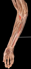
What muscles are in the anterior compartment of the arm? |
Flexors and Pronators |
|
|
What muscles are in the posterior compartment of the arm? |
Extensors and supinator (and Abductor pollicis longus) |
|
|
Palmaris Longus -- weak wrist flexor, median nerve |
|
|
Flexor carpi radialis - flexes wrist and abducts hand, median nerve |
|
|
Flexor carpi ulnaris - flexes wrist and adducts hand, ulnar nerve |
|
|
Flexor Digitorum Superficialis - flexes wrist, 2nd-5th MP joints and PIP joints, median nerve |
|
|
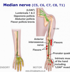
Flexor Digitorum Profundus - flexes wrist, 2nd-5th MP joints and PIP joints, median nerve (lateral 1/2) and ulnar nerve (medial 1/2) |
|
|
Flexor Pollicis Longus - Flexes MP joint of thumb, IP joint of thumb median nerve |
|
|
Pronator quadratus - Pronates forearm, median nerve |
|
|
Extensor carpi ulnaris - extends wrist, adducts hand, radial nerve |
|
|
Extensor digiti minimi - extends wrist, MP, and PIP joints of finger 5, radial nerve |
|
|
Extensor digitorum - extends wrist, extends 2-5 MP, PIP, DIP joints, radial nerve |
|
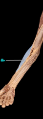
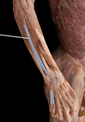
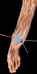
What two muscles is this showing? |
Extensor carpi radialis - extends wrist, abducts hand, radial nerve |
|
|
Abductor Pollicis Longus - Abducts thumb, radial nerve |
|
|
Extensor Pollicis Brevis - Extends MP joints of thumb, radial nerve |
|
|
Flexor Digitorum Profundus - flexes wrist, 2nd-5th MP joints and PIP joints, median nerve (lateral 1/2) and ulnar nerve (medial 1/2)
|
|
|
Flexor Pollicis Longus - Flexes MP joint of thumb, IP joint of thumb median nerve
|
|
|
Pronator Teres - Pronates forearm, median nerve |
|
|
Pronator quadratus - Pronates forearm, median nerve |
|

|
Extensor Carpi Radialis brevis - extends wrist, abducts hand, radial nerve |
|
|
Extensor Digitorum - - extends wrist, extends 2-5 MP, PIP, DIP joints, radial nerve |
|
|
Extensor Pollicis Longus - abducts thumb, radial nerve |
|
|
Abductor Pollicis Brevis - median nerve |
|
|
Flexor Pollicis Brevis- median nerve |
|
|
Abductor digiti minimi - ulnar |
|
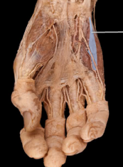
|
Flexor digiti minimi brevis - ulnar |
|
|
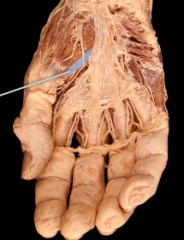
Lumbricals |
|
|
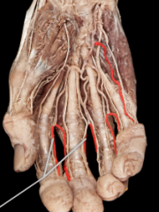
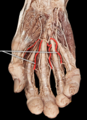
Proper Digital Arteries |
|
|
Common Digital Arteries |
|
|
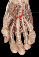
Superficial Palmar Arch |
|
|
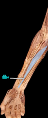
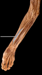
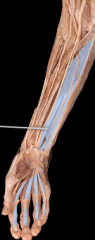
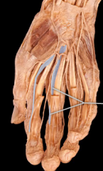
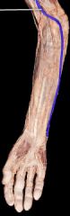
Basilic Vein |
|
|
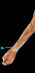
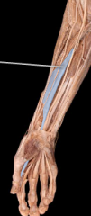
Cephalic Vein |
|
|
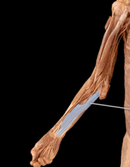
Cephalic Vein |
|
|
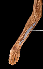
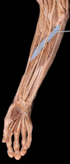
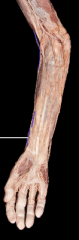
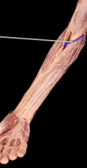
Median Cubital Vein |
|
|
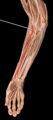
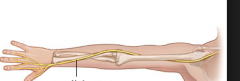
Median Nerve |
|
|
Brachial Artery |
|
|
Ulnar Artery |
|
|
Radial Artery |
|
|
Biceps Brachii on the forearm - Actions, Origin, Insertions, Innervation |
A: Flexes elbow, powerful supinator of forearm O: Supraglenoid tubercle of scapula (long head) and coracoid process of scapula (short head) I: Radial tuberosity Innervation: Musculocutaneous nerve |
|
|
Brachilalis - Actions, Innervation |
A: Primary flexor of elbow Innervation: Musculocutaneous nerve |
|
|
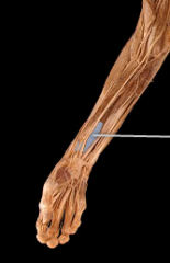
Brachioradialis - Actions, Insertions, Innervation |
Actions: Flexes elbow Insertion: Styloid process of radius Innervation: Radial Nerve |
|
|
Triceps Brachii on forearm - Actions, Origin, Insertions, Innervation |
A: Primary extensor of the elbow O: Infraglenoid tubercle of scapula Insertion: Olecranon process of ulna Innervation: Radial Nerve |
|
|
Pronator teres - Actions, Origin, Insertions, Innervation |
A: Pronates forearm O: Medial epicondyle of humerus I: Lateral radius Innervation: median nerve |
|
|
Flexor Carpi Radialis - Actions, Origin, Insertions, Innervation |
A: Flexes wrist and abducts hand O: Medial epicondyle I: Base of 2nd and 3rd metacarpals Innervation: Median Nerve |
|
|
Palmaris Longus - Actions, Origin, Insertions, Innervation |
A: Weak wrist flexor O: Medial epicondyle I: Palmar apopneurosis Median Nerve |
|
|
Flexor Carpi Ulnaris - Actions, Origin, Insertions, Innervation |
A: Flexes wrists and adducts hand O: Medial epicondyle I: Medial carpal and metacarpal bones Ulnar nerve |
|
|
Flexor Digitorum Superficialis - Actions, Origin, Insertions, Innervation |
Actions: Flexes Wrist, 2nd-5th MP and PIP O: Medial Epicondyle I: Middle phalanges of fingers 2-5 Median Nerve |
|
|
Flexor Pollicis Longus: Actions, Insertions, Innervation |
A: Flexes MP Joint of thumb I: Distal phalanx of thumb Median Nerve |
|
|
Flexor Digitorum Profundus - Actions, Insertions, Innervation |
Actions: Flexes wrist, 2nd-5th MP, PIP and DIP I: Distal phalanges of fingers 2-5 Innervation: Median Nerve (lateral 1/2) and ulnar nerve (medial 1/2) |
|
|
Pronator Quadratus - Actions, Innervation |
Pronates the forearm Innervated: Median Nerve |
|
|
Extensor Carpi Radialis Longus - Actions, Innervation |
Extends wrist, abducts hand Radial Nerve |
|
|
Extensor Carpi Radialis Brevis - Actions, Origin, Innervation |
Actions: Extends wrist, abducts hand O: Lateral epicondyle of humerus Radial Nerve |
|
|
Extensor Digitorum - Actions, Origin, Innervation |
Actions: Extends wrist, extends 2-5 MP, PIP, DIP O: Lateral epicondyle Radial Nerve |
|
|
Extensor Digiti minimi - Actions, Origin, Innervation |
Actions: Extends wrist, MP and PIP of digit 5 O: Lateral epicondyle Radial Nerve |
|
|
Extensor Carpi Ulnaris - Actions, Origin, Innervation |
A: Extends wrist, adducts hand O: Lateral epicondyle Radial Nerve |
|
|
Abductor Pollicis Longus - Actions, Innervation |
ACtion: Abduct thumb Radial Nerve |
|
|
Extensor Pollicis Brevis - Actions, Innervation |
Extends MP joint of thumb Radial Nerve |
|
|
Extensor Pollicis Longus - Actions, Innervation |
Extends MP and IP joint of thumb Radial Nerve |
|
|
Extensor indicis - Actions, Innervation |
Extends MP, PIP, DIP joints of finger 2 Radial Nerve |
|
|
Supinator - Actions, Innervation |
Supinates forearm Radial Nerve |
|
|
What muscles of the hand does the median nerve innervate? |
|
|
|
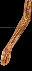
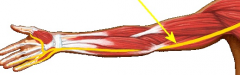
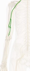
Ulnar Nerve |
|
|
Radial Nerve |
|
|
Musculocutaneous Nerve |
|
|
Dorsal Scapular Nerve: runs posteriorly to the rhomboids and levator scapulae |
|
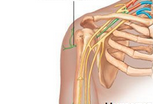
Name the nerve |
Suprascapular nerve: runs posteriorly towards the scapula. It runs through the suprascapular notch to reach the supraspinatus, then through the spine of the scapula to reach the infraspinatus |
|
|
Lateral Pectoral Nerve: Approaches pectoralis major. |
|
|
Medial brachial and medial antebrachial cutaneous nerve |
Branch distally to the medial pectoral nerve of the medial cord. They run to the skin of the medial arm and forearm. It can be difficult to tell them apart. |
|
|
Medial Pectoral Nerve |
The nerve is usually the first branch from the medial cord and it runs through the pectoralis minor. innervating it on its path to the pectoralis major. |
|
|
Long Thoracic Nerve - runs tight to the thoracic wall and innervates the serratus anterior |
|
|
Thoracodorsal Nerve |
This nerve runs just lateral to the long thoracic nerve on it's way to the latissimus dorsi muscle. |
|
Name the three branches |
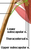
Subscapular: Subscapularis and teres major |
|
|
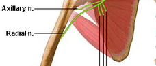
Axillary Nerve: This nerve branches from the posterior cord and runs laterally toward the deltoid. |
|
|
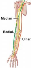
Radial: Runs on the posterior side of the arm Median: Middle nerve formed in the "M" Ulnar: Most medial branch, continue along the medial side of the arm and passes around the medial epicodyle of the humerus. |
|
|
Musculocutaneous Nerve: The musculocutaneous nerve supplies the coracobrachialis muscle in the arm and then runs between the biceps brachii and the brachialis muscles. |