Reading...
![]()
Play button
![]()
Play button
![]()
Use LEFT and RIGHT arrow keys to navigate between flashcards;
Use UP and DOWN arrow keys to flip the card;
H to show hint;
A reads text to speech;
24 Cards in this Set
- Front
- Back
+ Femur Text
|
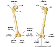
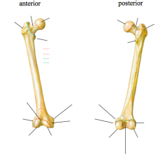
Femur
1. head 2. neck a. clinical • site for fracture…traumatic or pathological 3. greater trochanter 4. lesser trochanter 5. adductor tubercle 6. medial/lateral condyles 7. medial/lateral epicondyles 8. patellar surface 9. intercondylar fossa (notch) 10. popliteal fossa |
|
+ Tibia and Fibula Text
|
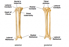
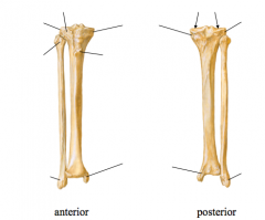
Tibia
1. medial/lateral condyles 2. tibial tuberosity 3. medial/lateral plateaus 4. medial malleolus Fibula 1. head of fibula 2. lateral malleolus |
|
+ Foot Text
|
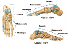
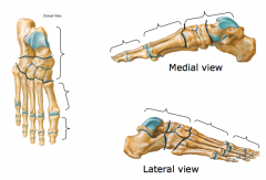
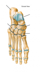
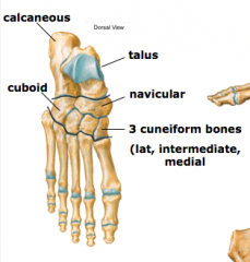
Foot
1. tarsals - calcaneous, talus, cuboid, cuneiforms (3), navicular 2. metatarsals 3. phalanges – 2 for great toe; 3 for other four toes |
|
|
|
|
+ Hip Joint Text
|
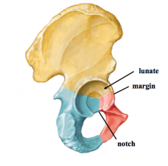
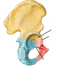
Hip joint
A. Femoral head articulates with acetabulum 1. acetabulum – margin, lunate, acetabular notch 2. acetabular labrum 3. head of femur – fovea capitis 4. clinical anatomy a. THR (total hip replacement) b. Childhood conditions • Legg Calve Perthes • Transient synovitis • SCFE (slipped capital femoral epiphysis) c. Dislocation of hip • Trauma – posterior dislocation is MC….often d/t MVA’s • Children – CHD (congenital hip dislocation)….newborn |
|
|
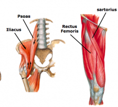
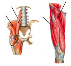
hip flexion
a. iliopsoas b. rectus femoris c. sartorius |
|
|
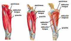
hip adduction
a. pectineus b. adductor longus c. adductor brevis d. adductor magnus e. gracilis |
|
|
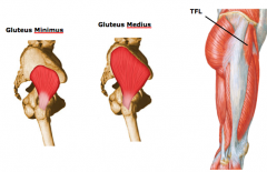
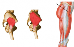
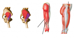
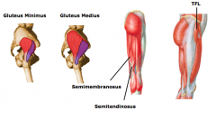
hip abduction
a. gluteus minimus b. gluteus medius c. TFL (tensor fascia lata) |
|
|
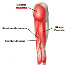
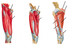
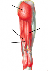
hip extension
a. gluteus maximus b. biceps femoris (long head) c. medial hamstrings • semimembranosus • semitendinosus |
|
|
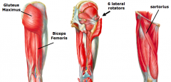
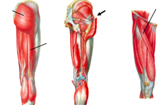
external rotation of hip
a. “six lateral rotators” or short external rotators • piriformis • superior gemellus • obturator internus • inferior gemellus • quadratus femoris • obturator externus b. gluteus maximus c. biceps femoris d. sartorius |
|
|
internal rotation of hip
a. gluteus minimus b. gluteus medius c. TFL d. medial hamstrings • semimembranosus • semitendinosus |
|
|
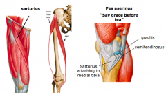
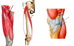
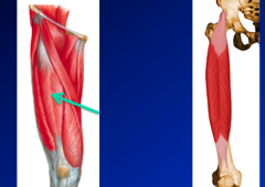
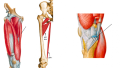
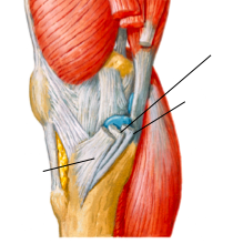
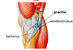
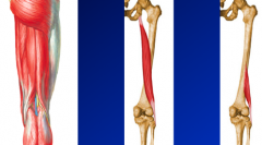
Sartorius
P = ASIS and superior part of notch inferior to it D = superior part of medial surface of tibia N = Femoral N (L2 and L3) A = Flexes, abducts, and laterally (externally) rotates thigh at hip; flexes leg at knee joint, |
|
|
Rectus Femoris
P = AIIS and ilium superior to acetabulum D = Base of patella and by ligament to tibial tuberosity N = Femoral (L2, L3, and L4) A = extends knee joint; also slightly flexes the hip |
|
|
Vastus Lateralis
P = Greater trochanter and latral lip of linea aspera of femur D = Base of patella and by ligament to tibial tuberosity N = Femoral (L2, L3, and L4) A = extends knee joint Vastus Medialis P = Intertrochanteric line and medial lip of linea aspera of femur D = Base of patella and by ligament to tibial tuberosity N = Femoral (L2, L3, and L4) A = extends knee joint Obliquus (VMO) distal fibers which primarily help to prevent patella to track laterally Vastus Intermedius P = Anterior and lateral surfaces of body of femur D = Base of patella and by ligament to tibial tuberosity N = Femoral (L2, L3, and L4) A = extends knee joint |
|
|
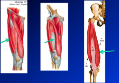
Adductor Longus
P = body of pubis inferior to public crest D = middle 1/3 of linear aspera of femur N = obturator N, branch of ant division (L2, L3, and L4) A = adducts thigh |
|
|
Adductor Brevis
P = body and inferior ramus of pubis D = pectineal line and proximal part of linea aspera of femur N = Obturator N (L2, L3, and L4) branch of ant division A = adducts thigh and some flexion as well |
|
|
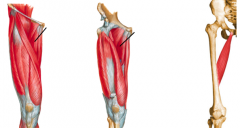
Gracilis
P = body of inferior ramus of pubis D = superior part of medial surface of tibia N = Obturator N (L2 and L3) A = adducts thigh, flexes leg, and helps to rotate leg medially |
|
|
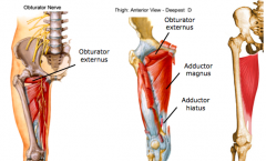
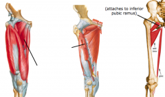
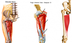
Adductor Magnus
P = adductor part: inferior ramus of pubis, ramus of ischium; hamstring part: ischial tuberosity D = adductor part: gluteal tuberosity, linea aspera, medial supracondylar line; Hamstrings part;adductor tubercle of femur N = adductor part: obturator N (L2, L3, and L4), branches of posterior division ; Hamstrings part: tibial part of sciatic nerve (L4) A = adducts thigh; adductor part: flexes thigh, Hamstrings part extends thigh |
|
|
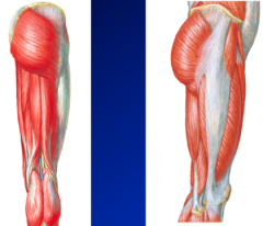
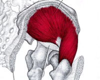
Gluteus Maximus (p551)
P = illium posterior to posterior gluteal line, dorsal surface of sacrum and coccyx, and sacrotuberous ligament D = Most fibers end in iliotibial tract that inserts into lateral condyle of tibia; some fibers insert on gluteal tuberosity of femur N = Inferior gluteal nerve (L5, S1, S2) A = Extends thigh (especially from flexed position) and assists in its lateral rotation; steadies thigh and assists in rising from a sitting position (also helps to stabilize knee due to its attachment onto the iliotibial tract which crosses the knee joint) |
|
|
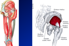
Gluteus Medius (p551)
P = External surface of ilium between anterior and posterior gluteal lines D = Lateral surface of greater trochanter of femur N = Superior gluteal nerve (L5, S1) A = Abducts and medially rotates thigh; keeps pelvis level when opposite leg is raised |
|
|
Gluteus Minimus (p551)
P = External surface of ilium between anterior surface of ilium between anterior and inferior gluteal lines D = Anterior surface of greater trochanter of femur N = Superior gluteal nerve (L5, S1) A = Abducts and medially rotates thigh; keeps pelvis level when opposite leg is raised |
|
|
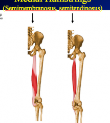
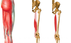
Semitendinosus
P = Ischial tuberosity D = Medial surface of superior part of tibia N = Tibial division of sciatic N (L5, S1, S2) A = Extend thigh; flex leg and rotate it medially with knee flexed Semimembranosus P = Ischial tuberosity D = Posterior part of medial tibial condyle; reflective attachment forms oblique popliteal ligament (to lateral femoral condyle) N = Tibial division of sciatic N (L5, S1, S2) A = Extend thigh; flex leg and rotate it medially with knee flexed |
|
|
|
|
|
Biceps Femoris (long head)
P = Ischial tuberosity D = lateral side of head of fibula; tendon is split by fibular collateral knee ligament N = tibial division of sciatic N (L5, S1, S2) A = Flexes leg and rotates it laterally when knee flexed; extends thigh at hip joint Biceps Femoris (short head) P = Linear aspera and lateral supracondylar line of femur D = lateral side of head of fibula; tendon is split by fibular collateral knee ligament N = Common fibular (peroneal) division of sciatic N (L5, S1, S2) A = Flexes leg and rotates it laterally (IT DOES NOT HAVE A DIRECT EFFECT ON THE HIP) |