![]()
![]()
![]()
Use LEFT and RIGHT arrow keys to navigate between flashcards;
Use UP and DOWN arrow keys to flip the card;
H to show hint;
A reads text to speech;
19 Cards in this Set
- Front
- Back
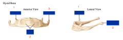
Parts of the Hyoid Bone :
-Body |
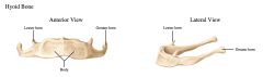
1. Lesser Horn 2. Body 3. Greater Horn |
|
|
Nuchal Ligament |
a ligament at the back of the neck that is continuous with the supraspinous ligament |
|
|
Supraspinous ligament |
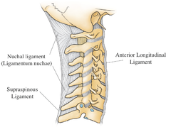
a ligament found along the vertebral column |
|
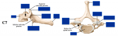
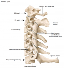
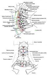
C7 Vertebra
-Transverse process -Transverse foramen -Articular facets -Body -Pedicles -Lamina -Spinous process |
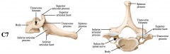
The C7 vertebra has the longest spinous process and is the first spine that is palpable (called vertebra prominent for this reason).
The spinous process of C7 is not bifid as are those of C2 through C6. |
|
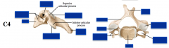
C4 Vertebra
-Transverse process -Transverse foramen -Articular facets -Body -Pedicles -Lamina -Spinous process |
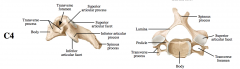
The spinous processes cervical vertebrae C2 through C7 are not palpable due to the presence of the nuchal ligament in the midline extending posteriorly from the cervical spines. |
|
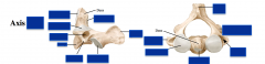
Axis : is it C1 or C2?
-Transverse process -Transverse foramen -Articular facets -Body -Pedicles -Lamina -Spinous process |
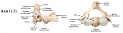
The axis has a dens (odontoid process) projecting superiorly from its body.
|
|
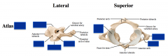
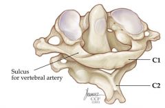
Atlas : Is it C1 or C2?
-Transverse process -Transverse foramen -Articular facets -Body -Pedicles -Lamina -Spinous process |
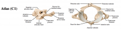
The atlas has no body or vertebral spine
The joint between the atlas and the skull, the atlanto-occipital joint, has articular surfaces that are concave and convex, respectively.
The primary movements at this joint are flexion and extension (as in nodding “yes”). |
|
|
Atlanto-axial joint :
-sites of articulation |
The joint between the atlas and the axis
Three sites of articulation:
1. The midline articulation between the anterior arch of the atlas and the dens of the axis allows for rotary movement (as in shaking your head “no”).
2. The two lateral parts of the atlanto-axial joint between the inferior and superior facets allow for flexion, extension and lateral bending, as do the remainder of the joints between the other cervical vertebrae. |
|
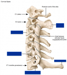
Cervical Spine :
-vertebral body -spinous process -transverse process -transverse foramina
|
-vertebral body -spinous process -transverse process -transverse foramina |
|
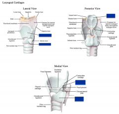
Laryngeal Cartilages :
-thyroid -cricoid -epiglottis -arytenoid |
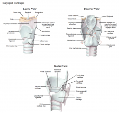
-thyroid
-cricoid
-epiglottis - The epiglottis is a flap of tissue that sits at the base of the tongue that keeps food from going into the trachea (windpipe) during swallowing. It is made of elastic cartilage tissue covered with a mucous membrane, attached to the entrance of the larynx. There are taste buds on the epiglottis.
-arytenoid - a pair of small three-sided pyramids which form part of the larynx, to which the vocal folds (vocal cords) are attached. These allow and aid in the vocal cords' movement. |
|
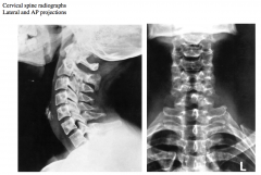
Note that while all cervical vertebrae are seen in the lateral projection, the atlas (C1) and axis (C2) are not seen in the AP projection. Why is this?
Identify the intervertebral discs and number the vertebral bodies.
In the lateral projection:
Draw a line indicating the approximate location of the skin on the anterior neck.
joints between the atlas and the axis?
What parts of the atlas are seen superior to the body and spinous process of the axis? |
The atlas and axis are not seen in the AP projection because of superimposition of the mandible.
|
|
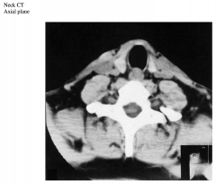
This CT is at the level of the T1 vertebra and includes the inferior parts of the lobes of the thyroid gland.
Identify the airway and the structure posterior to it.
Which vessels can be seen postero-lateral to the thyroid gland?
How do you know?
Which is medial, which is lateral?
Which superficial vein is seen?
What is its relationship to the muscles of the neck at this vertebral level?
Draw a line indicating the retropharyngeal space. |
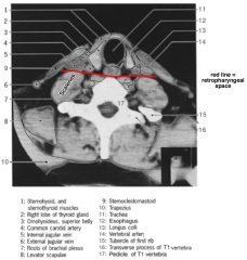
The inferior parts of the lobes of the thyroid gland are lateral to the trachea. Remember that the isthmus of the thyroid gland is usually anterior to the second or third tracheal rings and this section is inferior to the isthmus.
The structure posterior to the trachea is the esophagus (the laryngopharynx is posterior to the larynx).
The sternohyoid and sternothyroid muscles are anterior to the trachea.
The superior belly of the omohyoid can also be seen lateral to the thyroid gland.
The common carotid artery and internal jugular vein can be seen posterior and lateral to the thyroid gland. The bifurcation of the common carotid artery usually occurs between the thyroid cartilage and hyoid bone. The common carotid artery is medial to the internal jugular vein.
The external jugular vein is seen posterior to the sternocleidomastoid and anterior to the trapezius. The external jugular vein is anterior to the sternocleidomastoid more superiorly in the neck. |
|
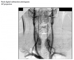
Identify the subclavian, common carotid and vertebral arteries.
What clues did you use to distinguish the common carotid from the vertebral arteries?
What other branch of the subclavian artery supplies the neck?
Does this branch come off the subclavian artery proximal or distal to the vertebral artery (this is not well seen in this arteriogram)?
Identify and name the one branch supplying the neck. |
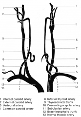
The vertebral arteries can be distinguished from the common carotid arteries based on their relationship to the vertebrae.
Note the location of the vertebral within the transverse foramina of the cervical vertebrae (they usually enter at C6, not C7). Furthermore, the common carotid artery can be seen bifurcating into the external and internal carotid arteries.
The vertebral arteries have no major branches in the neck.
The thyrocervical branch of the subclavian artery arises distal to the vertebral artery. The inferior thyroid branch of the thyrocervical artery also supplies the neck. |
|
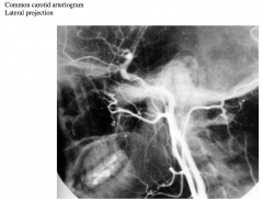
Distinguish the external and internal carotid arteries.
What clue did you use to distinguish one from the other?
Some of the branches of the external carotid artery can be seen.
The occipital, facial and lingual arteries are distinct, however, the distal part of the facial artery is not seen. |
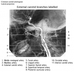
The external carotid artery can be distinguished from the internal carotid artery because the internal carotid artery has no branches in the neck. |
|
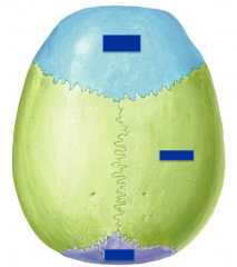
Superior surface of the cranium:
-Frontal Bone -Parietal Bone -Occipital Bone
What are the names of the sutures between these bones? |
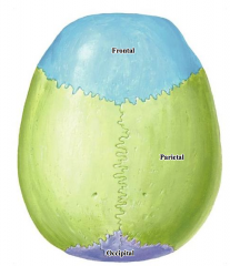
Sagittal suture – between the parietal bones
Coronal suture – between the frontal and parietal bones
|
|
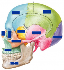
Lateral surface of the cranium:
-Frontal Bone -Parietal Bone -Occipital Bone -Temporal Bone -Sphenoid Bone -Zygomatic Bone (with zygomatic arch) -Lacriminal Bone -Nasal Bone -Maxilla Bone -Mandible Bone
What bones meet at an area called the pterion?
What is the external acoustic meatus? This meatus is in what bone?
|
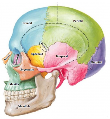
External Acoustic Meatus = opening of the ear. It is within the temporal bone.
Zygomatic Arch - formed by a portion of both the temporal and zygomatic bones
Pterion = joining of the frontal, parietal, temporal and sphenoid bones. |
|
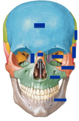
Facial surface of the cranium:
-Frontal Bone -Zygomatic Bone -Nasal Bone -Maxillary Bone -Mandible Bone -Sphenoid Bone -Temporal Bone -Parietal Bone -Lacriminal Bone
of the orbit, in addition to the sphenoid, ethmoid and lacrimal bones (a tiny contribution of the palatine bone is not seen in this model).
-What bones make up the rim of the orbit?
Note the opening for the nose.
-Which bones form the piriform aperture (anterior nasal opening)?
-What bones make up the cheek bones? |
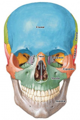
Rim of the orbit = formed by the frontal, zygomatic and maxillary bones
Piriform Aperture = the anterior nasal opening. It is formed by the maxillary and nasal bones
The cheekbones are composed of maxillary and zygomatic bone. |
|
|
Inferior surface of the cranium:
-Temporal Bone -Sphenoid Bone -Palatine Bone -Vomer Bone -Maxillary Bone
What are the surfaces that articulate with the atlas?
Examine the temporal bone and identify the mastoid process, mastoid notch and styloid process. Which of these is most superficial, which is most deep?
How does this explain the relationship of the muscles in the posterior part of the submandibular triangle of the neck? |
Foramen Magnum is in the occipital bone
Occipital Condyles articulate with the atlas
Mastoid process - lateral to the mastoid notch and the
Styloid process - anterior and medial to them
This explains why the sternocleidomastoid is the most superficial muscle, why the posterior belly of the digastric is immediately deep to it and why the stylohyoid is the most medial of the muscles with a more anterior attachment. |
|
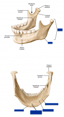
Mandible:
-Body -Ramus -Angle -Mylohyoid line -Submandibular fossa -Digastric fossa -Inferior mental spine. |
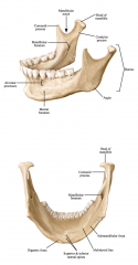
See Picture |